Major Infectious Diseases
Porcine Circovirus Associated Diseases (PCVAD)
Clinical importance
Porcine circovirus is an ubiquitous virus, present in swine-raising countries over the world. There are 2 types of virus: Porcine circovirus type 1 is non-pathogenic and has been identified as a cell culture contaminant whereas Porcine Circovirus type 2 (PCV2) is the one affecting pigs worldwide. Within type 2, there are 5 identified subtypes: a, b, c, d, and e. PCV2a and PCV2b are responsible for most of the associated clinical signs seen in the US. There are 3 main Porcine Circovirus Associated Diseases: Post-Weaning Multisystemic Wasting Syndrome (PMWS), the Porcine Dermatitis and Nephropathy Syndrome (PDNS) and a reproductive form. The virus can affect sows as well as nursery and growing pigs. The clinical importance of PCV2 has decreased with the introduction of effective vaccination protocols.
Etiology and Transmission
PCV2 is a non-enveloped virus with circular DNA. There are multiple ways by which a pig can get infected by PCV2. Piglets can get infected by the sow in utero since PCV2 can cross the placenta. Piglets can also ingest viruses through milk and colostrum. Boars tend to shed the virus in their semen. However, the most common route of transmission is oronasal as pigs shed the virus in their saliva and nasal secretions. Indeed, pigs can carry the virus for weeks after infection. Due to its non-enveloped nature, PCV2 is quite resistant in the environment and pigs can get infected via fomites.
However, it is important to differentiate infection from disease! Clinical signs do not necessarily develop after infection. PCVAD are multifactorial diseases, triggered by stress conditions: high pig density, poor ventilation, weaning stress, and co-infections with other pathogens are all risk factors.
PCV2 replicates in lymphocytes and is spread around the body through macrophages. It is a major cause of immunosuppression in swine and therefore, a great primary pathogen for opportunistic and secondary infections.
Associated symptoms
Many PCV2 infections are subclinical or may contribute to decreased growth rate without any other clinical signs.
Post-weaning Multisystemic Wasting Syndrome
The common age for developing PMWS is nursery to early growing phases, time of new environmental stresses combined with declining maternal antibodies. As the name suggests, PMWS characteristic symptom is wasting and failure to thrive. Pigs affected by this disease have growth retardation and lose so much weight that the spine becomes marked. Other symptoms include dyspnea, pale skin, with rough haircoat. In naïve herds, mortality can rise as high as 20%.
Porcine Dermatitis and Nephropathy Syndrome
Pigs in nursery and growing-finishing phases can be affected by PDNS but the prevalence is usually low, around 1%. Pigs are lethargic and anorexic. Their skin display red to purple, circular, crusty, multifocal lesions on the hind legs, progressing towards the abdomen. In the most severe cases, mortality can follow.
Reproductive symptoms
PCV2 can cause late-term abortions, still-births and weak born-alive piglets in sows, especially if they are naïve against the virus.
Infection by PCV2 always leads to the development of Porcine Circovirus Associated Diseases (PCVAD). True or False?
- True
- False
Associated lesions
Macroscopic lesions
PMWS
The most typical lesions associated with PMWS are extremely enlarged lymph nodes. The lungs will be rubbery, with intense interlobular edema. Liver can be enlarged as well. The kidneys may show white spots caused by interstitial nephritis. A side effect of the wasting and decreased feed intake is gastric ulceration.
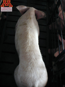
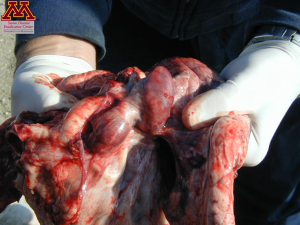
Emaciated pig due to PMWS (left) and severely enlarged mediastinal lymph nodes (right) | Source: Dr. Carlos Pijoan
PDNS
The characteristic lesion of PDNS is necrotizing skin, usually red to purple in color. The lesions can be raised and vary widely in size. As the disease progresses the lesions will be covered by a crust and eventually fade. In dead pigs, kidneys are enlarged displaying petechiae and/or multifocal areas of necrosis (white spots) on their surface.
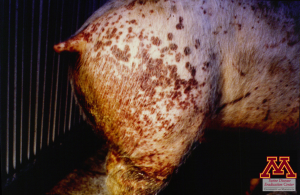
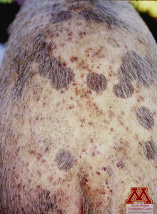
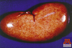
Skin lesions (left and middle) and kidney lesions due to PDNS | Source: Dr. Carlos Pijoan
Reproductive disease
Abortions and still-birth are seen in this presentation of the disease. At necropsy, the piglets show enlarged heart and liver.
Microscopic lesions
PMWS
In the affected lymph nodes, multinucleated, non-functional, gigantic cells associated with depletion of lymphocytes B and T are common. The virus can be found in inclusion bodies in the cytoplasm of lymphocytes. The lungs show signs of interstitial pneumonia.
PDNS
Upon microscopic evaluation, the skin lesions have been determined to be caused by necrotizing vasculitis. In the kidneys, fibrino necrotizing glomerulitis can be seen.
Reproductive disease
PCV2 tends to multiply in high-replication rate cells. In fetuses, this means that the virus has a tropism towards the heart which shows signs of necrosis in the myocardium.
What is the most common macroscopic lesion associated with PDNS?
- Rubbery edematous lungs
- Circular crusty skin lesions
- Button-ulcers in the spiral colon
Diagnosis
Serology tests are available but due to the widespread vaccination against PCV2, the results are difficult to interpret. PCR testing of oral fluids, serum samples gives a good indication of the status of the farm but infection is not equal to disease in the case of PCV2!
PMWS
Infection by PCV2 does not necessarily mean that the disease is happening. Remember that it is multifactorial. Therefore, 3 components need to be present to establish a PMWS diagnosis.
- Clinical signs have been observed: wasting, pale pigs
- Histopathological lesions are found in lymphoid tissues
- PCV2 has been detected in the lesions of the tissues
The samples to submit for a PMWS diagnosis are lymph nodes, lung, liver, kidney and spleen tissues both fresh and fixed. The tissues can then be tested by PCR for a quick response or through ImmunoHistoChemistry or In Situ Hybridization to ensure the presence of the virus within the lesions.
PDNS
PDNS diagnosis is made by clinical observation associated with histological lesions of vasculitis in the skin and nephritis in the kidney. The best samples to submit are tissues.
Reproductive disease
Diagnosis of reproductive disease associated with PCV2 is based on clinical signs, myocarditis lesions in the aborted fetuses with the presence of the virus in the lesions.
Differential diagnosis
Differential diagnosis for PCVAD includes PRRS for the reproductive form, the wasting pigs and the dyspnea, influenza for the respiratory form, erysipelas for the reproductive form and the skin lesions, African swine fever and classical swine fever due to the similar lesions on the kidneys.
What parameters do you need to establish a diagnosis of Post-weaning Multisystemic Wasting Syndrome?
- Clinical signs, histology lesions in the lymph nodes, presence of PCV2 within the lesions
- Histology lesions in the lymph nodes, positive PCR testing on serum
- Clinical signs, and positive serology on serum
Treatment, Prevention and Control
There is no treatment available against PCV2. Control measures include commercial vaccines which have been proven to be very effective. Currently, in the US, the vast majority (>90%) of the piglets are vaccinated against PCV2 before weaning. However, PCVAD are multifactorial and improved management on the farm can make a big difference. Some measures that can be taken are all-in, all-out management, single-source of the nursery, increased age at weaning, lower pig density, good ventilation, and control of co-infections, especially PRRS. As usual, thorough cleaning and disinfection of the facilities is important.
