Enteric System: Pre-Weaning Pigs
Coccidiosis
Clinical importance
Coccidiosis refers to the disease caused by any type of coccidia infection. In swine, most of the cases are caused by Isospora suis. Coccidiosis is ubiquitous and causes diarrhea in pre-weaning pigs. The impact of the disease is mainly related to the slow growth of the infected piglets and poor quality of the piglets at weaning. Mortality is rare.
Etiology and transmission
Isospora suis is an intracellular parasite, specific to pigs. Its life cycle lasts roughly 7 days, 2 of which are in the environment. Pigs get infected by ingesting the oocyst form of the parasite which then colonizes enterocytes in the small intestine (jejunum and ileum) to continue its development and ultimately reproduce by creating new oocysts. Oocysts are shed in the feces and sporulate into the highly resistant form in 1 to 2 days. This form survives for a long time in the environment, contaminating farrowing crates, mats, and ultimately, the udder of the sow by contact. Therefore, the main route of transmission is indirect infection through the environment.

Associated symptoms
Signs of this disease typically present in pigs between 7 and 11 days of age. Remember that the full cycle takes 7 days, 5 of which happens in the intestine: Day-1 diarrhea cannot be caused by coccidia.
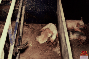
Piglets showing clinical signs of coccidiosis | Source: Dr. Carlos Pijoan
Diarrhea is the main symptom of coccidiosis and has often a yellowish, pasty appearance. Piglets may also appear dehydrated, grow more slowly and develop a rough hair coat. Mortality is rare and due to complications, usually secondary bacterial infections. Coccidiosis is not as common in nursery-age pigs. A few individuals may start shedding due to the stress of weaning and contaminate the group. Clinical signs are less severe.Adult animals do not present any clinical signs and rarely shed.
Associated lesions
Macroscopic lesions
The main macroscopic lesion seen is inflammation of the small intestine that becomes more intense with the level of infection. There are rarely any other gross lesions, even in very severe cases.
Microscopic lesions
The villi of intestinal epithelial cells are commonly damaged by coccidiosis. This is the cause of diarrhea, as the cells can no longer absorb fluids properly.
Diagnosis
The quickest and easiest way to diagnose coccidiosis is to observe the oocysts in fecal samples examined either by fecal flotation or fecal smear. Pigs that have been showing clinical signs for 2 to 3 days are the most likely to be shedding and constitute the best sample. If collecting fecal samples, focus on the solid part of the stool which contains a higher concentration of oocysts than the liquid one. When viewed under a microscope I. suis oocysts have a characteristic feature known as “hazy bodies” between the sporont and the oocyst wall (Insert picture?)
Treatment, Prevention and Control
Anticoccidial treatments are widely used in commercial herds, molecules like toltrazuril and ponazuril are very effective but are used extra-label. Every piglet in the litter receives an oral dose at processing to prevent clinical signs and to decrease the environmental pressure by limiting the shedding. Colostrum does not provide adequate protection against the parasite. Thorough cleaning and disinfection of the farrowing rooms between groups is critical but very challenging as the oocysts are hardy and survive several months in the environment. Some reports suggest steam cleaning to be more effective to eliminate the oocysts.
