Respiratory System
Enzootic pneumonia
Clinical importance
Mycoplasma hyopneumoniae initiated pneumonia is a concern to the swine industry world-wide. First isolated in 1965, M.hyopneumoniae continues to gain recognition as a primary cause of respiratory disease. The bacteria also contributes to secondary infections of the upper respiratory system by suppressing immune response and destroying the mucociliary apparatus responsible for preventing infection from other bacteria. The main concerns from this disease are reduced appetite, and the ability to amplify other pathogens. Pigs get infected early in life but Mycoplasma colonization is slow, therefore they only show clinical signs much later, in the growing-finishing phase. M.hyopneumoniae is ubiquitous around the world and very common on commercial operations in the United States.
Etiology and transmission
Mycoplasmas are very small bacteria that are lacking a cell wall. M.hyopneumoniae typically spreads via the oral-nasal route, but it has also been demonstrated to transmit via aerosols. Sows, especially young parity ones, shed bacteria in their nasal secretions and infect piglets of their litter which in turn transmit the bacteria to their penmates. Infected animals can carry the bacteria for up to 254 days, which is almost the lifetime of a market hog.
Mycoplasma colonization begins by binding to the cilia of the epithelium in the respiratory airways. M.hyopneumoniae presence leads to the loss of the cilia, inhibiting the mucociliary apparatus responsible for clearing bacteria, dust and other debris from the respiratory system. Opportunistic bacteria such as H. parasuis, S. suis, P. multocida, and A. pleuropneumoniae, which are commensal of the upper respiratory system, are then able to reach the alveoli and create lesions. Their journey is made easier by M.hyopneumoniae suppressing macrophages, the first line of defense of the immune system. Once there, the various bacteria cause the infection known as enzootic pneumonia.
Enzootic pneumonia is the term for this process of coinfection with M.hyopneumoniae as primary pathogen and opportunistic/secondary bacteria as secondary pathogens.
Associated symptoms
Not all M.hyopneumoniae infections result in pneumonia. Development of symptoms is often dependent on the secondary bacterial pathogens. There are two forms of the disease: epidemic and more commonly endemic. The endemic form of the disease manifests as a persistent, dry cough that can last as little as two weeks or as long as the entire growing period. This cough is most noticeable when pigs are roused from a lying position. Fever, decreased appetite, and dyspnea can be seen in more severe cases and are due to the secondary pathogens. Because this disease makes the pigs less active and willing to eat, mycoplasmal pneumonia has also been associated with reduced daily weight gain, and decreased efficiency. Mortality due to enzootic pneumonia is usually low unless the affected herd was naive.
Enzootic pneumonia is challenging to control because pigs get infected in their first weeks of life but express clinical signs in finishing stage. True or False?
- True
- False
Associated lesions
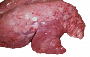
Macroscopic lesions
The most common macroscopic lesions seen in mycoplasmal pneumonia is consolidation of the cranioventral lobes of the lungs. The lobes are firm, red-purple in color, edematous and the delimitation between healthy and affected tissue is clear. In complicated enzootic pneumonia cases, the alveoli can be swollen, and mucus can block portions of the airways.
Microscopic lesions
Cell congestion around the airways is often seen with enzootic pneumonia. These cuffs are typically made up of lymphocytes. The alveoli and airways are filled with fluids and macrophages.
Clinical signs and lesions associated with enzootic pneumonia are very similar to the ones caused by another respiratory pathogen. Which one?
- Actinobacillus pleuropneumoniae
- Mycoplasma hyorhinis
- Swine influenza A
Diagnosis
In acute, severe cases, when pigs are coughing a lot, diagnosis of Mycoplasma hyopneumoniae can be done ante-mortem by collecting oral fluids and test the samples via PCR. Culture is almost never done because the process is fastidious and M.hyopneumoniae is prone to be overgrown by other mycoplasma.
In chronic, endemic cases, which represent most cases, diagnosis can be done by taking oro-pharyngeal swabs (i.e. by swabbing the soft palate and the tonsils) and testing it by PCR as well. A bronchoalveolar lavage has good sensitivity as well but it is more difficult to do antemortem. Lung tissue samples including airway sections are appropriate to submit as long as the pig was not treated with antibiotics.
Very recently, gene sequencing has been developed to identify various strains of M.hyopneumoniae and differentiate between re-infection and introduction of a new strain.
Serology tests can be used by there is a concern with the interpretation of the tests. First, the majority of piglets are vaccinated within their first month of life and the tests tend to have low sensitivity and specificity.
Differential Diagnostic
When facing a case with dry hacking cough, swine influenza, Bordetella bronchiseptica (and Ascaris suum in outdoor pigs) must also be considered.
What is the best antemortem test to diagnose endemic enzootic pneumonia?
- Culture of nasal swabs
- PCR on laryngeal swabs
- Serology on serum
Treatment, Prevention and Control
Group antibiotic treatment through the water, when the disease first enters the herd is the preferred method. Individual pigs showing more severe signs can be treated by intramuscular injections to help control secondary bacteria. Antibiotics may not, however, heal lesions or totally eliminate the organism from the respiratory tract. Also important to note, M.hyopneumoniae does not have a cell wall, so beta-lactams such as penicillin, amoxicillin or cephalosporins are not effective. Tetracyclines, quinolones and macrolides are usually effective. Whole cell vaccinations are effective and widely used in weaning pigs. Providing a clean environment with good ventilation and appropriate pig density can ensure that air quality is not an additional risk factor for pneumonia.
Eradication protocols have been successful in create negative herds. Usually the technique called herd closure is done It consists in stopping the introduction of new animals (gilts) in the sow herd and treating all of the sows against Mycoplasma hyopneumoniae to eliminate it from the environment and the pigs as much as possible. When all of the sows have stopped shedding (at least 254 days later), the herd is monitored to see if the weaned piglets are negative. If so, then new naive gilts are introduced. The technique has been effective, showing great results in finishing pigs which became more resistant to respiratory infections.
National Animal Health Monitoring Survey (NAHMS). 2000. www.aphis.usda.gov/animal_health/nahms/swine/index.shtml#swine2000
Why is penicillin not effective against enzootic pneumonia?
- M.hyopneumoniae has no cell wall
- M.hyopneumoniae has a resistance plasmid
- M.hyopneumoniae has no ribosomes
