Reportable and Transboundary Diseases
Classical Swine Fever
Clinical Importance
Also called hog cholera, Classical Swine Fever is a disease in the reportable list of the OIE (World Organization for Animal Health). Any country infected by the virus is banned from export of any pork product, leading to disastrous consequences on the swine industry economy. For example, pork exports represent $4.6 billion in revenues, driving economic sustainability for hog farmers. Contrary to ASF, classical swine fever is highly contagious and spread quickly. Domestic pigs and wild boars are equally affected by the disease
Etiology and Transmission
Classical Swine Fever virus belongs to the Pestivirus genus along with Bovine Viral Diarrhea (BVD) virus. It is an enveloped, RNA virus therefore prone to mutations. 3 genotypes have been identified. Genotype 1 has been isolated in South America and Russia, genotype 2 in Europe and genotype 3 in Asia. The main path of transmission is oro-nasal, directly from secretions or blood of contaminated pigs, indirectly through fomites or ingestion of infected tissues. The virus then replicates in the tonsils and spreads to peripheral lymph nodes through the blood. Classical Swine Fever virus is immunosuppressive as it replicates in monocyte-macrophages cells and vascular endothelial cells, causing hemorrhages. The virus can cross the placenta and infect the fetuses, leading to abortion or to viremic piglets at birth.
Associated symptoms
Depending on the strain of the virus, clinical signs can be moderate to severe.
In both the acute and chronic forms, pigs are apathetic, anorexic, and show signs of dyspnea and constipation followed by diarrhea. The difference is that in the milder form, pigs die in 2 to 3 months instead of days. The clinical signs are non-specific making the diagnosis of Classical Swine Fever challenging.
Associated lesions
Macroscopic lesions
In the more acute form, the carcass is hemorrhagic with petechiae on the skin, kidneys and lymph nodes. Infarcts on an enlarged spleen are a strong indicator but this lesion is not consistent. The ulceration of the tonsils can also be a telling sign if present. Do not forget to check them.
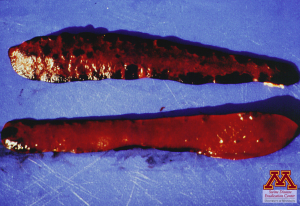
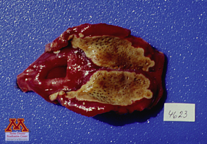
Spleen infarcts (left) and areas of necrosis on tonsils (right) due to infection by Classical Swine Fever | Source: Dr. Carlos Pijoan
Microscopic lesions
Histopathology shows depletion in lymphoid tissues. Microthrombosis, hemorrhages and destruction of vascular endothelial cells can be seen in hemorrhagic organs like the kidneys.
Diagnosis
The best samples to submit are serum, and tissues such as tonsils, spleen, lymph nodes and kidneys. Those samples can be used for virus isolation as well as for PCR testing.
Differential diagnosis
Clinical signs are non-specific which makes the differential list extensive. It should include: African Swine Fever, PRRS, Salmonella choleraesuis, erysipelas and PDNS.
Treatment, Control, and Prevention
Classical Swine Fever is endemic in several parts of the world such as Russia or Africa. There is no treatment against Classical Swine Fever and so the best defense is to prevent its introduction in the farm. Tight biosecurity measures should be put in place. Vaccines are available but trade agreements require that CSF-free countries not to use them. Should an outbreak happen in a naive country, the infected herds and the neighboring farms would be culled to stop the spread of the infection. Wild boars are commonly infected in Europe and represent a risk of introduction.
