Enteric System: Pre-Weaning Pigs
Clostridia: Clostridium perfringens and Clostridioides difficile
Until recently (2020), Clostridioides difficile was named
Clostridium difficile. The resources are in the process of
being updated. Thank you for your patience.
Clinical importance
Two main species of Clostridia can affect pigs: Clostridium perfringens and Clostridioides difficile. They both affect piglets before weaning causing enteric issues with various degrees of mortality. Within the Clostridium perfringens species, 2 types routinely cause disease in pigs, Clostridium perfringens type A and Clostridium perfringens type C. Type C is the more severe of the two, while type A causes a similar, but milder, version of Type C disease. Clostridium perfringens is a worldwide disease that causes high mortality in young piglets. It is difficult to treat, but vaccinations can help prevent it in a herd.
Clostridioides difficile is extremely common cause of pre-weaning diarrhea US herds. They tend to proliferate after antimicrobial treatments when the intestinal flora has been eliminated. In pre-weaned piglets with enteritis in the United States around 66% have been demonstrated to have Clostridioides difficile present.
Etiology/Cause
Clostridia are either strict or aerobic-tolerant anaerobic gram-positive rods that can form highly-resistant spores.
Clostridium perfringens type C penetrates the absorptive cells of the small intestine or colonizes lesions induced by other pathogens such as coronaviruses for example. In adult pigs, Clostridium perfringens may be present in the intestine without causing any clinical signs. Therefore, piglets first become infected through the sow’s feces and then contaminate each other, through the feco-oral route. Clostridium perfringens competes with other intestinal bacteria, gets amplified in the intestine which leads to clinical signs. Both C.perfringens type C and type A can sporulate making them extremely resistant in the environment.
Clostridioides difficile is part of the normal flora of the swine colon. Piglets get contaminated by the sow’s feces. However, contrary to C. perfringens type C, the bacteria itself is not sufficient to declare clinical signs. They usually are a response to antimicrobial treatment, such as penicillin, disrupting the normal flora of the gut. Clostridioides difficile can also sporulate and be very resistant to changes in the environment.
How do piglets get infected with Clostridia?
- In the birth canal
- In utero, through the placenta
- By contact with the sow’s feces
Associated symptoms
Clostridium perfringens
Clostridium perfringens can begin to cause disease in piglets within the first 12 hours of life. Type A and type C cause different clinical presentations.
Clostridium perfringens type C
There are four forms of clinical presentation induced by Clostridium perfringens type C: peracute, acute, subacute, and chronic.
- Peracute form
The most characteristic clinical sign associated with the peracute form is hemorrhagic diarrhea staining the rear end of the animal followed by death within 12 to 36 hours of life. Piglets get apathetic, weak and may present a low body temperature around 95F. - Acute form
Clinical signs are very similar to the peracute form, with the main difference being that the piglets may survive for 1 to 2 days after the peak of clinical signs. Therefore, bloody diarrhea that may contain necrotic debris is present as well as the staining of the perineal area. Piglets are also too weak to nurse and suffer from dehydration. Mortality is still very high in this case. - Subacute form
Piglets originally appear healthy, but over a period of five to seven days they become weak, dehydrated and show signs of yellow to clear diarrhea with small pieces of necrotic tissue. Most piglets will not survive longer than one week. - Chronic form
In the chronic form, piglets go to alert and healthy looking to progressively emaciated and dehydrated in a matter of 5 to 6 days. Yellow to clear diarrhea similar to the subacute form can be noticed. Mortality is uncommon but piglets tend to be euthanized later on because they never catch up to their unaffected penmates.
Clostridium perfringens type A
Type A infection results in diarrhea in piglets less than 48 hours of life that lasts about five days. Piglets are a coarse haircoat and may show some stain of the perineal region. The diarrhea is brown and sticky looking. Piglets recover, but do not ever reach the same weight as unaffected pigs.
Clostridioides difficile
Symptoms in piglets usually occur during the first week of life. As the flora in the gut matures, Clostridioides difficile is easily replaced, so disease rarely occurs in pigs older than two weeks. Affected piglets display light to dark yellow diarrhea associated with mild mortality.
What pathogen is associated with clinical signs after an antibiotic treatment?
- Clostridium perfringens type C
- Clostridium perfringens type A
- Clostridium difficile
Associated lesions
Clostridium perfringens
Macroscopic lesions
Severly hemorrhagic jejunum and ileum is the most noticeable sign during the necropsy of piglets dead from the peracute form of the disease. Clostridia are anaerobic and produce gas when multiplying, therefore bubble can be seen in the intestinal wall.
In the acute form hemorrhagic intestines with a thickened intestinal wall are also seen. Urate crystals commonly accumulate in the kidneys.
In the subacute and chronic forms, adherences between sections of the intestinal tract can be seen. The intestinal wall is thickened, and a necroptic membrane is found in the lumen.
Piglets affected by Clostridium perfringens type A show thin-walled intestines without hemorrhagic content.
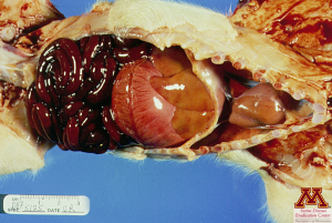
Hemorrhagic intestines due to Clostridium perfringens type C | Source: Dr. Carlos Pijoan
Microscopic lesions
The common microscopic lesion seen across almost all forms is necrosis of the jejunal villi. Inflammation may be seen in the submucosa and other deeper layers of the intestinal wall, in the most acute forms of the disease. Piglets affected with C.perfringens type A show little specific lesions: they may or may not show necrosis of the villi but can show heavy colonization of the small intestine.
Clostridioides difficile
Macroscopic lesions
Macroscopic lesions include edema of the mesocolon (which shows as a dilatation of the connective tissue and separation of each of the loops of the spiral colon) and urates in the kidneys. Contents in the colon will also be yellow and watery.
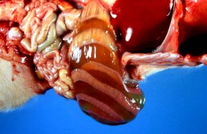
Mesocolon edema in a piglet | Source: Dr. Marie Culhane
Microscopic lesions
The colon shows typical lesions of scattered foci of suppuration associated with an increased number of neutrophils. Suppurative colitis looks like an eruptive volcano under the microscope and is specific to Clostridioides difficile. There also may be damaged villi in the small intestine.
Which pathogen causes hemorrhagic diarrhea followed by death in 12 to 36 hours?
- Clostridium perfringens type C
- Clostridium perfringens type A
- Clostridium difficile
Diagnosis
Clostridium perfringens
In the peracute and acute forms, clinical presentation and necropsy are usually sufficient to make a diagnosis. Other forms can also be presumptively diagnosis with typical clinical signs and lesions. A quick method to definitively diagnose a Clostridium perfringens infection is to examine a smear of the intestinal contents for gram-positive rods. Most commonly, an anaerobic culture is done to differentiate between type A and type C in the chronic form. PCR can be done to identify the genes responsible for the toxin production.
Clostridioides difficile
The best way to diagnosis Clostridioides difficile is to run an ELISA on colonic contents to detect the toxins. A culture can also be ran, but the bacteria can be difficult to grow.
Differential diagnosis
Few other diseases cause enteritis in such young piglets, so ruling out E.coli, rotavirus and coronaviruses is necessary.
Which pathogen causes microscopic lesions in the shape of an “eruptive volcano”?
- Clostridium perfringens type C
- Clostridium perfringens type A
- Clostridium difficile
Treatment, Prevention and Control
Clostridium perfringens
Once the clinical signs are present, prognosis is poor especially in the most acute forms. The focus is then to protect the unaffected piglets. Antitoxin or antibiotics such as amoxicillin can be given parenterally as metaphylactic measures. Sows get vaccinated 2 to 3 weeks before farrowing to develop immunity and protect piglets through the ingestion of colostrum. Vaccines usually are a solution of toxoid from C.perfringens type C. Cleaning and disinfecting the environment is important but difficult since Clostridia can sporulate.
Clostridioides difficile
Antimicrobial treatment is used to limit the occurrence of clinical signs. Changing management practices to avoid disrupting intestinal the flora is enough to prevent the development of the disease. Autogenous vaccines for sows are available.
For which pathogen is the culture of intestinal content, the most appropriate diagnostic method?
- Clostridium perfringens type C in peracute form
- Clostridium perfringens type A
- Clostridium difficile
