Nervous and Locomotor Systems
Osteochondrosis
Clinical importance
Osteochondrosis is extremely common in the USA and is estimated to affect almost 100% of the population of finishing pigs. The vast majority of the pigs are subclinical and does not express any associated lameness. There is no known treatment for osteochondrosis in pigs.
Etiology
Osteochondrosis is a multifactorial disease defined as a focal or multifocal failure in endochondral ossification that occurs in the growth plate and the articular-epiphyseal cartilage complex.
There are three different stages of osteochondrosis:
- Osteochondrosis latens: at this stage, a failure in the vascularisation of the growth plate and articular-epiphyseal cartilage complex happens
- Osteochondrosis manifesta: the devascularized zone of the cartilage fails to ossify and therefore becomes more fragile
- Osteochondrosis dissecans: a trauma hits the fragilized zone and causes a rupture of the articular cartilage surface due to the lack of ossification
Osteochondrosis is non infectious but it is aggravated by high growth rates. An imbalance in dietary vitamins and minerals is also considered a risk factor. Excessive Calcium, Phosphorus and Zinc or deficiency in vitamins A, D, and C as well as Copper and biotin have been shown to influence the disease. Most importantly, a trauma, even minor, is necessary to get the pig from a subclinical to clinical presentation of the disease. Osteochondrosis affects pigs between 16 and 20 weeks of age most commonly.
Associated symptoms
Pigs affected with subclinical osteochondrosis do not present noticeable clinical signs.
Osteochondrosis is non infectious so it is an affection of the individual pig rather than of a population.Lameness, abnormal gait, reluctance to stand or move are seen in a clinical presentation of the disease.
Associated lesions
Macroscopic lesions
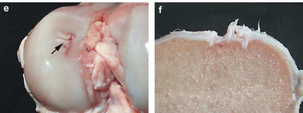
Subclinical osteochondrosis is not seen at a macroscopic level until the condyles are cut through and a default of ossification is noticed. This would require dedicated equipment, often unavailable in swine farms. At the osteochondrosis dissecans stage, when the pig is affected clinically, a fracture of the articular cartilage surface can be seen. Osteochondrosis is more likely to affect long bones such as the humerus and the femur, therefore, the most affected joints are the elbow and stifle.
Microscopic lesions
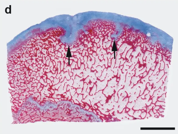
Under the microscope, an infolding of the cartilage into the bone as well as a disruption of the bone structure can be observed.
Diagnosis
The diagnostic is usually done through the clinical presentation and the macroscopic lesions seen at necropsy.
Differential diagnosis
Any cause of lameness in finishing pigs should be considered a differential for osteochondrosis. Bone fracture, erysipelas, Mycoplasma hyosynoviae and Mycoplasma hyorhinis should be considered.
Treatment, Prevention and Control
There is no treatment for osteochondrosis. The clinical presentation of the disease is painful and should be a welfare concern. Prevention of clinical signs is managed by avoiding any cause of trauma. Housing conditions, especially flooring should be under scrutiny. When the first cut of pigs goes to market and the remaining animals have more room to move around and play, an increase in lameness due to osteochondrosis can be observed.
