Main Body
18. Agents and Actions of the Autonomic Nervous System
Physiology of the Autonomic Nervous System
Why is this topic important?
- many drugs have autonomic side effects
- many drugs act on autonomic receptors to treat a number of pathologies such as:
- Alzheimer’s Disease (PD3 fall)
- Angina (PD2 fall)
- Asthma (PD2 spring)
- Benign Prostatic Hyperplasia (PD3 fall)
- Cardiac Dysrhythmias (PD2 fall)
- COPD (PD2 spring)
- GERD (PD1 spring)
- Heart Failure (PD2 fall)
- High Blood Pressure (PD2 fall)
- Incontinence (PD3 fall)
- Impotence (PD3 fall)
- Schizophrenia (PD3 fall)
Organization of the Nervous System
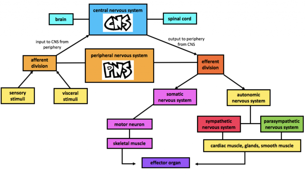
- Central Nervous System [CNS] – composed of brain and spinal cord
- Peripheral Nervous System [PNS] – composed of nerves outside of the brain and spinal chord including the afferent division (sends messages to CNS) and efferent division (sends messages away from the CNS)
- Autonomic System [ANS] – involuntary nervous system composed of two divisions; the sympathetic [SNS] and parasympathetic nervous systems
- Somatic Nervous System [SoNS] – voluntary nervous system that controls via skeletal muscles
- Effector Organs – organs on which nerves from the autonomic and somatic nervous systems act
Communication in the Nervous System
Neurotransmitters, or chemical messengers, allow for cell-to-cell communication within the nervous system. Two important neurotransmitters are involved in the activity of the autonomic system: acetylcholine [ACh] and noradrenaline, more commonly known as norepinephrine [NE].
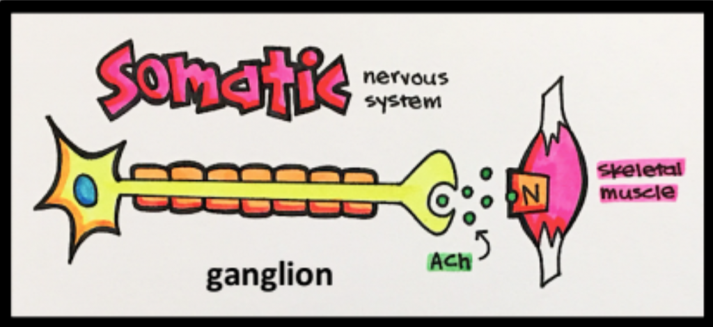
The image below is an example of cell-to-cell communication. Once depolarized, the presynaptic neuron releases ACh, which goes on to stimulate the nicotinic receptor [NAChR] on the postsynaptic neuron. Reminder: NAChR is an ion channel coupled receptor.
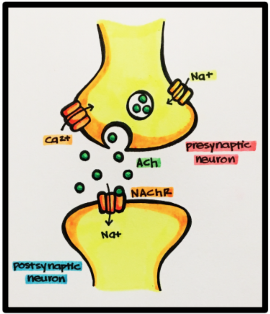
Ganglion – A group of nerve cell bodies located in afferent and efferent nerves. The somatic and autonomic nervous systems communicate via ganglia.
Efferent Division
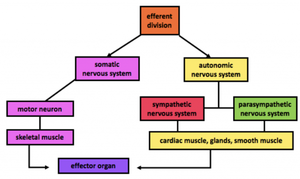
Somatic Nervous System
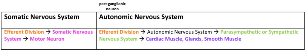
The somatic nervous system [SoNS] is our voluntary nervous system. It is composed of sensory neurons [afferent] or nerves that deliver a message to the CNS and motor neurons [efferent – think EXIT] that deliver a message from the CNS to skeletal muscles. Skeletal muscle contraction is under our voluntary control.
Autonomic System [ANS]
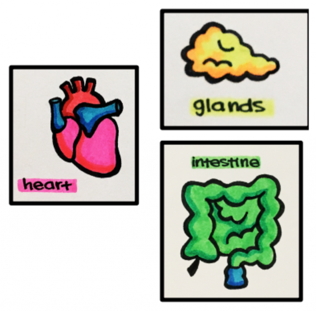
The autonomic system is our involuntary nervous system and is composed of two divisions; parasympathetic nervous system and sympathetic nervous system. Most organs receive dual parasympathetic [PNS] and sympathetic innervation [SNS]. These two divisions are complementary to one another and often result in the opposite effects upon stimulation. You can think of the sympathetic system as the “accelerator” and the parasympathetic system as the “brake.”
 |
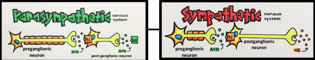 |
| The SoNS involves one nerve ganglion that communicates with the skeletal muscle. | The ANS involves two steps of neurotransmission; one at the ganglia (where acetylcholine serves as the neurotransmitter for both the sympathetic and parasympathetic nervous systems) and another at the innervated organs. For the latter, acetylcholine and norepinephrine are released from postganglionic parasympathetic and sympathetic neurons, respectively. |
Parasympathetic Nervous System [PNS]
Parasympathetic Nervous System – “REST AND DIGEST”
The PNS can also be thought of as the “D” division – defecation, digestion, and diuresis.
Most organs/tissues are innervated with parasympathetic ganglia.
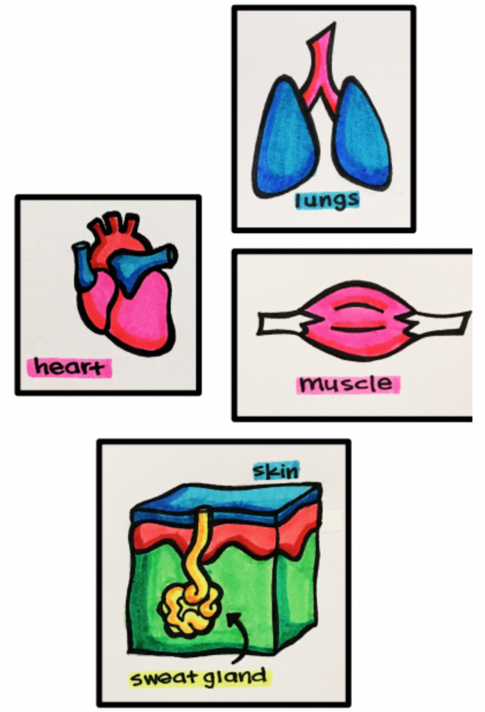
EXCEPTIONS – most blood vessels and all sweat glands only have sympathetic innervation.
What does your body need when at rest?
- Decreased cardiac output (compared to sympathetic) – lower oxygen demand when at rest
- Energy storage (glycogenesis, lipogenesis) – lower energy demand at rest
- Increased digestion – increased GI motility and secretions
- Waste elimination – defecation and urination
Parasympathetic Neurons
Pre-ganglionic and post-ganglionic parasympathetic neurons release acetylcholine [ACh]. The pre-ganglionic nerve releases ACh, which then stimulates nicotinic receptors [N]. The post-ganglionic neuron also releases ACh, however, it stimulates muscarinic receptors [M] located on the end organs. Reminder: muscarinic receptors are GPCRs [G-protein coupled receptors].
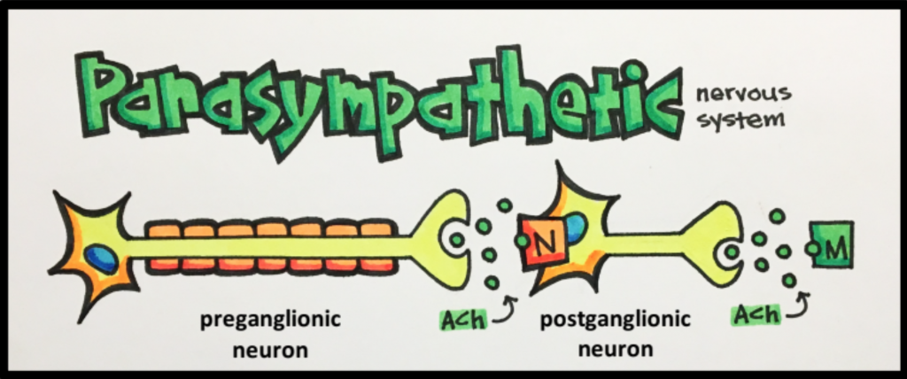
Acetylcholine & PNS Receptors
Acetylcholine interacts with two types of receptors:
- Nicotinic receptors [N] – ion channels located on ganglia
- Muscarinic receptors [M] – GPCRs located on effector/end organs
Sympathetic Nervous System [SNS] – “fight or flight”
The SNS can also be thought of as the “E division” – embarrassment, emergency, exercise, and excitement.
Most organs/tissue are innervated with sympathetic ganglia.
EXCEPTION – the ciliary smooth muscle of the eye only has parasympathetic innervation
What does your body need to do when in fight or flight situation?
- Alertness – think clearly in emergent situation
- Bronchodilation – increased oxygen necessary for brain and muscles to function well
- Blood shunted to muscles and organs – need muscles and organs like the brain to function well
- Decreased digestion – body does not need to exert energy on digestion during a fight or flight response
- Increased cardiac output – improved blood and oxygen delivery
- Production of energy (fatty acid release, glycogenolysis) – important for skeletal muscles to perform
- Prevention of waste elimination – body does not need to exert energy on elimination in emergency
- Sweat – help maintain homeostasis
Sympathetic Neurons
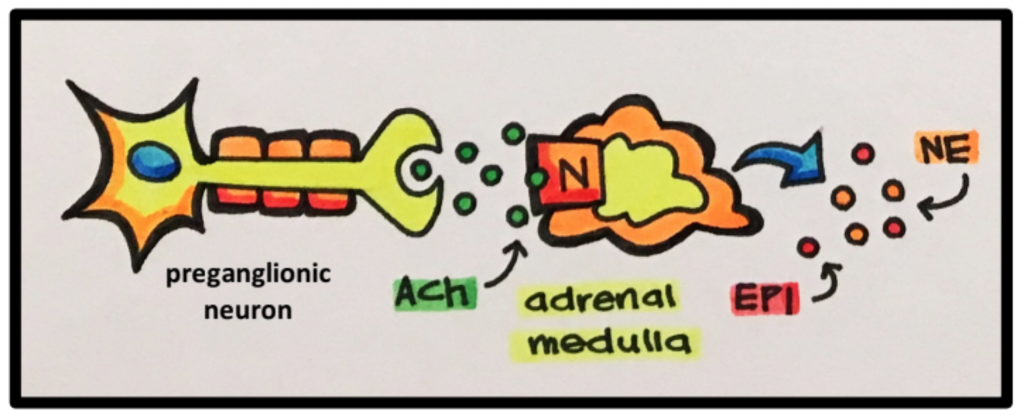
Pre-ganglionic sympathetic neurons release acetylcholine [ACh] and post-ganglionic sympathetic neurons release norepinephrine [NE]. ACh will then go on to stimulate the post-ganglion sympathetic neurons to release NE. Then NE will go onto stimulate adrenoceptors located on a variety of effector organs and tissue such as cardiac muscle, smooth muscle, and glands.
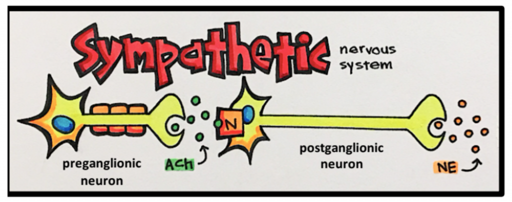
EXCEPTION – acetylcholine [ACh] is released by post-ganglionic sympathetic neurons innervating sweat glands (as opposed to NE), which means that ACh receptors (as opposed to adrenoceptors) have to be blocked in order to decrease sweating.
Reminder: parasympathetic ganglia do not innervate sweat glands.
Norepinephrine
Norepinephrine [NE] interacts with two types of receptors:
- Alpha-adrenergic receptors [alpha adrenoceptors] – GPCRs located on effector organs/tissue
- Beta-adrenergic receptors [beta adrenoceptors] – GPCRs located on effector organs/tissue
