Main Body
15. Nuclear Receptors
Nuclear receptors are receptors located inside the cell. These receptors are found either in the cytoplasm (Type I) or the nucleus (Type II) of a cell. Examples include: estrogen, glucocorticoids, thyroid hormone T3 or vitamins D and A. Receptor stimulation of any intracellular receptor primarily results in altered gene transcription.
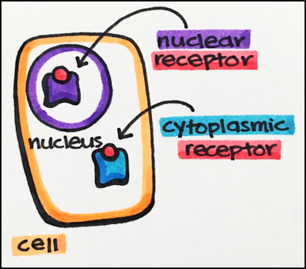
- Type I • intracellular receptors located in the cytoplasm of a cell. These receptors are translocated to the nucleus after stimulation by an agonist.
- o Examples androgen [AR], glucocorticoid [GRa], mineralocorticoid [MR], and progesterone receptors [PR].
- Type II • intracellular receptors located in the nucleus of a cell, even in the absence of agonists.
- Examples • peroxidsome proliferator [PPAR a,ß,g, sigma], retinoic acid [RAR a,ß,g], thyroid receptors [TR a,ß].
Nuclear Receptors • a subtype (Type II) of intracellular receptors that directly alter gene transcription. Nuclear receptors [NR] are a superfamily of transcription factors. They are modulated by and bind small lipophilic molecules (partial or mixed agonists) with low affinity. Some NRs do not have a known ligand at this point in time and are therefore known as Orphan Receptors.
Structure • monomeric proteins with similar general structure
- N-terminal Domain • Activation function 1 [AF1] site binds other transcription factors to modify the binding or activity of the receptor itself. These transcription factors are specific to a given cell.
- Core Domain • It is at this domain that DNA recognition and binding of hormone response elements [HREs] occur. The domain is made of two zinc fingers (cysteine/histidine rich loops in the amino acid chain that are held in a particular conformation by zinc ions).
- o HREs • portion of gene regulated by NRs that also result in receptor dimerization.
There are a number of nuclear receptors that produce a wide range of physiological functions. These receptors and their resultant functions will be discussed throughout the curriculum.

Type I Nuclear Receptors
- Androgen Receptors [AR] Endogenous AR agonists include testosterone and dihydrotestosterone [DHT]. These ligands bind AR in the cytosol of a cell and then translocate to the nucleus in order to alter transcription leading to protein synthesis, cell growth, formation of gonads, development of secondary sex characteristics and more.
- Glucocorticoid Receptors [GR] Cortisol is one of the most a common pharmaceutical targets as a endogenous GR agonist. Cortisol and other glucocorticoid hormones are stress hormones essential to maintaining homeostasis for a number of physiological processes such as metabolism, immune function, skeletal growth, cardiovascular function, reproduction and cognition. Upon binding, GR agonists can result in either activation or repression of transcription (depending on the particular isoform). For more details, see the link below:https://www.ncbi.nlm.nih.gov/pmc/articles/PMC4084612/
- Progesterone Receptors [PR] Upon binding PR in the cytosol, progesterone results in activation of transcription affecting a number of biological processes including the menstrual cycle.
Type II Nuclear Receptors
- Peroxisome Proliferator-Activated Receptor Alpha Receptors [PPAR]. Upon stimulation, these receptors lead to an increase in lipoprotein lipase (an enzyme that catalyzes the breakdown of lipoproteins) resulting in decreased hepatic triglyceride [TG] production. Fibrates, a drug class that targets these receptors, are PPARa agonists.
Their primary action is to decrease TG blood levels, however, they also help increase HDL (“good fat”), lower LDL (“bad fat”), improving a patient’s lipid profile and providing improved cardiovascular protection.

RXR • Retinoid X Receptor
PPRE • Peroxisome Proliferator Response Element
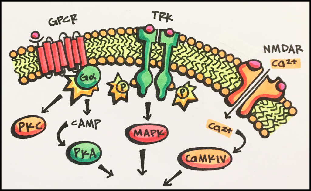
- Thyroid Hormone Receptors [THR a,ß] Upon stimulation, these receptors play a role in the rate of metabolism, neural development and cholesterol metabolism. Hypothyroidism, reduced TR stimulation, leads to slowed metabolism and presents as general fatigue and lethargy, weight gain, slowed heart rate, feeling cold and more. Conversely, hyperthyroidism, increases TR stimulation, leads to increased metabolism and presents as weight loss, increased heart rate, feeling hot and more. Nuclear Receptor-mediated transcriptional regulation can be modulated by several signaling pathways; this is a great example of convergence. Convergence is when multiple pathways lead to the same effect and in the case of nuclear receptors the effect is transcription. These pathways can be initiated by either both external and internal signals involving GPCRs, ion channels, TRKs as well as several other receptors.

Overview of Receptor Types
| Enzyme-Linked Receptors | G-Protein-Coupled Receptors | Ligand-Gated Ion Channels | Intracellular Receptors | |
|---|---|---|---|---|
| Location | cell membrane | cell membrane | cell membrane | cytoplasm or nucleus |
| Effector | protein kinases | channel or enzyme | ion channel | gene transcription |
| Coupling | direct
|
G-protein | direct | via DNA |
| Structure | single transmembrane helix linking extracellular receptor domain to intracellular enzyme (often kinases) domain.
Hormones bind to a receptor dimer to result in activation. |
7 trans-membrane helices with intracellular G-protein-coupling domain
|
oligomeric assembly of 4-5 subunits surrounding a central pore
|
monomeric structure with separate receptor and DNA-binding domains
|
| Examples | insulin, growth factors, cytokine receptors | muscarinic acetylcholine receptors, adrenoceptors | nicotinic acetylcholine receptors, GABAA receptor | estrogen receptors, vitamin A receptors |


