5.3 Inside Roots
Learning objectives
By the end of this lesson you will be able to:
- Explain how roots elongate and increase in diameter via primary and secondary meristems.
- Identify the tissues in the root that originate from the root meristems and the cells into which they eventually mature.
- Distinguish between monocot and dicot roots.
- List the functions of the cells in plant roots.
Root review
Please read the OpenStax pages summarizing root biology. This resource reinforces several of the topics addressed below, so you may read it either before or after you complete this chapter. You don’t need to memorize the information there; use it to reinforce the content below.
The functions of roots include:
- Anchoring the plant in the soil (and stabilizing the soil).
- Supporting the upright growth of the plant.
- Providing a site for the symbiotic relationship of the plant with particular beneficial fungi and bacteria.
- Absorbing water and dissolved minerals from the soil.
- Storing nutrients like starch for subsequent use by the plant (tap roots and tuberous roots are examples of geophytes that store nutrients in roots).
Angiosperm (flowering) plants are often classified by whether they rely on a primary root system an adventitious root system. A plant doesn’t necessarily have only primary or just adventitious roots. One of these systems, however, will be dominant. Whether a root is considered primary or adventitious depends on whether the root traces back to the radicle (embryonic root) or arises from (normally underground) stem tissue.
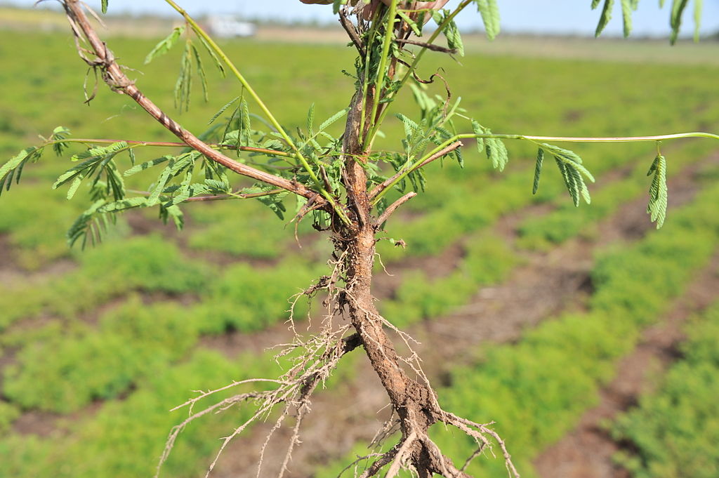
Tap root (or primary root) system
Tap or primary roots arise as a continuation of the embryonic radicle tissue and persist into maturity. Secondary roots (also called lateral roots) arise from the primary root, and tertiary roots arise from the secondary. This primary –> secondary –> tertiary root formation is the usual rooting system for dicots. Next time you see a dandelion or other weed that doesn’t look like a grass, yank it up and look at the root system. The main root you see (assuming that it didn’t break off when you pulled it up) is probably a tap root. The photo on the right, above, demonstrates this point. It shows a young taproot system with many secondary roots branching off the primary root.
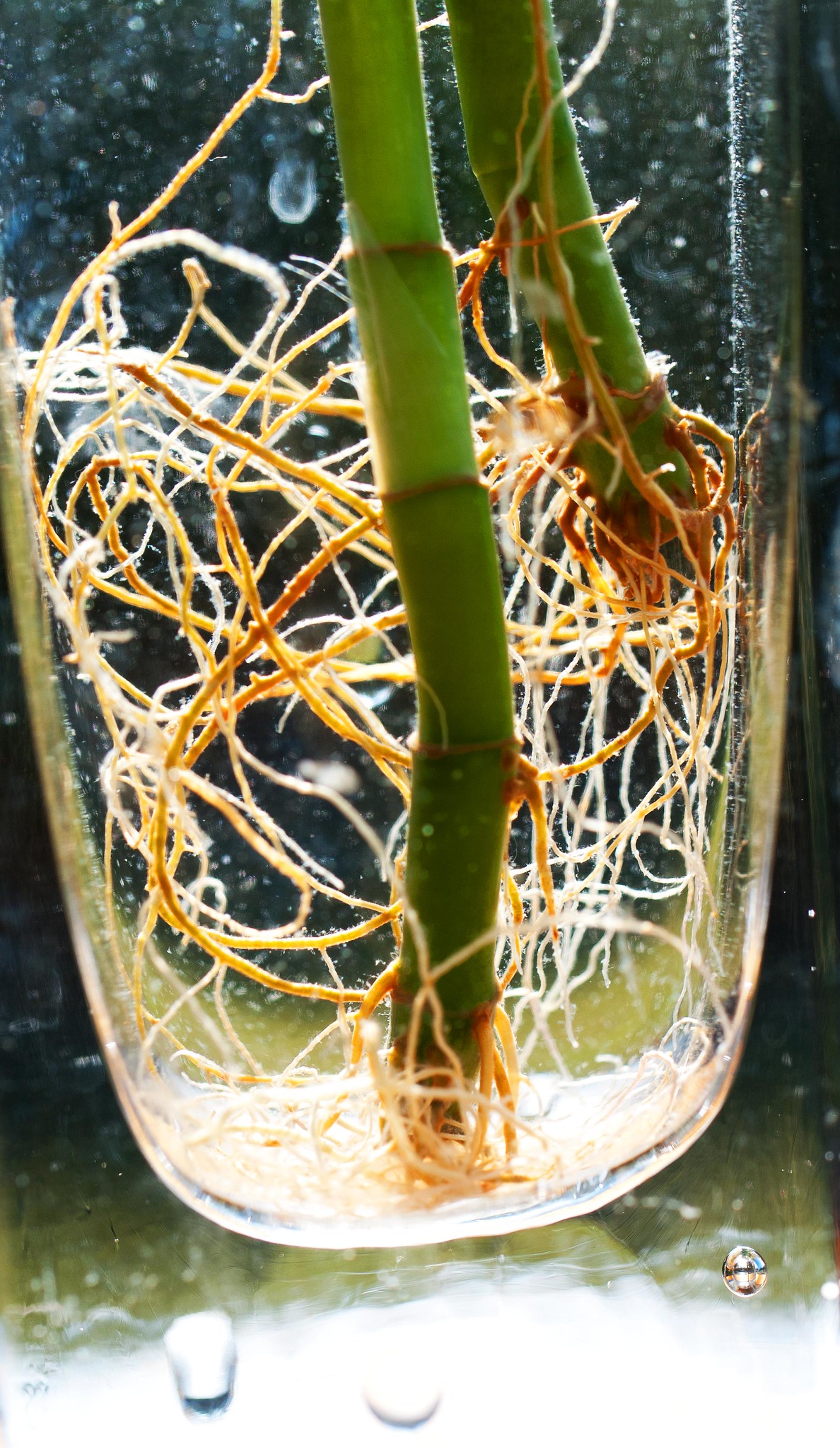
Adventitious or fibrous root system
In plants of this type, the primary root, which originates from the radicle, weakens prior to maturity and new, vigorous, adventitious roots arise from stem tissue. Adventitious roots may grow from nodes or might arise from the internodes. They originate from parenchyma cells in the cortex of the stem. Dig up a clump of turfgrass and look at the roots. Turfgrass has a fibrous root system, as does corn.
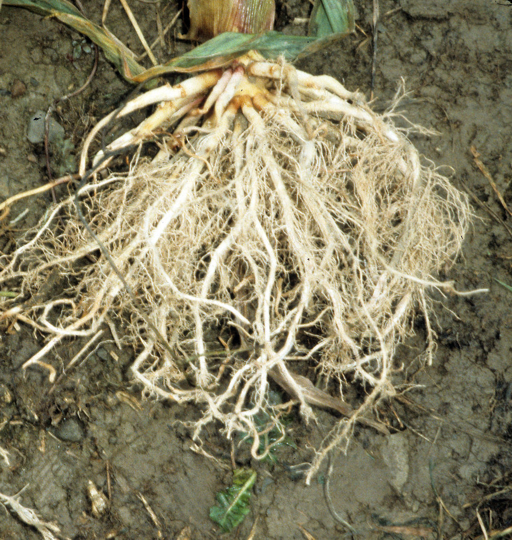
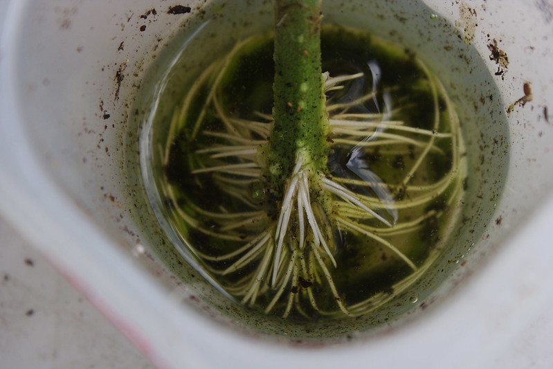
As noted above, although adventitious roots originate from the stem they don’t have to emerge from a node. While it’s not apparent in the photo, the adventitious corn roots you see above do trace back to a node. In contrast, in the photo of a tomato stem, below, we see emergence of adventitious roots from both node and internodal regions of the stem.
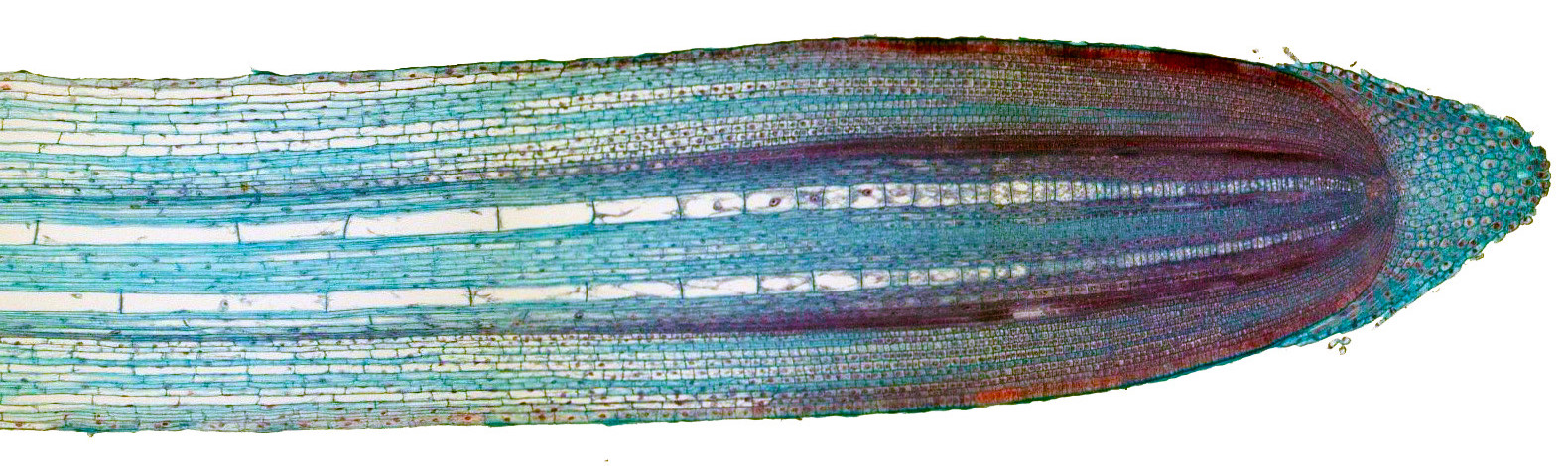
Internal root structure
Watch this video to take a closer look at root structure (6:21):
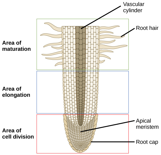
Root cap
Shaped like a thimble, this structure covers the tip of the root and provides protection as the root grows into the soil. These parenchyma cells are produced by the root’s meristem which is just behind the root cap. The outer cells of the root cap are continuously worn away through contact with the soil, and new cells are added to the inner portion.
In addition to protecting the interior of the root, the cap secretes a mucilage which stabilizes the water content of the surrounding soil, ensuring longer-lasting nutrition to the root system and making for easier root probing. Finally, the root cap contains statocytes, specialized cells that help the plant sense gravity and grow accordingly. These cells are full of starchy organelles which settle at the lowest part of the cell and encourage growth in that direction. If the root cap, with these statocytes, is removed, a plant may grow in random directions because it has lost the ability for gravitropism (growth in response to gravity).
Root meristem
The cells here divide rapidly via mitosis to form new cells. New cells are laid down toward the root cap to replace those worn away during root growth, and also laid down away from the root cap. These new cells laid away from the root cap elongate and then mature into more specialized root tissues.
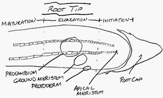
Region or zone of elongation
In this region, the cells produced by the root meristem undergo rapid elongation — they expand in length and volume. Root growth is the result of two processes: new cell production by the root meristem, and subsequent elongation of those new cells. This growth pushes the root further into the soil and also expands the diameter of the root. Within the region of elongation just behind the meristem you will find the following undifferentiated tissues:
- Protoderm — new cells laid down toward the exterior of the root which will mature to become the root dermal tissue (primarily epidermis cells).
- Procambium — new cells in the central part of the root which will mature to become the vascular tissue (xylem, phloem, and vascular cambium), labeled in the illustration above this section as the vascular cylinder.
- Ground meristem — the new cells lying between the protoderm and procambium that will mature to become the cortex tissue (primarily parenchyma cells).
Region of differentiation (also called the region of maturation)
Here the root becomes thicker, and secondary or lateral roots are initiated. In this region the protoderm, procambium, and ground meristem cells undergo differentiation into the specialized cells associated with the dermal, vascular, and cortex tissues, as noted above.
Root-hairs begin to form in the region of differentiation; these are fine outgrowths of epidermis cell walls and membranes, and increase the area of absorption of the root.
Review questions
- What two processes result in root growth?
- Protoderm will differentiate or mature into what type of tissue? How about procambium? And ground meristem?
- Do adventitious roots arise from stem tissue or from the primary root? Are they always found emerging from nodes or from internodes, or does it depend on the type of plant?
Dicot roots vs. monocot roots
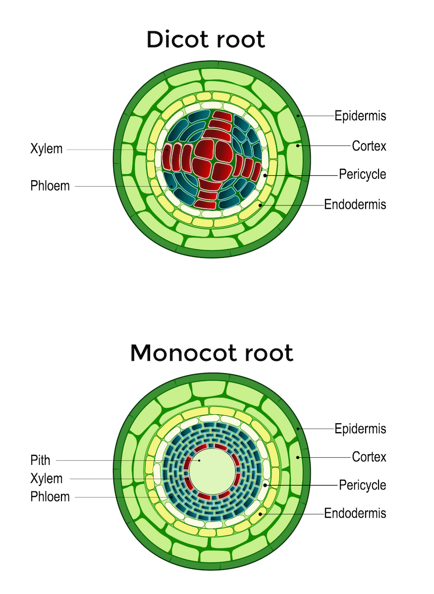
Let’s take a look at the root another way. If we were to cut a cross section of the root just above the root hairs in the zone of differentiation, you would find that the cells have differentiated to form the following tissues:
Epidermis – a one-cell thick layer of cells surrounding the root. These cells do not have the cutin or waxy covering that you will find in stem epidermis cells because roots don’t need to keep moisture inside the root. Quite the opposite, the roots need to be permeable to moisture. Root hairs arise from the epidermal cells.
Cortex – The cells inside the epidermis make up the cortex. Cortex cells in the root usually consist of parenchyma cells with numerous intercellular spaces through which water can move. However, the innermost cells of the cortex, called the endodermis, are rectangular in shape with thicker walls, are stacked close together, and have a band-like deposit of waterproof suberin that wraps around each cell like caulk in the intercellular spaces. This thick band of suberin is called the Casparian strip and it closes up the intercellular space between cells. The Casparian strip forms a barrier to the water that has been moving freely through intercellular spaces and forces the water to instead move internally through a layer of cells before it gets into the xylem. That way, the cells can exert some regulation on water passage into the root vascular system.
Let’s talk a bit more about water movement in roots. Some water moves from soil to root through the root hairs, which you will recall are extensions of the root epidermis cells. Because the water is moving into a cell, the water must pass through the cell membrane, which is a semipermeable membrane that exerts some control over what comes into and goes out of the cell. The water then moves from cell to cell through the cortex to the vascular bundle. Because it is moving within cortex cells it is not constrained by the Casparian strip because the Casparian strip is a band around the outside of the innermost layer of cortex cells. This type of water movement through the cortex is called symplastic movement. Symplastic means from cell to cell.
A second type of water movement through the cortex is called apoplastic movement. Apoplastic water movement occurs outside of the cells. Water squeezes between the cortex cells until it reaches the Casparian strip, and then if it wants to go any further into the plant it must move into the cortex cells through the cell membrane. Once in the cortex cells, it continues to the vascular bundle through symplastic movement. We’ll get more into this later when we look closer at water and plants.
Summary of cortex function:
- Allows for the diffusion of water, minerals, and oxygen from the root hairs inwards.
- Stores food reserves, especially starch.
- The endodermis, with the aid of the Casparian strips, facilitates the regulated apoplastic movement of water from cortex to xylem.
When talking about stem cortex tissue, we noted it was made up of three cell types: parenchyma, collenchyma, and sclerenchyma. Just above we said root tissue cortex is made up of parenchyma cells. What about the other two cell types? The roots aren’t in need of stiffening the way that an upright above ground stem needs in order to resist gravity and wind, so they don’t need the cell types (collenchyma and sclerenchyma) that provide that stiffening in the stem cortex.
Vascular Cylinder or Stele – The vascular cylinder begins just inside the endodermis.
The first set of cells you encounter as you move into the vascular cylinder is a single layer of tightly packed cells called the pericycle. The pericycle cells retain the ability to divide and produce new cells. This layer of cells is the source of lateral roots. The image to the right shows an example of lateral root initiation from the pericycle. You may recall that earlier I mentioned that adventitious roots in fibrous root systems arise from cortex tissue. Here I’m talking about a tap root system where the laterals arise from the pericycle cells in the vascular tissue.
The root vascular cylinder also contains xylem and phloem. In roots of dicotyledonous plants (plants whose seeds have two cotyledons) the xylem is in the center of the root, typically in the form of a thick-armed “X”, with phloem in the spaces between the arms. Not all dicots have the xylem in an obvious X shape…in some cases it looks a bit more round than a distinct X (as with the carrot taproot).
Dicot roots
Watch this video for a detailed description of dicot roots (5:19)
Monocot roots
Watch this video for a detailed description of monocot roots (2:10)
In monocot roots (plants whose seeds have one cotyledon) the xylem is in a ring surrounded by phloem with pith in the very middle.
Review questions
- What are the differences between endodermis and pericycle in their locations and functions?
- How could you tell the difference between a monocot and dicot root based on the vascular cylinder?
- What layer of cells provides a barrier to intercellular water movement?
- Why don’t you typically find collenchyma and sclerenchyma in the cortex tissue of the root?
Root that forms from the embryonic radicle.
Roots that emerge from the stem rather than roots.
Root that forms off of the primary root.
Root system where the radicle grows and then rapidly slows or completely halts in growth. Once this happens roots will emerge above the radicle and from the stem tissue located below the soil.
Thimble-shaped mass of cells that covers and protects the root apical meristem from rocks, dirt, and pathogens.
Specialized cells that help the plant to sense gravity and grow accordingly.
Growth in response to gravity.
Cell division where a cell divides into two identical daughter cells.
New, primarily epidermis, cells laid down toward the exterior of the root which will mature to become the root dermal tissue.
New cells in the central part of the root that will mature to become the vascular tissue (xylem, phloem, and vascular cambium).
The outermost layer of cells in the plant.
Also known as the ground meristem, is found just inside the epidermis and extends toward the interior of the stem and root, and is made up of three types of cells: parenchyma, collenchyma, and sclerenchyma.
