14.1 Gametogenesis
Learning objectives
By the end of this lesson you will be able to:
- Identify where sporangia are found in the angiosperm flower.
- Describe how the male gametophyte is formed.
- Describe how the female gametophyte is formed.
- List which cells unite during double fertilization.
- Explain how the embryo goes through developmental stages that are identified according to the developing embryo’s shape.
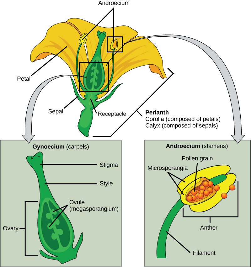
Recall, as shown below, that the flower is made up of a compressed, four-node stem called the receptacle that supports four whorls of modified leaves: calyx (sepals), corolla (petals), androecium (stamens), and gynoecium (carpels). The two whorls nearest the tip of the receptacle, the androecium and gynoecium, contain structures housing the (micro- and mega-) sporangia, where meiosis takes place and the gametophyte generations develop.
Watch this video for a chalkboard demonstration of gametogenesis (4:49)
Sporangia
The sporangia of angiosperm plants are found in two places within the flower depending, on whether they lead to a female or male gametophyte. The sporangium in the anther (where the male gametophyte will be formed) is called the microsporangium. The sporangium in the ovary (where the female gametophyte will be formed) is called the megasporangium. Diploid microspore mother cells in the microsporangium and diploid megaspore mother cells in the megasporangium divide by meiosis to form four haploid micro- or mega- spores. These spores initiate the gametophyte phase. Two (male) to three (female) mitotic divisions later, the mature micro- and mega- gametophytes have been formed. Note how few divisions take place to form the gametophyte after the meiotic division of the microspore or megaspore mother cell, and that those two or three additional divisions are mitotic divisions. One round of meiosis is sufficient to generate haploid cells. From there on the divisions are mitotic, but in this case mitosis starts with one haploid cell and generates two haploid cells.
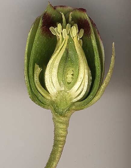
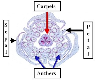
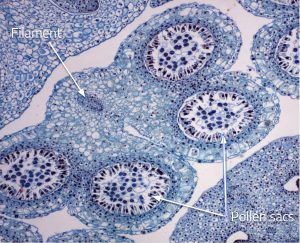
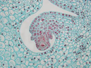
The images above provide a visual orientation to the micro- and megasporangia. In the photo, the bud in the horizontal cross section shows the three chambers in the fused carpels as well as a number of anther cross sections. In the center of the vertical cross section of the flower bud you’ll see a carpel with ovary, locule (interior chamber), and ovule within the locule. Also note the anther cross sections containing pollen.
Microsporogenesis
Watch this video for a chalkboard demonstration of microsporogenesis (5:35)
The illustration below shows the development of the male gametophyte within the anther. The process, called microsporogenesis, or male gametogenesis, starts in the top left hand corner. Study these steps and be able to draw them. Red “2n” or “n” notations indicate the ploidy of the nuclei and blue notations show where the mitotic cell divisions occur. To this point we’ve learned that mitosis starts with one diploid cell and results in two diploid cells. We’ll modify that now to say that mitosis starts with a cell and results in two cells that are exact copies of the original cell. If the starting cell is diploid, there will be two diploid copies. If the starting cell is haploid, there will be two haploid copies.
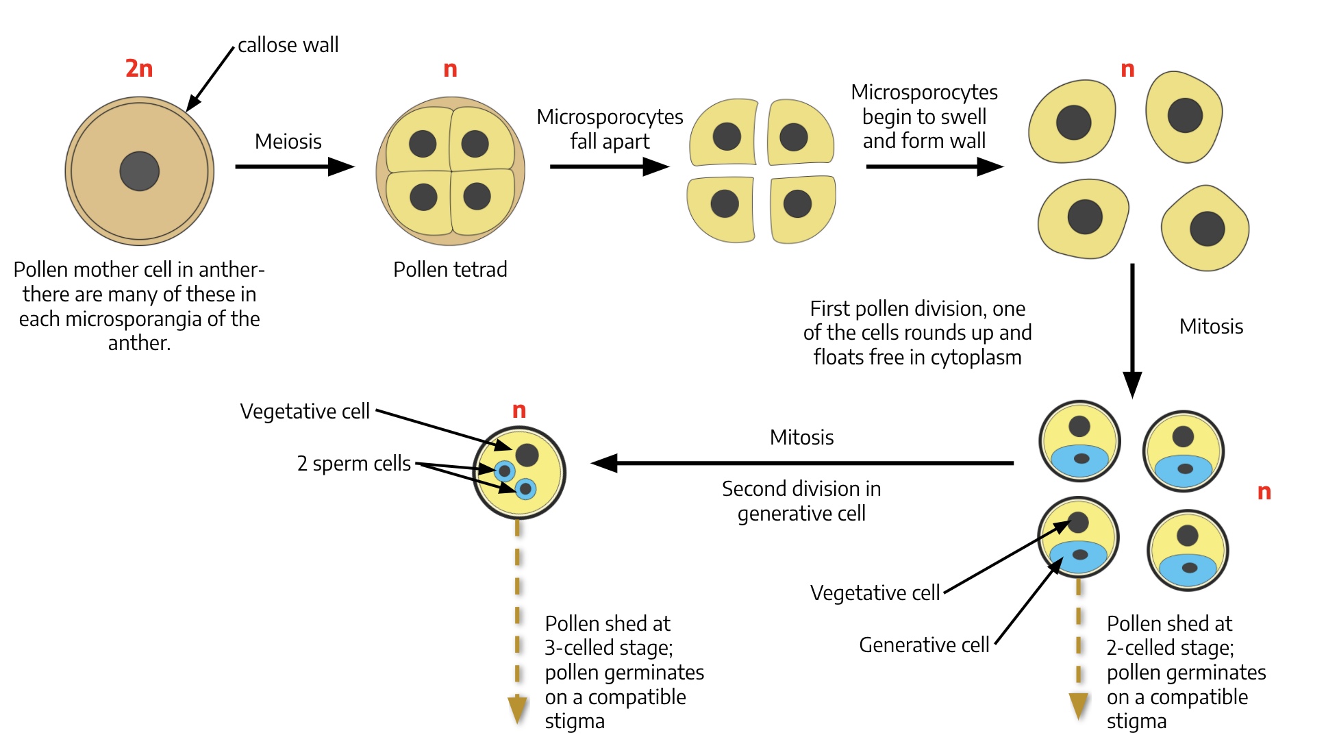
The result of microsporogenesis, shown on the bottom row of the illustration above, is either a male gametophyte that is a two-celled pollen grain (with vegetative and generative cells), where the generative cell will later undergo another mitotic division to produce two sperm cells (shown to the right of the box), or, following mitosis of the generative cell and shown to the left of the box, a three-celled pollen grain with a vegetative (or tube) cell plus two sperm cells.
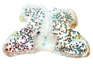
Below is a cross section of young anther showing the developing pollen grains in four chambers.
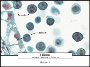
Below are tetrads of cells following meiosis of the microspore mother cell. Note the sets of four cells still attached together.
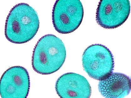
Below are nearly mature, two-celled pollen grains with the start of an exine coat. The exine coat is made up of protein and other compounds deposited by the inside wall of the anther, and it forms a protective coat around the three-celled microgametophyte.
In this micrograph the exine coat looks like rough, geometrically shaped plates on the surface of the pollen. The exine coat patterns are characteristic of the species and can be used to identify the species of plant that produced the pollen.
Below is a mature, dehiscing (split and releasing pollen) anther.
Review questions
- Where are the microsporangia located?
- Where is the mature male gametophyte in the Pollen Development and Growth illustration above?
- Starting with the microspore mother cell, how many meiotic and mitotic cell divisions are required to produce the mature male gametophyte?
Megasporogenesis
Watch this video for a chalkboard demonstration of megagametogenesis (4:28)
The illustration below shows the development of the female gametophyte, a process is called megasporogenesis or female gametogenesis. Study this diagram and be able to draw it.
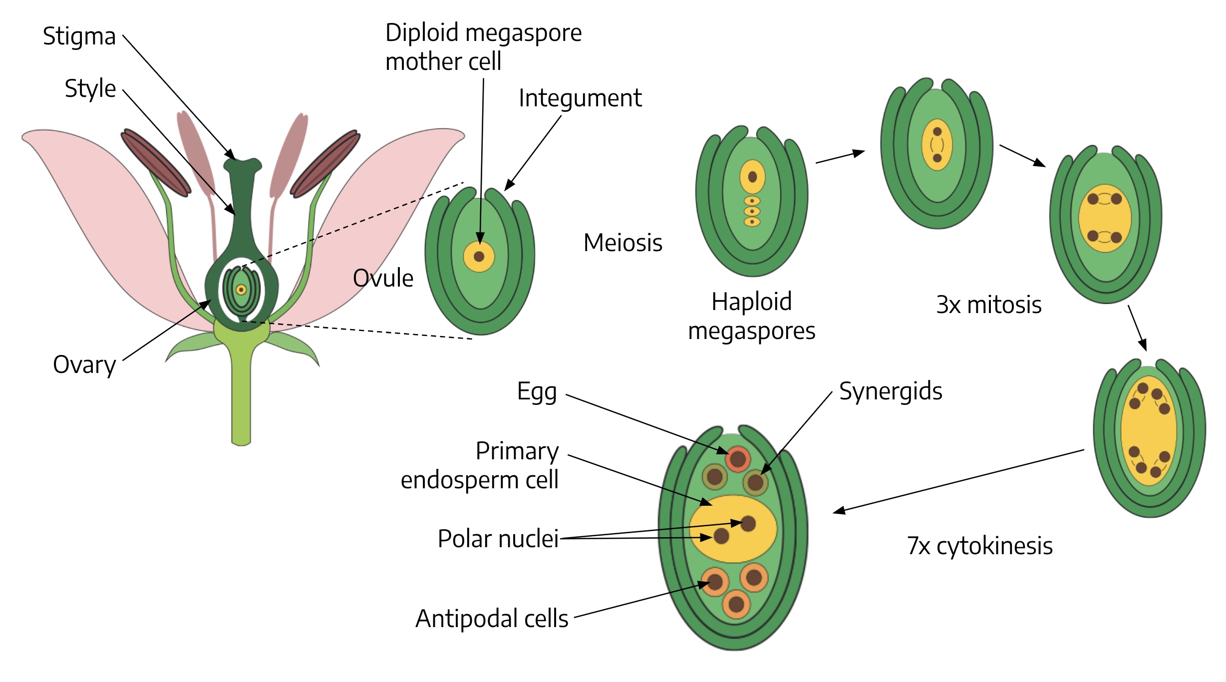
The chambers within the ovary are the locules, and within the locules are the ovules. The ovule wall is made up of integument tissue, and it is from this integument tissue that diploid megaspore mother cells form. A diploid megaspore mother cell undergoes meiosis and initially produces four haploid megaspores. Three of the four spores formed through meiotic division of the megaspore mother cell disintegrate, as indicated by the “X” through three of the spores in the illustration. The lone surviving megaspore subsequently undergoes three mitotic divisions to form eight haploid cells that are held within the ovule in an embryo sac. These eight cells comprise the female gametophyte.
Only six of the nuclei are surrounded by their own cell membrane after the three rounds of mitosis. The two remaining nuclei are surrounded by one cell membrane. These two nuclei in one membrane become the polar nuclei and contribute two of the three nuclei to the endosperm tissue, with the other nucleus contributed by a sperm cell.
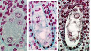
The megaspore mother cell is the swollen cell shown in the micrograph below. Note the linear tetrad of haploid megaspores resulting from meiosis of the megaspore mother cell. Three will disintegrate and one will undergo three rounds of mitosis.
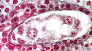
The four cells in the embryo sac in the left-hand image below are from two mitotic divisions of the one surviving megaspore. After one more mitotic division (center image below), the embryo sac, or female gametophyte, contains 7 cells (with 8 nuclei), and their arrangement is shown in the rightmost image below. The egg and two synergids are positioned on the bottom, two polar nuclei are shown in the center, and three antipodal cells are positioned at the top.
Review questions
- Where are the megasporangia located?
- Where is the mature female gametophyte in the Development of the Female Gametophyte illustration above?
- Is the female gametophyte diploid or haploid? Is the integument tissue diploid or haploid?
- Starting with the megaspore mother cell, how many meiotic and mitotic cell divisions are required to produce the mature female gametophyte?
Fusion or fertilization
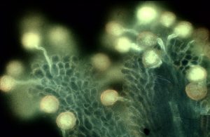
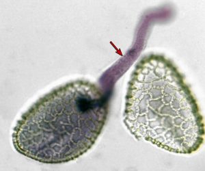
In the image below, pollen has fallen on the stigma and some grains are germinating. The next image shows the germination of pollen and tube growth in an artificial medium.
Think of the pollen tube as an extension of the cell membrane and cell wall of the pollen grain. The pollen tube isn’t a long string of cells, but is one long skinny cell with two or three nuclei (one vegetative and either one generative or two sperm depending when the generative cell undergoes mitosis). The tube grows down the style through the intercellular (between cell) spaces rather than through cells. Pollen tube growth is an active area of study, with some work showing that the style cells are important for providing nutrients to the pollen tube and also apparently provide directional guidance to tube growth.
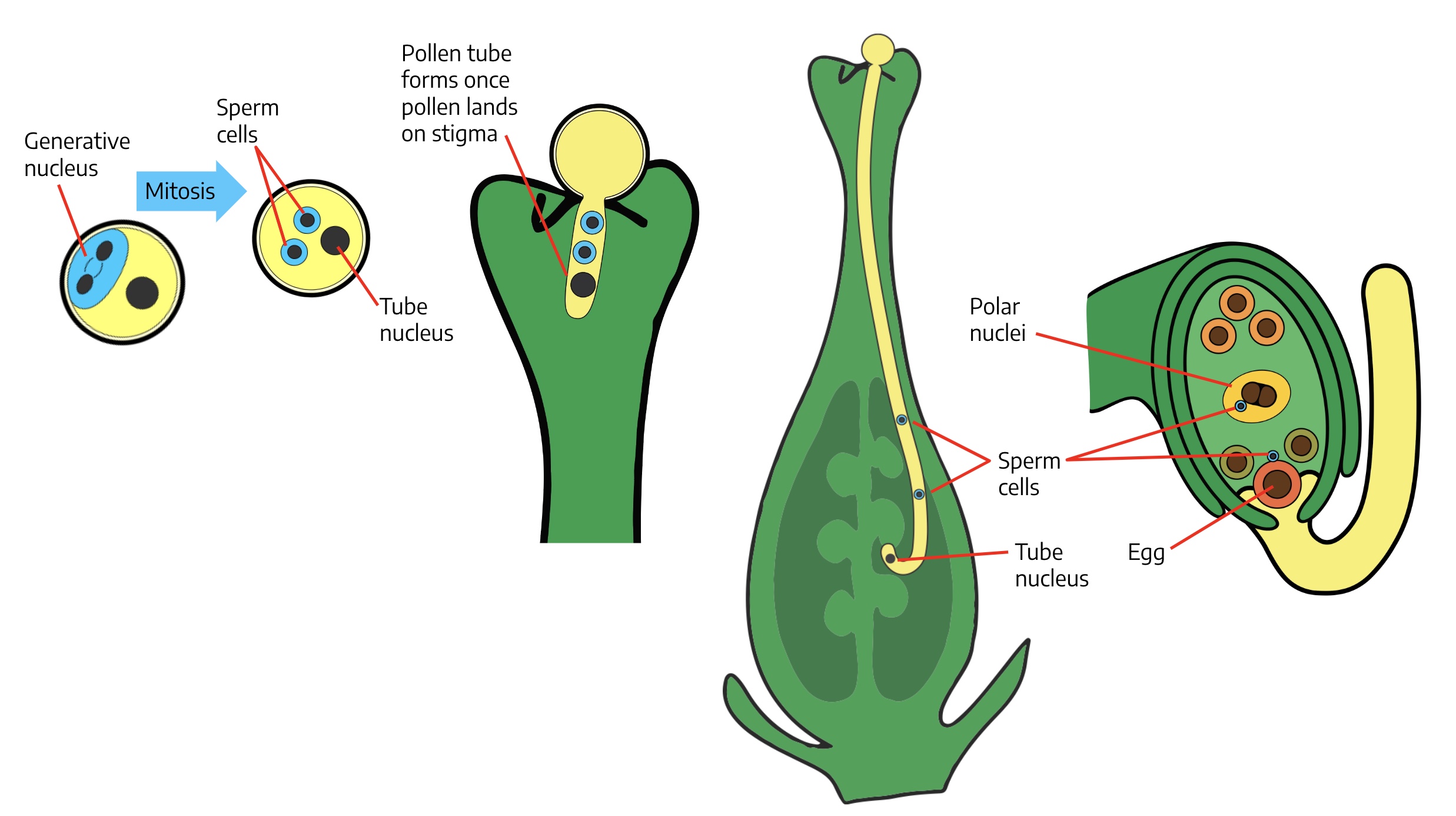
In the illustration below we see a generative nucleus in a pollen grain. This subsequently undergoes mitosis to form two sperm cells. As noted earlier, in some species this happens during pollen development before it is shed from the plant; in other species it occurs in the pollen tube. The image on the far left below identifies all of the cells in both the female and male gametophyte: in the female gametophyte are three antipodal cells at the top of the ovule, the polar nuclei in the middle, and at the bottom are two synergid cells flanking the egg. There are eight cells or nuclei in total.
In the illustration, you can see the three cells within the pollen tube: two sperm and one tube or vegetative nucleus. The tube finds its way to the embryo sac through the micropyle and ruptures, and the sperm are delivered to the egg and polar nuclei. The rightmost section of the illustration is a closeup of the ovule and shows double fertilization. One sperm nucleus has fused with the egg nucleus to form a 2n zygote. The other sperm nucleus has fused with the two polar nuclei to form the 3n endosperm.
Embryo growth
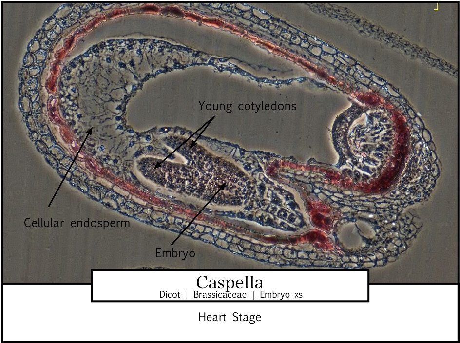
Embryo development is initiated when the zygote divides once mitotically to form an apical and a basal cell. The basal cell undergoes several additional mitotic divisions to form a suspensor that anchors the apical cell to the nucellus tissue on the inner surface of the maturing ovule wall (seed coat). Next, the apical cell begins to divide mitotically to form the embryo. The embryo goes through distinctive phases that look like particular shapes. This image shows the early embryo heart shape (second from right), and the torpedo shape (far right) stages. The lobes will become cotyledons. Between the lobes is the stem or shoot apical meristem, and at the base near the suspensor is the root apical meristem.

Below is a heart stage embryo. Note the endosperm developing in another part of the embryo sac.
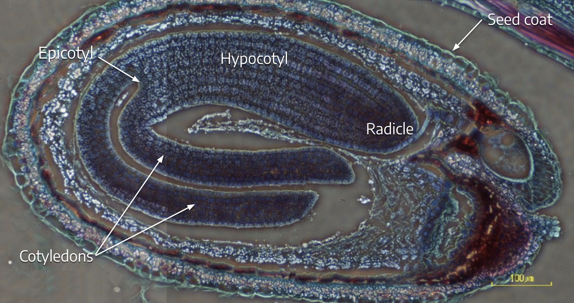
Finally, a later stage embryo with cotyledons readily apparent. This lesson has brought you from flower to seed.
Review questions
- How many nuclei are in the pollen tube?
- Where is the egg cell of the embryo and what does it do?
- A heart-shaped embryo has two swollen lobes that give it the shape of a heart. What will those lobes become when the embryo is mature?
- You may recall that the ovule is attached to the placenta of the ovary by a stalk called the funiculus. What is the difference between the funiculus and the suspensor that is produced by mitotic divisions of the basal cell?
Place in the anther where the male gametophyte will be formed.
Cells that form the ovary wall. Nucellus cells on the interior of the ovule wall develop into megaspore mother cells.
Nucleus in the immature male gametophyte that will later divide by mitosis to produce two sperm cells.
Cells flanking the egg cell in the mature female gametophyte.
Nucleus in the male gametophyte that is associated with pollen tube growth.
Produced by multiple mitotic cell divisions of the embryo’s basal cell; the suspensor anchors the apical cell of the embryo to the ovule wall.
