15.2 The first two weeks
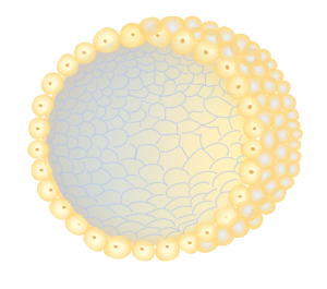
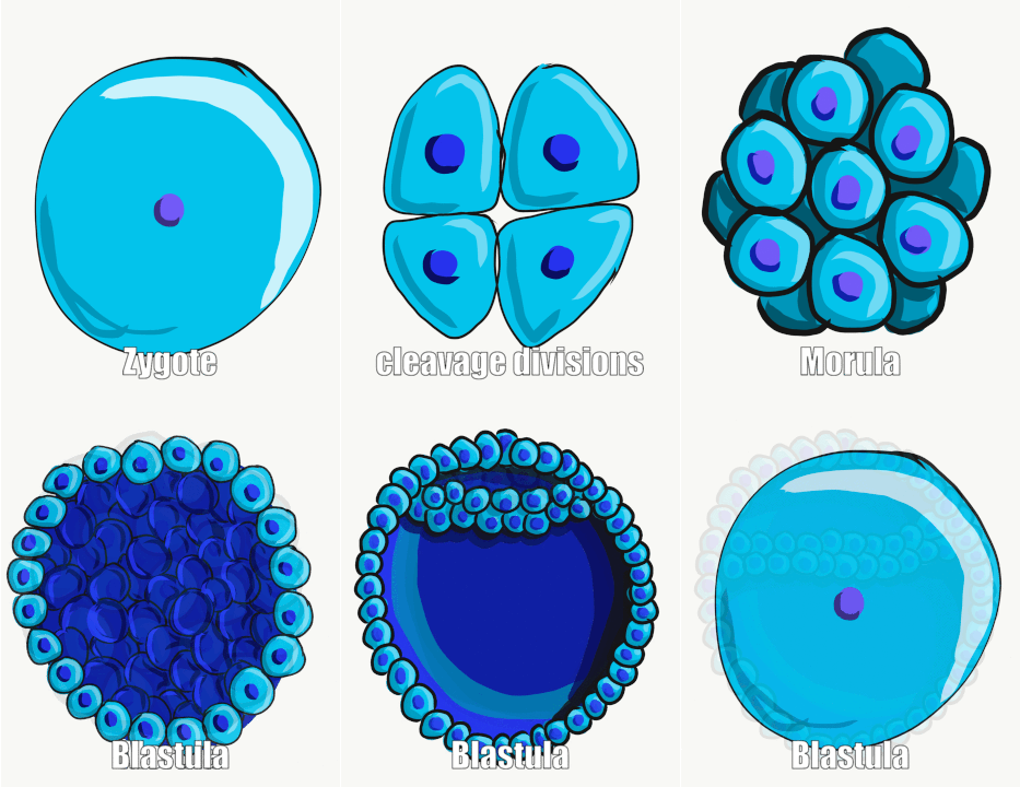
As we discussed in chapter 14, once the genetic material from a sperm fuses with the genetic material from an egg, a zygote is formed. This zygote will now begin to divide from one cell to two, from two cells to 4, etc. During this time the embryo[1] begins to move down the oviduct toward the uterus. Around day 2-3 post-fertilization the developing embryo is a ball of cells called a morula. Around day 4-5 after fertilization the egg becomes a blastula, which is a hollow ball of cells. At this stage it enters the uterus and will “hatch” out of a tough outer covering called the zona pellucida. Once hatched (1-2 weeks post-fertilization) the embryo will interact with the uterine lining and may implant, or imbed itself in the uterine lining. Only about 50% of embryos successfully implant in the uterine lining (the rest are carried out of the body with the next onset of menstruation).


Once implantation has occurred, a pregnancy is established and circulating levels of human chorionic gonadotropin (HCG) increase. HCG is the hormone that is detected in urine by over-the-counter pregnancy tests. This hormone also (as discussed in chapter 13.8) signals the corpus luteum (the collapsed group of cells from which an oocyte emerged) to continue to produce progesterone. Progesterone prevents the shedding of the uterine lining that would normally happen during menstruation. The corpus luteum continues to produce progesterone for the first 7-10 weeks of pregnancy, after which the developing placenta takes over. What is often confusing when talking about pregnancy stages is that, while implantation is happening only about 2 weeks post-fertilization, the calculation of stage of pregnancy is done from the date of the first day of the last menstrual cycle (first day of the period). Thus, a physician would describe a person who has a newly established pregnancy as 4 weeks pregnant (even though the first two weeks of the “pregnancy” were before fertilization occurred!).

The outer cells of the blastocyst that are imbedded in the uterine wall will differentiate into the placenta, and the inner cells will eventually develop into the fetus. At this stage the cells of the developing embryo are identical and pluripotent (i.e., from those cells, any of the parts of the human body can develop). In fact, if an embryo splits in two prior to this time, the result can be two fully formed babies that are genetically identical (these are monozygotic or identical twins).
Check Yourself
Read More
For detailed explanation of the stages of embryonic and fetal development, see: https://embryology.med.unsw.edu.au/embryology/index.php/Main_Page
- Embryo is a general term for the early stages of the developing organism. The terms zygote, morula, and blastocyst refer to more specific developmental stages. Once a developing human reaches around 12 weeks it is referred to as a fetus. ↵

