3.2 Parts of the Nervous System
Learning Objectives
By the end of this section, you will be able to:
- Describe the difference between the central and peripheral nervous systems
- Explain the difference between the somatic and autonomic nervous systems
- Differentiate between the sympathetic and parasympathetic divisions of the autonomic nervous system
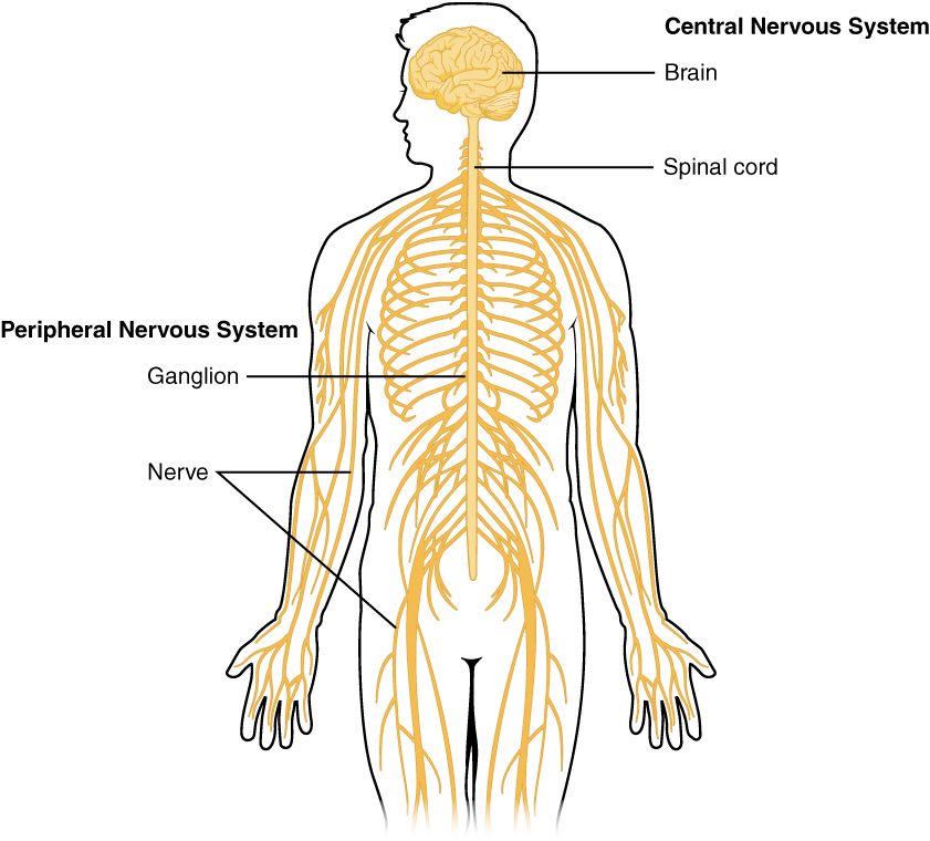
The nervous system can be divided into two major subdivisions: the central nervous system (CNS) and the peripheral nervous system (PNS), shown in Figure 1. The CNS is comprised of the brain and spinal cord; the PNS connects the CNS to the rest of the body. In this section, we focus on the peripheral nervous system; later, we look at the brain and spinal cord.
*
Peripheral Nervous System
The peripheral nervous system is made up of thick bundles of axons, called nerves, carrying messages back and forth between the CNS and the muscles, organs, and senses in the periphery of the body (i.e., everything outside the CNS). The PNS has two major subdivisions: the somatic nervous system and the autonomic nervous system.
The somatic nervous system is associated with activities traditionally thought of as conscious or voluntary. It is involved in the relay of sensory and motor information to and from the CNS; therefore, it consists of motor neurons and sensory neurons. Motor neurons, carrying instructions from the CNS to the muscles, are efferent fibers (efferent means “moving away from”). Sensory neurons, carrying sensory information to the CNS, are afferent fibers (afferent means “moving toward”). Each nerve is basically a two-way superhighway, containing thousands of axons, both efferent and afferent.
The autonomic nervous system controls our internal organs and glands and is generally considered to be outside the realm of voluntary control. It can be further subdivided into the sympathetic and parasympathetic divisions (Figure 2). The sympathetic nervous system is involved in preparing the body for stress-related activities; the parasympathetic nervous system is associated with returning the body to routine, day-to-day operations. The two systems have complementary functions, operating in tandem to maintain the body’s homeostasis. Homeostasis is a state of equilibrium, in which biological conditions (such as body temperature) are maintained at optimal levels.
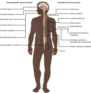
The sympathetic nervous system is activated when we are faced with stressful or high-arousal situations. The activity of this system was adaptive for our ancestors, increasing their chances of survival. Imagine, for example, that one of our early ancestors, out hunting small game, suddenly disturbs a large bear with her cubs. At that moment, his body undergoes a series of changes—a direct function of sympathetic activation—preparing him to face the threat. His pupils dilate, his heart rate and blood pressure increase, his bladder relaxes, his liver releases glucose, and adrenaline surges into his bloodstream. This constellation of physiological changes, known as the fight or flight response, allows the body access to energy reserves and heightened sensory capacity so that it might fight off a threat or run away to safety.
While it is clear that such a response would be critical for survival for our ancestors, who lived in a world full of real physical threats, many of the high-arousal situations we face in the modern world are more psychological in nature. For example, think about how you feel when you have to stand up and give a presentation in front of a roomful of people, or right before taking a big test. You are in no real physical danger in those situations, and yet you have evolved to respond to any perceived threat with the fight or flight response. This kind of response is not nearly as adaptive in the modern world; in fact, we suffer negative health consequences when faced constantly with psychological threats that we can neither fight nor flee. Recent research suggests that an increase in susceptibility to heart disease (Chandola, Brunner, & Marmot, 2006) and impaired function of the immune system (Glaser & Kiecolt-Glaser, 2005) are among the many negative consequences of persistent and repeated exposure to stressful situations.
Once the threat has been resolved, the parasympathetic nervous system takes over and returns bodily functions to a relaxed state. Our hunter’s heart rate and blood pressure return to normal, his pupils constrict, he regains control of his bladder, and the liver begins to store glucose in the form of glycogen for future use. These processes are associated with activation of the parasympathetic nervous system.
*
Section Summary
The brain and spinal cord make up the central nervous system. The peripheral nervous system is comprised of the somatic and autonomic nervous systems. The somatic nervous system transmits sensory and motor signals to and from the central nervous system. The autonomic nervous system controls the function of our organs and glands, and can be divided into the sympathetic and parasympathetic divisions. Sympathetic activation prepares us for fight or flight, while parasympathetic activation is associated with normal functioning under relaxed conditions.
*
Glossary
- autonomic nervous system
- controls our internal organs and glands
- central nervous system (CNS)
- brain and spinal cord
- fight or flight response
- activation of the sympathetic division of the autonomic nervous system, allowing access to energy reserves and heightened sensory capacity so that we might fight off a given threat or run away to safety
- homeostasis
- state of equilibrium—biological conditions, such as body temperature, are maintained at optimal levels
- parasympathetic nervous system
- associated with routine, day-to-day operations of the body
- peripheral nervous system (PNS)
- connects the brain and spinal cord to the muscles, organs and senses in the periphery of the body
- somatic nervous system
- relays sensory and motor information to and from the CNS
- sympathetic nervous system
- involved in stress-related activities and functions
