95 Ulceration Upset!
Sarah Schmidt; Elizabeth Wal; and Amelia Adelman
Gastric Ulcers in Horses: Glandular vs. Non-Glandular Ulcers
What is the difference between glandular and non-glandular ulcers, what are clinical signs and common preventatives for gastric ulcers in horses?
Learning objectives
- Explain the general anatomy and flow of food through the equine digestive tract
- Differentiate between glandular and non-glandular ulcers in horses.
-
Describe the anatomical differences between glandular and non-glandular regions of the horse’s stomach.
-
Explain why the non-glandular region is more susceptible to ulcers compared to the glandular region.
-
Identify the roles of stress and medication in the development of glandular and non-glandular ulcers.
-
-
Identify the clinical signs of gastric ulcers in horses.
-
Discuss the importance of early detection and treatment of gastric ulcers to maintain a horse’s health and performance.
-
-
Understand common preventatives for gastric ulcers.
-
Evaluate preventative strategies for gastric ulcers and determine which would be most effective in various management scenarios.
-
Lesson
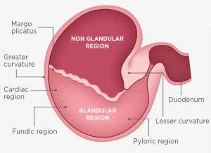
Gastric ulcers are a common and significant health concern in horses, affecting their overall well-being and performance. The horse’s stomach is divided into two distinct regions: the glandular and non-glandular areas, each with its unique structure and function. Understanding the differences between these two areas is crucial because it helps explain why some horses develop ulcers more easily in one region than the other.
The glandular region, located at the bottom of the stomach, produces mucus and bicarbonate to protect itself from the acidic environment. In contrast, the non-glandular region, found at the top, lacks these protective features, making it more vulnerable to stomach acid. This anatomical difference is one reason why non-glandular ulcers are more common in horses.

Gastric ulcers can lead to various clinical signs such as weight loss, poor appetite, and behavioral changes, which can severely impact a horse’s health and performance. Fortunately, understanding the causes and implementing effective preventative measures can reduce the risk and improve a horse’s quality of life.
In this lesson, we will explore the differences between glandular and non-glandular ulcers, identify the clinical signs of gastric ulcers, and learn common strategies to prevent them. Through engaging activities and assessments, you’ll gain the knowledge needed to recognize and help prevent this condition in horses.
Glandular Ulcers:
- Location: Lower portion of the horse’s stomach, which secretes protective mucus.
- Features: Less common than non-glandular ulcers but can be more severe.
- Causes: Often related to long-term stress, prolonged use of non-steroidal anti-inflammatory drugs (NSAIDs), or compromised blood flow.
Non-Glandular Ulcers:
- Location: Upper portion of the stomach, lacking protective mucus.
- Features: More common because this region is less protected and exposed to stomach acid.
- Causes: Related to feeding practices, infrequent feeding, high-concentrate diets, and stress.
Clinical Signs:
- Weight loss
- Poor appetite or change in eating habits
- Behavioral changes (e.g., irritability, discomfort)
- Mild to moderate colic symptoms
- Decreased performance
Common Preventatives:
- Providing access to continuous forage to buffer stomach acid.
- Reducing stress through consistent turnout and socialization.
- Limiting the use of NSAIDs or using gastroprotective drugs when NSAIDs are necessary.
- Ensuring a proper balance of roughage and grain in the diet.
- Offering supplements that support stomach health (e.g., antacids, coatings).
Video 1: describes broad overview of the physiology, causes, and clinical signs of equine gastric ulcers. Credit for video to ADM Protexin Ltd. Youtube channel.
Activities
Organize the characteristics based on if they are relating to glandular ulcers, non-glandular ulcers or both.
Color in one of these two pages:
- equine digestive tract to trace how food flows. (Taken from Cornell University Animal Science Department’s “Horse Guts and Math”).
- equine stomach to differentiate between the glandular and non-glandular sections. (Taken from Monique Warren’s “Preventing Equine Gastric Ulcers- How Forage Buffers Acid”).
Complete this crossword
Assessment
Short answer: What are the most common clinical signs of gastric ulcers in horses, and how can they be prevented?
Further exploration
The UMN Page on Stomach Ulcers in Your Horse
The UC Davis Page on Equine Gastric Ulcer Syndrome

