108 Doggy Diarrhea! Instructor guide
Timothy Magdall; Skylar Milne; and Natalya Wells
Doggy Diarrhea! Exploring Canine Hemorrhagic Gastroenteritis and the Relationship between Diarrhea and the GI Tract
Timothy Magdall, Skylar Milne, and Natalya Wells
Instructional Guide
Students will explore a case study of a canine patient with hemorrhagic gastroenteritis to learn the causes, diagnostics, and treatment of diarrhea. Learners will also apply this information to how it relates to the gastrointestinal (GI) tract. The lesson will include a short reading followed by learning activities and a self-assessment quiz at the end.
Intended Grade Level
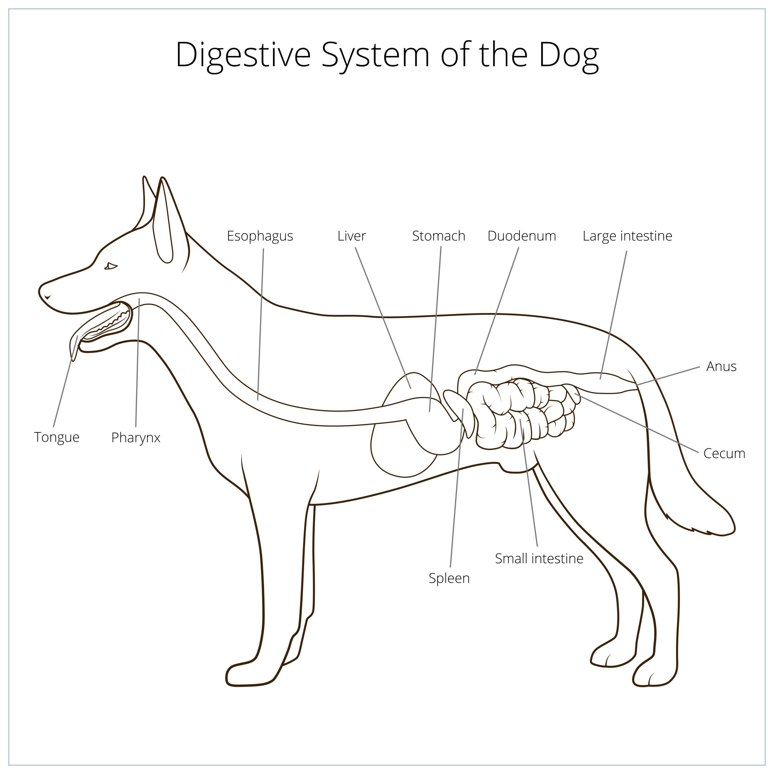
High school students (9th-12th grade). Students should be familiar with the general anatomy of the GI tract, such as the esophagus, stomach, and intestines. In addition, knowledge of the basic functions for the main organs involved in the digestive system is needed.
Minnesota State Standard addressed in this lesson:
| HS-LS1-2. | Develop and use a model to illustrate the hierarchical organization of interacting systems that provide specific functions within multicellular organisms. |
|---|
Learning Objectives
- Students will be able to explain the general anatomy and pathway of digesting food in the canine gastrointestinal tract.
- Students will be able to differentiate between digested and fresh blood in feces.
- Students should be able to describe how diarrhea affects the normal function of the digestive tract.
- Students can identify clinical signs for hemorrhagic gastroenteritis and appropriate treatment for dogs.
Lesson Format
This lesson should be taught to a typical classroom size (20-25 students). Students can either follow the material on their own computers or be provided printed copies with the instructions projected in front of the entire class. Activities may require a printer and colors.
- First, students will read over the background knowledge section on the GI anatomy of canines and how diarrhea affects its normal function through the lens of a case study where a patient has hemorrhagic gastroenteritis. This can be done independently or in a large group setting between the students. (15 minutes)
- Next, students will complete a crossword puzzle on the material learned and optional coloring pages may be provided. They can either work in small groups or independently. (25 minutes)
- The assessment will be an individual short quiz at the end of the lesson. (5 minutes)
Lesson Background
The gastrointestinal track is a continuous passageway from the mouth to the anus and has different sections based on unique functions. The mouth does the initial breakdown of the food through chewing, in addition to enzymes in the saliva. The esophagus moves the food or digesta (changes name after entering GI tract) from the mouth to the stomach where digestive enzymes and acid breakdown the particles further. The intestines continue the enzymatic breakdown and absorb nutrients and water from the digesta. The rectum then stores the digested food until the body is ready to excrete it. The GI tract is a very complex system that balances between breaking down food and absorbing the nutrients and water for the body to use. When things go wrong the GI tract, the body usually reacts with either vomiting or diarrhea.
The intestines are broken down in between two main sections: the small intestines and the large intestines. The main purpose of the small intestines is to mix the digesta with digestive enzymes and work to absorb excess water. The structure of the intestinal wall with specialized cells increases the efficiency of nutrient absorption, such as villi. These increase the surface area for absorption, and as surface area goes up, the amount of nutrients and water absorbed goes up. Villi are made up of specialized cells called epithelial cells that are themselves made up of little finger-like projections called microvilli. The epithelial cells have goblet cells interspersed between them that secrete mucus. Mucus protects the lining of the intestines from harmful microorganisms, acid, and digestive enzymes. The small intestines are the longest part of the GI tract.The large intestines are much shorter than small intestines. Large intestines continue absorption of nutrients and water that were not absorbed in the small intestine.
Gastroenteritis is an umbrella term meaning inflammation of the lining of the stomach and intestines. Hemorrhage is the term for loss of blood from the body. Putting these two terms together gives us the subject of our case study: hemorrhagic gastroenteritis, otherwise known as Acute Hemorrhagic Diarrhea Syndrome (AHDS). The symptoms of this disorder are vomiting, abdominal pain, lethargy (lack of energy), dehydration, and most notably, bloody, mucoidal diarrhea. The exact causes for AHDS are unknown but is predicted to be associated with dysbiosis. This can occur if the dog eats something that it does not normally eat or should not eat (inedible objects). Some dogs may develop no symptoms, some may develop mild cases of diarrhea.
Typically, diarrhea is thought of the way the body attempts to clear the intestines of something harmful. Physiologically, it is predicted that during AHDS, the body is reacting to the bacteria (C. perfringens) by excreting water and mucus into the intestines to try to restore the microbiome. The leakage of extra fluids into the intestines also brings red blood cells. This information should help students understand why then the diarrhea can appear bloody.
The color of the stool is clinically important since it can give a clue to the veterinarian as to where in the GI tract the blood may be coming from. Red blood in stools indicates that it is “fresh”, undigested blood – indicating the blood is coming from the lower GI tract closer to the rectum. Black material in the stool may indicate digested blood that is coming from higher up in the GI tract, further from the rectum.
There is no cure or medications for AHDS specifically. Therefore, treatment often focuses on treating the symptoms. This may include giving fluids, medications to protect the lining of the GI tract, pain medications, probiotics, and some antibiotics. These treatments are done to keep the patient comfortable, hydrated, and sustained nutritionally until their body is able to resolve the cause of the hemorrhagic diarrhea.
Activities
- Students should now begin the activities portion of the lesson.
- These activities can be completed by students in small groups or individually.
- The activities portion includes a crossword with material-relevant terms that students should understand by the end of this lesson.
- After the crossword is completed, students will start the coloring activity, highlighting the different parts of the gastrointestinal tract.
- Students will have an option to do a drag-and-drop activity to label an image of their coloring sheet with the correct parts of the GI tract.
Common misconceptions and challenge points
Hemorrhagic gastroenteritis can be a difficult topic to understand, especially for students. Below are some common misconceptions within this lesson.
- All blood diarrhea means hemorrhagic gastroenteritis.
- When learning about this topic, students might take away that all bloody diarrhea is a sign of hemorrhagic gastroenteritis. This is incorrect.
- Bloody diarrhea can be a result from many different things. Common problems indicated by bloody diarrhea can be parasites, dietary indiscretion, or other systemic illnesses.
- Mucous in the stool always indicates a disease.
- Some students might think that mucous in the stool always indicates a problem, but this is incorrect.
- Small amounts of mucous in the stool is normal and is an essential part of digesting food through the GI tract.
- However, excess amounts of mucous within the stool can indicate disease.
- Making this distinction to students can help to clear confusion with the purpose of mucous.
- Diarrhea is always harmful to the animal.
- Thinking that diarrhea is always harmful to the animal and should be stopped is false.
- While diarrhea can indicate bowel issues and underlying health problems, it is important to understand that diarrhea serves a purpose.
- Diarrhea is sometimes used by the animal’s body to expel harmful substances from the GI tract.
Assessment
After students finish reading the background knowledge section and completing the associated activities, they will take an individual short quiz. This will assess whether they are meeting all four learning objectives pertaining to their knowledge of canine GI tract anatomy, digested versus fresh blood in feces, the affects of diarrhea on the GI tract, and clinical signs/treatment for hemorrhagic gastroenteritis.
Further exploration
For students who are interested in veterinary medicine, below are some engaging resources and activities to support their learning:
- The Dog’s Digestive System – My Pet Nutritionist
- Systems – University of Georgia
- Resources For Aspiring Vets – Vet Set Go
- I want to be a veterinarian – VIN Foundation
Other chapters within this textbook may be interesting for students. Some options can be found linked below:
