138 Multiple GI Tracts? Jeez!
Mary Holmes; Elizabeth Hurley; and Jenna Striffler
Learning objectives
- Students will compare and contrast monogastric, ruminant, and avian digestive tracts.
- Students will identify why different digestive systems are advantageous for different species.
- Students will explain which digestive systems correspond to different diets and why.
Lesson
While every animal eats and eliminates, the stuff that happens in the middle varies by species. Veterinarians need a working knowledge of this variety. The main types are monogastrics (humans and dogs) and ruminants (cattle, goats). There are even pseudo-ruminants (alpacas and llamas). Birds are basically their own category; it is hard to fly with a full gastrointestinal tract!
Read these descriptions and watch the videos below to learn about these wild differences. Try your knowledge out by completing the wordsearch and the coloring pages. Finally, complete the quiz to the best of your ability. Your instructor has the answer key!
Monogastrics
Oral Cavity
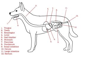
Digestion for monogastrics starts in the oral cavity, which includes the lips, teeth, and tongue. The lips help in prehension, or acquiring the food into the mouth. Once in the mouth, saliva and chewing help to break down the food. The teeth mash the food into smaller pieces, increasing the surface area for enzymes to act on. The saliva lubricates the food so that it can be swallowed. Some monogastrics such as humans contain salivary amylase in their saliva, which starts the digestion process of carbohydrates. However, dogs and cats do not contain salivary amylase in their saliva. Food is swallowed and moves into the esophagus where it is then delivered into the stomach.
Stomach
Monogastrics are defined by their single-chambered stomach. The stomach is where protein digestion begins in monogastrics. Inactive pepsinogen is produced by chief cells. Hydrochloric acid is released by the parietal cells which converts pepsinogen into pepsin. Acid and pepsin then break proteins down into polypeptides. Some fat digestion takes place in the stomach due to the presence of gastric lipase, but it is not the main source of fat digestion. The food then moves into the small intestine through the pyloric sphincter.
Small Intestine
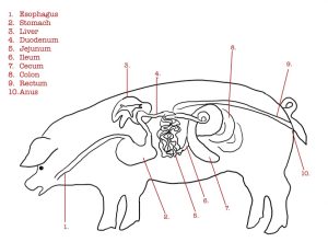
The small intestine is the major site of digestion and includes three regions: the duodenum, jejunum, and ilium. The first part is the duodenum, which receives the partially digested food from the stomach. Food then moves through the jejunum and ilium, where nutrients continue to be absorbed. During this process, bile from the gallbladder is secreted into the small intestine to emulsify fats to digest them. The pancreas also releases enzymes to help break down the food for digestion. All the parts work together to break down carbohydrates, fats, and proteins into their simplest form to be digested. The small intestine also has special hair-like projections called villi. These structures increase the surface area of the small intestine to maximize absorption and digestion. The leftover contents are then moved to the large intestine.
Large Intestine
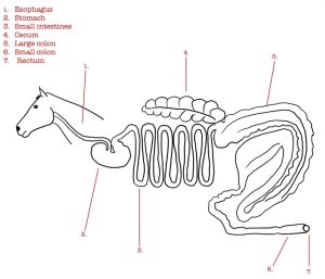
The large intestine continues to absorb water and vitamins and prepares feces to be excreted. In carnivores and omnivores, this process is fairly simple. However, hindgut fermentors such as rabbits and horses do things a little differently. These animals eat primarily grasses, which the animal itself cannot digest. Instead, they rely on microbial colonies to break down cellulose into volatile fatty acids, which the individual can then use. Because this process is different than other monogastrics, hindgut fermenters rely on an additional part of the digestive tract that is less pronounced or absent in other monogastrics: the cecum. The cecum is the site of billions of microorganisms that can break down the structural fibers that compose most of these animals’ diets. Because the cecum is so large in hindgut fermenters, it slows down the process of digestion, which allows the microbes more time to break down the grasses that are ingested. The process of digestion ends the same in both hindgut fermentors and other monogastric animals, which is the excretion of waste through the anus. It should be noted that pigs have an elongated, spiral colon that allows for some digestion of cellulose.
Why Are Monogastrics Designed This Way?
Monogastrics are incredibly diverse in their anatomy, so there is not one answer as to why these animals have a monogastric digestive system. Much of it can be attributed to evolution and the diet of each species. For carnivores and omnivores, a monogastric system is the most effective for breaking down proteins that can be used for energy. They also consume very little, if any, structural fibers, so they do not need as extensive of a digestive system as ruminants. The outlier in this feature is the hindgut fermenters, who primarily consume structural fibers. The reasons these animals are monogastric can also attributed to evolution. The most likely explanation is that the ancestors of the hindgut fermenters could only rely on fibrous food materials, like grasses and hays. However, they might not have been able to effectively digest these structural fibers. Somewhere along the way, hindgut fermenters developed a cecum that allowed them to derive energy from the food sources that were readily available to them. This left us with monogastrics that primarily consume structural fibers.
Ruminants
Mouth
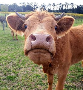
Ruminants use their mouth and tongue to graze by wrapping their tongues around the plant and pulling to tear the forage. Ruminants have a hard pallet that lacks top incisors but have bottom incisors. Premolars and molars match between the upper and lower jaw that are used to crush and grind plant material during initial chewing and rumination. The ruminant will chew forages to about 1-1.5″ during the initial eating. Once the ruminant is not actively eating or drinking, it will regurgitate the partially digested feed to be grind down to small pieces. Ruminants need to salivate to add saliva to the rumen. Saliva serves to provide liquid for the microbes in the stomach, recirculates nitrogen and minerals, and buffers the rumen. Saliva also starts the breakdown of fat and starch using enzymes such as lipase and amylase.
Esophagus
The esophagus functions bidirectionally in ruminants, allowing them to regurgitate their cud for further chewing, if necessary. The process of rumination or “chewing the cud” is when forage and other feedstuffs are forced back to the mouth for further chewing and mixing with saliva. This cud is then swallowed again and passed into the reticulum.
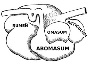
Stomach
Ruminants have 1 stomach with 4 chambers – the rumen, the reticulum, the omasum, and the abomasum.
Rumen
The rumen can be found on the left side of the animal and is the largest compartment. It has several sacs, and an adult cow can hold 25 gallons. The rumen acts as a storage or holding vat for feed. The rumen is compared to shag carpet making an environment for bacteria to thrive in. Fermentation happens in the rumen as the environment favors the growth of microbes. These microbes digest or ferment feed within the rumen and make volatile fatty acids (VFAs). The rumen absorbs most of the VFAs from fermentation. Fermentation also produces 30 to 50 quarts of gas per hour in cattle. Cows must release this gas to avoid bloating causing the cow to belch.
A good blood supply to the rumen walls improves absorption of VFAs and other digestion products. Tiny projections (papillae) line the rumen, which increases the rumen’s surface area and the amount it can absorb.
Reticulum
This pouch-like structure is closer to the head and heart in the thorax of the body. The reticulum is known for its honeycomb walls. Heavy or dense feed and metal objects eaten drop into this compartment. “Hardware disease” can be caused by these metal objects penetrating the chambers and organs which causes loss of yield and potentially death. A magnet can be given orally and attract these metal objects in the reticulum for the entirety of the cow’s life. Many dairy farms have a magnet as part of their vaccination protocol.
Omasum

This globe-like structure contains projects of skin like pages in a book to increase surface area for absorption. Water absorption is increased in the omasum causing the feed found here to be drier than in previous chambers.
Abomasum
Also known as the true-stomach, the abomasum is the most similar chamber to that of the monogastric. These glands release hydrochloric acid and digestive enzymes, needed to break down feeds.
Small Intestines
The small intestine is similar to other digestive tracts with having the three parts – duodenum, jejunum, ileum. The gallbladder and pancreas secrete enzymes that further break down food that are then absorbed using villi. The villi are fingerlike projections that increase surface area. After nutrients are absorbed, they enter the blood stream and lymphatic system. Most of the digestion happens in the small intestine.
Cecum
The cecum is found where the small intestine and large intestine meet. While it aids in digestion of undigested fibers, the exact importance remains unknown.
Large Intestine
Some digestions happens here, but the main priority of the large intestine is the absorption of water.
Young Ruminants
Young ruminants have an esophageal groove that allows milk to bypass the rumen and reticulum. The microbes in the rumen would ferment the milk causing it to curdle and can cause colic. The rumen will not develop until the young are introduced to grains and forages.
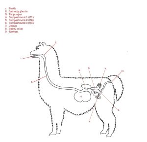
Ruminants vs Pseudo ruminants
Pseudo ruminants have a 3-chamber stomach – missing the omasum. These are called C1, C2, and C3. They still go through the steps of rumination and the rest of the digestive system is similar. Examples of pseudo ruminants include the camelid family such as llamas and alpacas.
Why are Ruminants Designed This Way?
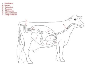
Ruminants are hoofed mammals, including cattle, sheep, and goats, with a unique digestive system that allows them to better use energy from fibrous plant material when compared with other herbivores. Unlike monogastrics, ruminants have a digestive system designed to ferment feedstuffs and provide precursors for energy for the animal to use. By better understanding how the ruminant digestive system works, veterinarians can work with livestock producers to better understand how to care for and feed ruminant animals.
Avian
Please note that when looking at the digestive systems of avian species, there can be a lot of changes between birds because they are different species. For more resources, please reach out to your professor for extra supplemental readings if you’re interested in learning more about the distinctions between species.
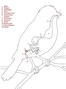
Oral Cavity

One of bird’s most definable traits are their beaks. Beaks – or bills for some species – vary in shape and size depending on the diet of the species. Unlike most mammals, birds don’t possess teeth or the ability to chew their food. Food is guided to the back of the mouth with their tongue, swallowed whole and broken down further in the bird’s digestive system. Some birds, primarily granivorous species, have salivary glands that secrete amylase to begin the digestive process.
Esophagus
Longitudinal folds allow birds to expand their esophagus to accommodate food being swallowed whole. Mucus from mucosal glands also help the food slide down the esophagus.
Crop
For many species, an expansion of the esophagus, called the crop, acts as storage. Birds can ingest large quantities of food during one feeding and store it in the crop until they are in a location where it’s safe to complete the digestion cycle. Emptying of the crop is tied to the gastric cycle. The structure of the crop is variable between species.
Stomach
Avian stomachs are comprised of two chambers: the proventriculus and ventriculus.
Proventriculus
The proventriculus is a glandular stomach and mimics the monogastric stomach or ruminant’s abomasum. Gastric glands secrete hydrochloric acid, pepsinogen, and mucus to aid in secretory digestion. The zona intermedia gastric is a narrow passage (isthmus) that separates the proventriculus and ventriculus. The proventriculus is variable in shape and size between species.
Ventriculus
The ventriculus, or gizzard, is the muscular stomach that contributes to mechanical digestion. Muscles and pressure work to grind and break down food. The interior is lined by koilin, a thick protein layer that preserves the gizzard from proteolytic enzymes and acid secreted by the proventriculus. The ventriculus is well developed and distinct in bird species that are granivores, herbivores, and insectivores whereas it is poorly developed in carnivorous species. Some species, like chickens, may eat small stones that will reside in the gizzard to aid in grinding food into smaller pieces.
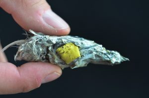
A unique adaptation of some carnivorous bird species is egestion, otherwise known as casting. Casting is the process of expelling nondigestible material, including fur and bones, through the oral cavity. Nondigestible material is contained in the gizzard. Once most nutrients have been absorbed from the food, the gizzard begins to contract stronger and more quickly 12 minutes prior to egestion. The nondigestible material condenses into a pellet and is pushed into the lower esophagus. Esophageal antiperistalsis moves the pellet cranially roughly 8 to 10 seconds before egestion where it is then expelled by the oral cavity.
Small Intestines
The small intestines facilitate enzymatic digestion and nutrient absorption of sugars, calcium, phosphorus, amino acids, and water. Structurally, avian small intestines are like monogastric and ruminant digestive systems in that there are three portions: the duodenum, jejunum, and ileum. Although variable between avian species, the length of the small intestine is typically much shorter than mammals. Due to the shorter size, duodenal refluxes reverse the motility of the duodenum and ilium, pushing ingesta back to the ventriculus to allow for further digestion. In contrast to mammals, bile flows into the duodenum via two ducts: the hepatocystic and right hepatoenteric duct.
Large intestine
The large intestine typically includes a paired ceca, colon/rectum, and cloaca. The ceca include a pair of sacs between the small and large intestine that absorbs water and facilitates bacterial fermentation, breaking down dietary fibers and proteins for energy. The paired ceca are emptied two to three times daily. Ceca are extremely variable in shape and size between species and absent in some.
The colon, or rectum, is shorter in birds than most mammalian species except for the ostrich. Except for defecation, antiperistalsis continually occurs in the colon. Unlike mammals that typically have numerous goblet cells and no villi in their colon, birds have large quantities of villi and very few goblet cells. Water absorption is the main function of the colon.
Whereas mammals have separate openings to excrete urine, fecal, and reproductive waste, birds have one opening and excrete all waste from there in the form of a mute. The cloaca has three incomplete chambers including the coprodeum, urodeum, and procteum. The reproductive and urine tracts empty into the urodeum. The procteum connects with the anus.
Why Are Birds Designed This Way?
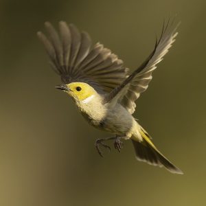
Birds possess lightweight beaks and a shorter digestive system to reduce their weight, making it easier for them to fly. Given their shorter digestive tract, refluxes and antiperistalsis help birds to absorb the maximum nutrients from their food. Birds also have faster metabolism to efficiently obtain enough energy for flight. The crop is advantageous for birds because it allows them to consume larger food portions. Birds can store food in their crop until they’re in a safe place to complete the digestion process. The digestive tracts of bird species vary to accommodate the different diets that birds possess.
Try this to check your understanding!
Coloring Book Pages
Download and/or print one of these coloring pages to see what you have learned over the course of this unit. Answers to these coloring pages are provided within the instructor guide.
Avian Digestive System Coloring Page
Assessment
Additional Resources
Diagrams created by the authors; CC for NC.

