11. Juvenile Cellulitis – Canine
Learning Objectives
- Know! Juvenile cellulitis, also known as puppy strangles, juvenile pyoderma, sterile granulomatous dermatitis and lymphadenitis, is a sterile pyogranulomatous condition usually seen in dogs less than 4 months of age. One or multiple puppies in the litter may be affected. Occasionally, it can be diagnosed in young adult dogs.
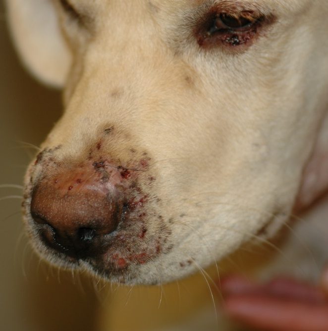
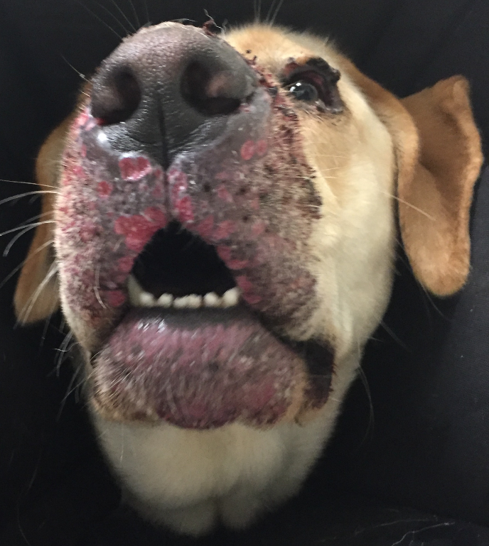
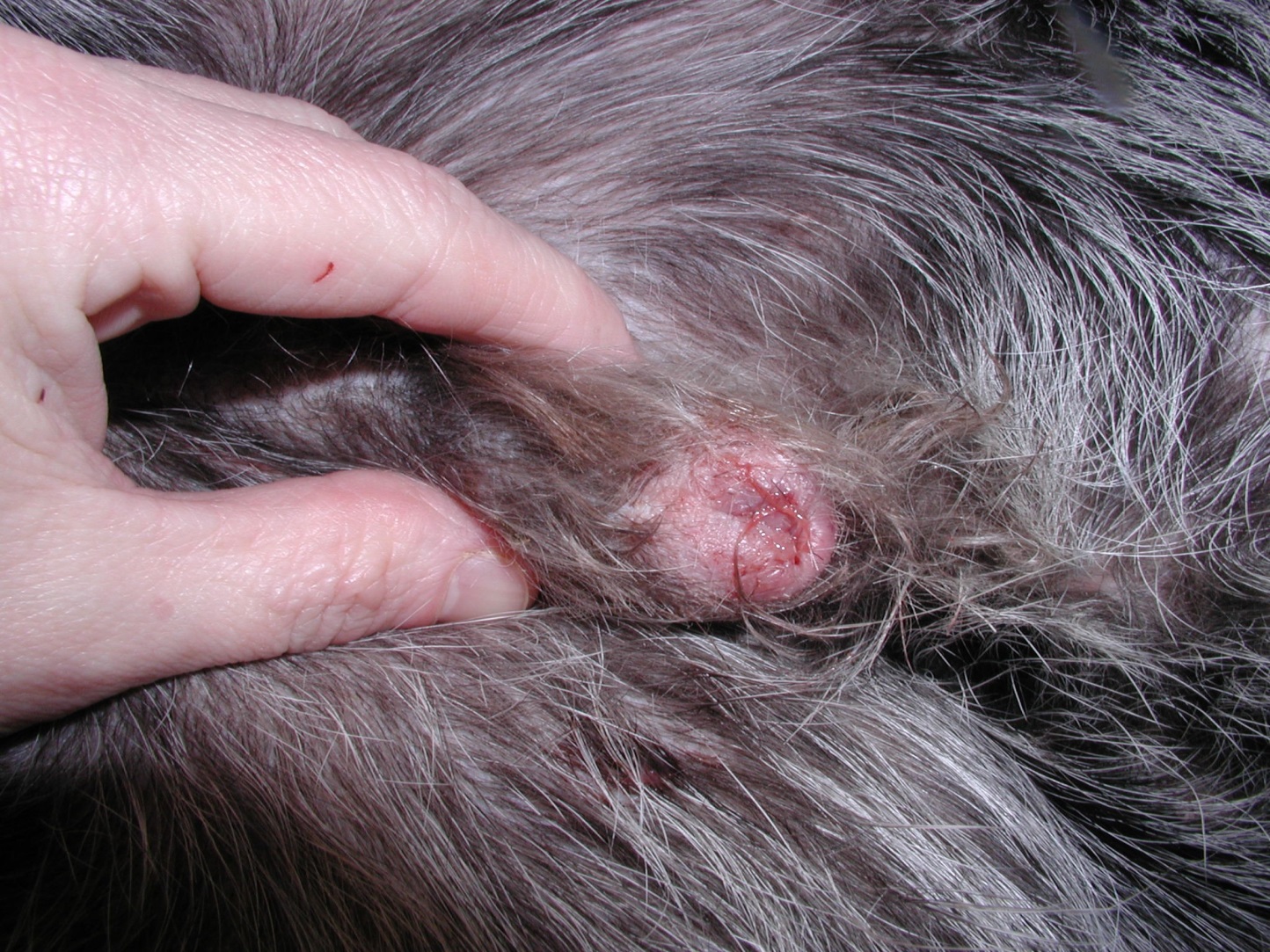
- Know! Lesions are initially characterized by ill-defined swelling that evolve to form papules, small nodules, and pustules localized to the pinnae, muzzle, chin, lips and peri-ocular area. Eventually, some lesions will form draining tracts and areas of erosions to ulceration develop. Marked lymphadenopathy (especially sub-mandibular), fever, depression and anorexia are noted.
- Remember! Some dogs may develop arthritis, lameness and paresis due to cerebrospinal fluid inflammation.
- Know! The diagnosis should be based on a characteristic history, clinical signs, cytological and histopathological findings. Avoid sampling open, draining lesions for cytology and histopathology.
- Remember! Always perform multiple skin scrapes to rule out demodicosis!
- Know! The first treatment is immunosuppressive doses of prednisone or prednisolone. The response should be noticed within 14 to 21 days. Severe or refractory cases may benefit from adding cyclosporine to the treatment regimen.
- Remember! Perform cytology of draining lesions to look for the presence of secondary bacterial infection. If secondary infection is present, add an antibiotic based on bacterial culture and susceptibility (e.g. cephalexin, cefadroxil or amoxicillin clavulanate).
- Remember to tell owners that lesions may heal with scarring.
-
General Considerations
- Juvenile cellulitis (also known as puppy strangles, juvenile pyoderma, and sterile granulomatous dermatitis and lymphadenitis) is an uncommon sterile pyogranulomatous skin disease of dogs that also affects peripheral lymph nodes and less commonly the joints.
- The condition is presumed to be immune mediated based on histopathological features and good response to immunosuppressive therapy.
- A heritable nature has been suggested because of increased occurrence in certain breeds and familial histories of disease.
- Several puppies or only one in the litter may be affected.
- The common history of vaccination before disease development makes vaccine antigens possible triggers.
- Juvenile cellulitis can spontaneously resolve within 1 to 3 months.
Important Facts
- Juvenile cellulitis is an uncommon sterile pyogranulomatous canine skin disease presumed to be immune mediated. It also affects peripheral lymph nodes and less commonly the joints.
- A heritable nature has been suggested because some breeds are more prone to develop the disease.
- The common history of vaccination before disease development makes vaccine antigens possible triggers.
- Juvenile cellulitis can spontaneously resolve within 1 to 3 months.
-
Signalment
- The dachshund, Labrador retriever, golden retriever, Gordon setter, Irish setter, Maltese and bichon frise dogs are overrepresented in various reports, but any breed can develop this disease.
- Most affected animals are 4 months old or younger, but juvenile cellulitis can affect older dogs. This has recently been confirmed in a retrospective study of 90 cases where the median age at disease onset was 3.54 years (range: 1-11.4 years, mean 4.29 years).
- No sex predilection has been reported.
-
Clinical Signs
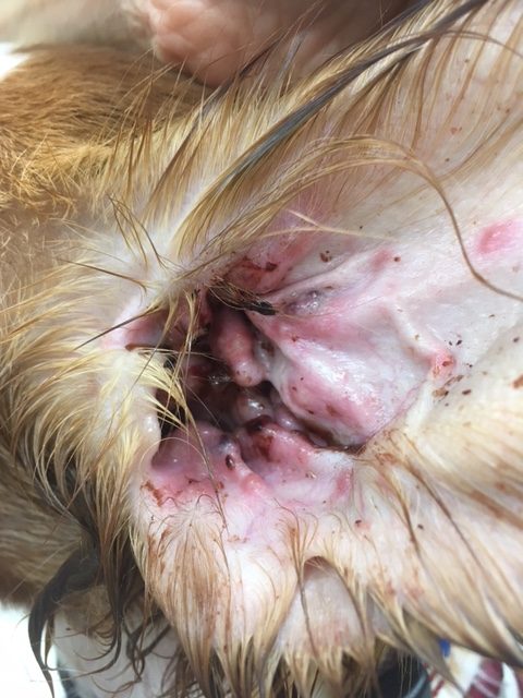
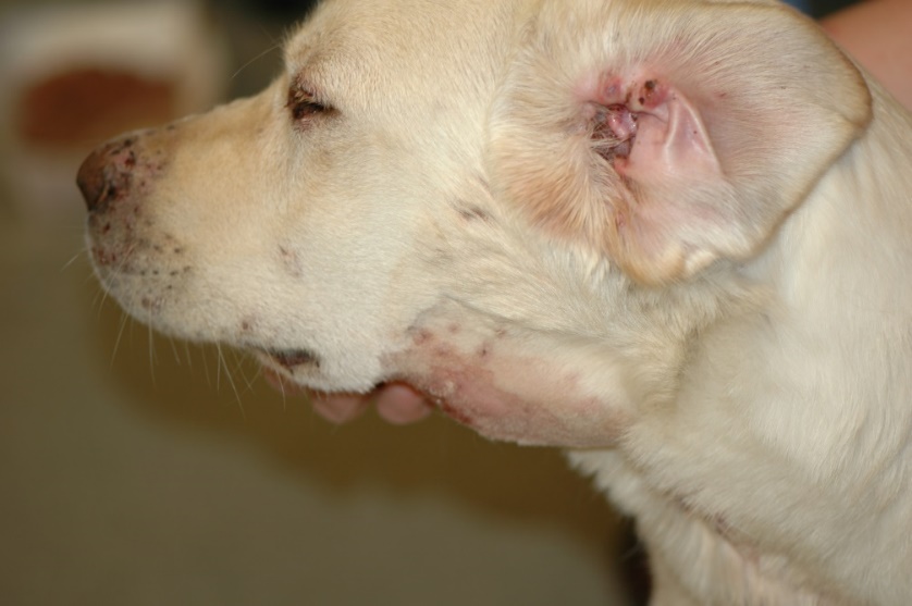
- The first sign in many cases is an acute facial swelling, which progresses within 24 to 48 hours to form papules and nodules on the concave pinnae, muzzle, chin, lips, periocular area, and eyelids. These lesions rapidly progress to abscessation, draining tracts and focal erosions and ulcerations. Vesicles and pustules can be also seen especially on the concave pinnae. Lesions on the pinnae often progress to the external ear canals causing otitis externa.
-
- Peripheral lymphadenopathy (especially mandibular lymph nodes but also prescapular or generalized) is a common clinical sign and may precede the skin lesions. Occasionally affected lymph nodes will abscess and drain. Rarely, dogs may present with mandibular lymphadenopathy as the only clinical sign.
-
- A few cases will develop nodules over the trunk, preputial and perineal areas. Some of these lesions may be associated with sterile granulomatous panniculitis and may eventually drain.
-
- Lesions of juvenile cellulitis are typically painful but not pruritic.
- Puppies are often febrile, depressed and anorexic. In the author’s experience, older dogs tend to have systemic signs more often and are typically pyrexic.
- Rarely, arthritis, lameness and paresis due to sterile inflammation of the joints and cerebrospinal fluid may occur.
- Permanent areas of alopecia and scarring may develop in chronic cases as deep lesions heal.
Important Facts
- Most affected animals are 4-month-old or younger, but juvenile cellulitis is also reported to occur in dogs from 1-11 years of age.
- Several puppies or only one in the litter may be affected.
- Diffuse swollen, papules, small nodules, vesicles or pustules are initially seen on the pinnae, muzzle, chin, lips and eyelids. Otitis externa often accompany the pinnal lesions.
- Lesions rapidly progress to abscessation and draining tracts.
- Some dogs may develop subcutaneous nodules of sterile pyogranulomatous panniculitis along the trunk, prepuce and perianal regions.
- Peripheral lymphadenopathy (especially mandibular) occurs and occasionally lymph nodes will abscess and drain.
- Puppies are usually febrile, depressed and anorexic. Older dogs tend to have systemic signs more frequently and are often pyrexic.
- Arthritis, lameness and paresis due to sterile inflammation of the joints and cerebrospinal fluid may occur.
- Chronic lesions may heal with scarring.
-
Diagnosis
- Differential diagnoses include angioedema (not associated with lymphadenopathy and systemic signs), demodicosis, deep pyoderma, sterile nodular panniculitis, sterile pyogranuloma/granuloma syndrome, and cutaneous drug reaction.
- Diagnosis is based on the pet’s signalment, detailed history, characteristic clinical signs, cytological examination of samples from draining lesions, intact pustules and nodules (show sterile pyogranulomatous inflammation), skin biopsy and response to immunosuppressive therapy.
- Skin scrapes should be performed in all cases to rule out demodicosis.
- Histopathological findings include a nodular to diffuse dermatitis and panniculitis composed of epithelioid macrophages and neutrophils.
- Bacterial cultures of samples taken from intact lesion yield no growth. However, they can be positive if samples are obtained from draining lesions, which often become secondarily infected.
Important Facts
- Diagnosis is based on the pet’s signalment, detailed history, clinical signs, cytological and histopathological findings and response to immunosuppressive therapy.
- Skin scrapes must be performed to rule out demodicosis.
-
Treatment
- Immunosuppressive doses of corticosteroids:
- Oral prednisolone/sone, at 2.0 mg/kg q 24h until remission is achieved (~2 to 3 weeks). Once lesions have resolved reduce the glucocorticoid dose slowly to prevent relapses and eventually discontinue therapy.
- Some dogs will respond better to oral dexamethasone at 0.2 mg/kg q 24h.
- Cyclosporine at 5.0 mg/kg q 24h given with prednisolone can be an option for more severe cases or cases not responding solely to glucocorticoids.
- A report showed success in treating six puppies with griseofulvin at 14.2 to 34 mg/kg q 12h. The reported efficacy was likely due to the immunomodulatory effect of griseofulvin.
- In a retrospective study including only young adult dogs of ≥ 1 year of age, the median time to remission independent of the treatment used was 28 days (range 12-116 days).
- If secondary bacterial infection is present, a first-tier antibiotic should be chosen based on culture and susceptibility such as, cephalexin, cefadroxil and amoxicillin clavulanate.
- Juvenile cellulitis usually responds completely to oral glucocorticoids with little chance of recurrence. Nevertheless, recurrence may occur if therapy is discontinued too soon. Close monitoring is needed to determine the proper time to discontinue therapy.
- Owners should be educated that some lesions may heal with scar formation.
- Immunosuppressive doses of corticosteroids:
Important Facts
- Immunosuppressive doses of glucocorticoids is required until remission (about 2-3 weeks).
- Cyclosporine combined with glucocorticoids may be required in severe cases or cases not responding only to glucocorticoids.
- First-tier antibiotic therapy based on bacterial culture and susceptibility should be added to the treatment regimen to manage secondary bacterial infections.
- Juvenile cellulitis usually responds completely with immunosuppressive therapy with little chance of recurrence. Close monitoring is needed to determine the proper time to discontinue therapy and avoid recurrence.
References
Inga A, Griffeth GC, Drobatz KJ et al. Sterile granulomatous dermatitis and lymphadenitis (juvenile cellulitis) in adult dogs: a retrospective analysis of 90 cases (2004-2018). Vet Dermatol 2019; DOI: 10.1111/vde.12820.
McKeever PJ, Nuttall T, Harvey RG. A Color Handbook of Skin Diseases of the Dog and Cat. 2nd edn. London: Manson Publishing Ltd., 2009; 159.
Miller WH, Griffin GE, Campbell KL. Muller & Kirk Small Animal Dermatology. 7th edn. St Louis: Elsevier Inc., 2013; 708-709.
Neuber AE, van den Broek AH, Brownstein D, et al. Dermatitis and lymphadenitis resembling juvenile cellulitis in a four-year-old dog. J Small Anim Pract 2004; 45:254-258.
Park C, Yoo JH, Kim HJ, et al. Combination of cyclosporine A and prednisolone for juvenile cellulitis concurrent with hindlimb paresis in 3 English cocker spaniel puppies. Can Vet J 2010; 51:1265-1268.
Park C, Jung Da, Park H. A case of juvenile cellulitis concurrent with hindlimb paresis in an English cocker spaniel puppy. Vet Derm 2004; 15:57.
White SD, Rosychuk RA, Stewart LJ, et al. Juvenile cellulitis in dogs: 15 cases (1979-1988). J Am Vet Med Assoc 1989; 195:1609-1611.
