20. Feline Proliferative and Necrotizing Otitis Externa and Dermatitis
Learning Objectives
- Know! Proliferative and necrotizing otitis externa and dermatitis (PNOED) is a rare condition of cats. Cats younger than 1 year of age are most often affected. In most cases, cats will have bilateral proliferative otitis externa, but some cats can have extra-auricular disease, which can be localized to a body region or generalized.
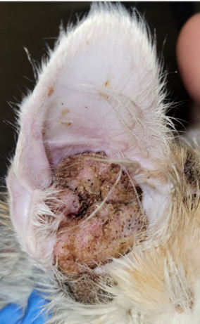
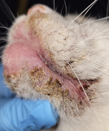
- Know! Lesions are characterized by coalescing papules, plaques and nodules that are covered with tightly adhered to fragile, yellowish to light brown to blackish crusts. Pruritus may or may not be present. Secondary infection can develop, especially in otic lesions.
- Know! The clinical diagnosis is based on the pet’s history and clinical signs. The otic disease is distinctive, which helps with the presumptive clinical diagnosis. Its proliferative aspect can resemble neoplasia. However, the young age of the animal at the time of disease development and the bilateral involvement, make neoplasia unlikely. Differential diagnoses for extra-auricular lesions include dermatophytosis, notoedric mange and pemphigus foliaceus. Skin biopsy is needed to confirm the clinical diagnosis. Cytology must be performed in all cases to investigate the presence of secondary infection, which is very common with the otic presentation.
- Know! Tacrolimus 0.1% ointment is the mainstay option for the otic presentation and extra-auricular disease localized to one body region. It may be impractical for generalized cases. Oral cyclosporine, glucocorticoid, or oclacitinib can be tried in cases of generalized extra-auricular disease or otic and localized extra-auricular cases refractory to tacrolimus 0.1% ointment. Some cases may spontaneously resolve.
- Remember! Monitor the patient closely for potential side effects when using immunomodulatory/immunosuppressive drugs.
-
General Considerations
- Proliferative and Necrotizing Otitis Externa and Dermatitis (PNOED) is a rare condition of cats.
- Most commonly, it affects the concave aspects of the pinnae and external ear canals but, in some cases, the disease develops in other parts of the body.
- Cats less than 1 year of age appear to be predisposed but PNOED can also affect older cats.
- The pathomechanism is not completely understood; however, there is some evidence to support the involvement of T-cells in disease development.
- Lesions typically respond well to immunomodulatory drugs, but spontaneous resolution can occur.
Important Facts
- Proliferative and Necrotizing Otitis Externa and Dermatitis is a rare condition of cats.
- It typically affects the concave aspect of the pinnae and external ear canals, but extra-auricular dermatitis has been reported.
- Cats < 1 year of age appear to be predisposed but adult cats can also be affected.
- There is some evidence to support the involvement of T-cells in disease development.
- In general, lesions respond well to immunomodulatory drugs, but spontaneous resolution has been reported.
-
Clinical Signs
- Otic lesions are characterized by coalescing erythematous papules, plaques and nodules covered with tightly adhered to fragile, yellowish to light brown to blackish crusts.
- In most cases, lesions affect the concave pinnae and the external ear canals. However, less commonly it can only affect the ear canals.
- Secondary bacterial and/or yeast otitis often occurs.
- Otic lesions are characterized by coalescing erythematous papules, plaques and nodules covered with tightly adhered to fragile, yellowish to light brown to blackish crusts.
-
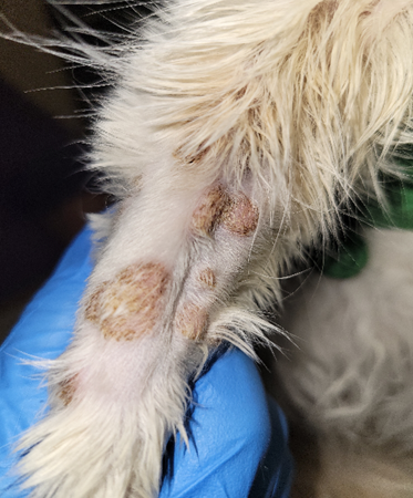
- Extra auricular involvement can occur and, typically, accompany otic lesions but the ears may not be involved in some cases. Lesions are similar to the ones developed in the ears.
- Lesions can develop in many parts of the body or can be localized to one body region.
- Cats with extensive extra-auricular involvement may present with systemic signs such as fever, hyporexia and lethargy.
- Secondary bacterial infection or overgrowth may develop.
- In chronic cases, the tympanic membrane can rupture leading to advancement of the material accumulated in the external canal to the middle ear. This material can provide a nidus for infectious organisms to grow and cause otitis media.
- Extra auricular involvement can occur and, typically, accompany otic lesions but the ears may not be involved in some cases. Lesions are similar to the ones developed in the ears.
-
- The crusts covering the otic and extra otic lesions are tightly adhered to fragile. The underneath skin may become eroded as tightly adhered crusts are removed.
- The disease can progress rapidly.
- Cats with PNOED may be pruritic.
Important Facts
- The concave aspect of the pinnae and external ear canals are more often affected but other parts of the body can also be affected and, in some cases, without otic involvement.
- Otic and extra auricular lesions are characterized by erythematous and coalescing papules, plaques and nodules, which are covered with adhered to fragile, light brown to blackish crusts.
- Systemic signs may be present in cats with extensive extra auricular involvement.
- Secondary infection occurs often.
- Pruritus may be present.
-
Diagnosis
- The otic lesions are distinctive, which helps with clinical diagnosis.
- The proliferative aspect of the otic lesions resembles neoplastic disease, at least in some cases. However, the young age of the animals at the time the lesions develop and the bilateral involvement, make neoplasia unlikely.
- Differential diagnoses for extra auricular lesions include dermatophytosis, notoedric mange and pemphigus foliaceus.
- Biopsy is needed to confirm a presumptive clinical diagnosis and rule out other causes.
- Key histopathological findings include marked epidermal and hair follicle epithelium hyperplasia associated with numerous and often coalescing, apoptotic keratinocytes. Parakeratotic hyperkeratosis is also present along the epidermis and in the hair follicle infundibulum.
- Cytology from ear canal samples and body lesions is important to investigate secondary infections.
- The otic lesions are distinctive, which helps with clinical diagnosis.
Important Facts
- The otic lesions are distinctive, which helps with clinical diagnosis.
- Biopsy is needed to confirm a presumptive clinical diagnosis and rule out other causes.
- Cytology from samples collected from the ear canals and body lesions is important to investigate secondary infections.
-
Treatment
- Tacrolimus 0.1% ointment applied once or twice daily has shown to work very well for cases with otic involvement.
- To facilitate medication delivery, the ointment can be diluted in 10 mL mineral oil or compounded as otic solution.
- In cases of generalized extra auricular disease or otic and localized extra auricular cases refractory to tacrolimus, the following systemic immunomodulatory/immunosuppressive drugs can be tried:
- Oral cyclosporine (microemulsion formulation) – 7-10 mg/kg q 24h. Few reported cases treated with oral cyclosporine, which showed questionable efficacy.
- Oral prednisolone – 1-2 mg/kg q 24h. The author’s experience indicates that it does not work well. Other glucocorticoids can be tried.
- Oclacitinib (Apoquel®) – 0.5 – 1.0 mg/kg q 12-24h. Very few reported cases treated with oclacitinib; however, the response was excellent.
- It is not labelled for use in cats and should be considered in cases refractory to tacrolimus, cyclosporine and/or prednisolone.
- These immunomodulatory/immunosuppressive therapies have potential and concerning side effects; therefore, the patient has to be monitored closely during therapy.
- Spontaneous resolution has been reported.
- Tacrolimus 0.1% ointment applied once or twice daily has shown to work very well for cases with otic involvement.
Important Facts
- Tacrolimus 0.1% ointment is the mainstay therapy for localized PNOED.
- Oral cyclosporine, glucocorticoid, or oclacitinib can be tried in cases of generalized extra auricular disease or otic and localized extra auricular cases refractory to tacrolimus 0.1% ointment.
- Monitor the patient closely for potential side effects when using immunomodulatory/immunosuppressive drugs.
- Spontaneous resolution has been reported.
References
Borio S, Massari F, Abramo F et al. Proliferative and necrotising otitis externa in a cat without pinnal involvement: video-otoscopic features. J Feline Med Surg. 2013;15: 353–356.
McAuliffe L, Sargent S, Locke E. Pathology in Practice. Proliferative and necrotizing otitis externa (PNOE), albeit with an unusual distribution, in a kitten. J Am Vet Med Assoc 2020; 257: 287-290.
Momota Y, Yasuda J, Ikezawa M, et al. Proliferative and necrotizing otitis externa in a kitten: successful treatment with intralesional and topical corticosteroid therapy. J Vet Med Sci 2017; 78: 1883-1885.
Mauldin EA, Ness TA, Goldschmidt MH. Proliferative and necrotizing otitis externa in four cats. Vet Dermatol 2007; 18: 370-377.
Panzuti P, Jongh O, Dony M, et al. Extra-auricular lesions of proliferative and necrotizing otitis externa in three kittens. Vet Dermatol 2021; 32:385-e110.
Stevens BJ, Linder KJ. Pathology in Practice. Proliferative and necrotizing otitis externa. J Am Vet Med Assoc 2012; 241: 567-569.
Vidémont E, Pin D. Proliferative and necrotising otitis in a kitten: first demonstration of T-cell-
Mediated apoptosis. J Small Anim Pract. 2010;51: 599–603.
