18. Feline Miliary Dermatitis
Learning Objectives
- Know! Feline miliary dermatitis is not a disease entity but a cutaneous reaction pattern associated with many feline dermatosis. The most common clinical presentation of fleabite hypersensitivity in cats is miliary dermatitis.
- Know! The lesions of miliary dermatitis consist of multiple, small and crusted erythematous papules. If self-inflicted alopecia is not present, the lesions can be identified through palpation. The distribution of the lesions can be a clue to the primary cause. For example, in the case of fleabite allergy, the lesions will be primarily on lumbar-sacral and base of the tail regions. Pruritus may or may not be present depending on the primary disease and will help with the development of a sensible list of possible causes.
- Know! It is easy to diagnose the miliary dermatitis lesions, but it can be challenging to identify the cause. A detailed history and thorough physical examination are needed to increase the chances of determining the underlying cause. Additional tests may include skin scraping, flea combing, cytology, food trial, and fungal culture. Prioritization of the possible causes will guide the clinician regarding the diagnostic tests to perform first. Biopsy findings are supportive of miliary dermatitis, but seldom yield a definitive diagnosis.
- Know! The successful management of miliary dermatitis is dependent upon identifying and controlling the primary causes.
-
General Considerations
- Miliary dermatitis is a multifactorial cutaneous reaction pattern of cats, and most cases are associated with fleabite allergies.
- All other causes of feline allergies including food allergy and feline atopic skin syndrome must also be considered as possible differential diagnoses.
- Parasitic disorders that may manifest with various degrees of miliary dermatitis include cheyletiellosis, otodectic acariasis, mosquito-bite hypersensitivity, pediculosis, demodicosis, and trombiculidiasis.
- Infectious agents that may may be associated with miliary dermatitis include dermatophytosis, bacterial folliculitis, and Malassezia dermatitis.
- Adverse cutaneous drug reactions can also manifest with signs of military dermatitis.
- In summary, almost every feline dermatosis can be associated with miliary dermatitis. Miliary dermatitis may be the predominant clinical sign of some diseases (e.g. fleabite allergy) or it may be a minor sign.
- Miliary dermatitis is a multifactorial cutaneous reaction pattern of cats, and most cases are associated with fleabite allergies.
Important Facts
- Miliary dermatitis is a multifactorial cutaneous reaction pattern of cats, and most cases are associated with fleabite allergies.
- Almost every feline dermatosis can be associated with miliary dermatitis to some degree.
-
Clinical Signs
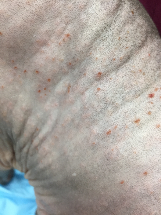
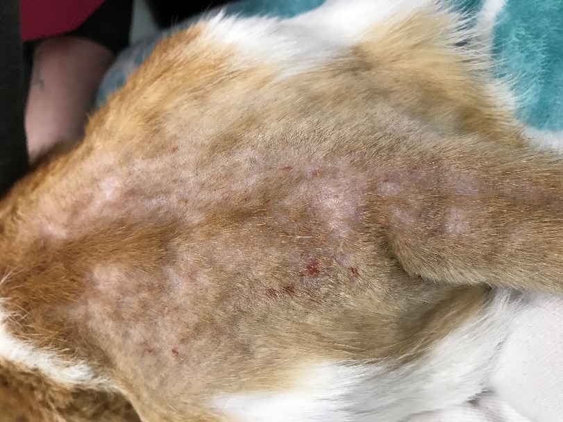
- The gross lesions of miliary dermatitis consist of multiple, small and crusted erythematous papules.
-
- In areas where the hair coat is intact, the papules are more easily palpated than noted and are variably distributed depending on the primary cause.
- Pruritus may or may not be present depending on the primary disease.
- Patchy or diffuse alopecia secondary to self-trauma is common, if pruritus is present.
Important Facts
- The lesions of miliary dermatitis consist of multiple, small and crusted erythematous papules.
- In areas where the hair coat is intact, the papules are more easily palpated than seen and are variably distributed depending on the primary cause.
-
Diagnosis
- It is not difficult to recognize lesions of miliary dermatitis. However, the challenging part is to diagnose the primary disease.
- A thorough history and physical examination are very important in helping identify the primary underlying disease causing miliary dermatitis.
- Presence or absence of pruritus will help prioritize the list of reasonable differential diagnoses.
- The distribution of lesions can be supportive of a specific underlying disease. For example, in the case of fleabite allergy, the lesions will be primarily localized on the lumbar-sacral region and base of the tail.
- Auxiliary tests may include skin scraping, flea combing, cytology, food trial, and fungal culture.
- A prioritized list of differential diagnoses will help select the needed tests for each patient.
- Biopsy findings are supportive of miliary dermatitis, but seldom will provide a definitive etiology.
- The inflammatory infiltrate concentrates primarily in the perivascular region and interstitial dermis, and eosinophils and mast cells predominate. Spongiotic eosinophilic pustules may be seen in the epidermis. Eosinophilic folliculitis and furunculosis can be present in severe miliary dermatitis lesions.
Important Facts
- It is not difficult to recognize lesions of miliary dermatitis. However, the challenging part is to diagnose the primary disease.
- History and physical examination findings such as presence of pruritus and distribution of lesions may be supportive of a specific cause of miliary dermatitis.
- Auxiliary tests may include skin scraping, flea combing, cytology, food trial, and fungal culture.
- A prioritized list of differential diagnosis will help select the needed tests for each patient
-
Treatment
- The successful management of miliary dermatitis is dependent upon identifying and controlling the primary cause.
Important Facts
- The successful management of miliary dermatitis is dependent upon identifying and controlling the primary cause.
References
Hobi S, Linek M, Marignac G, et al. Clinical characteristics and causes of pruritus in cats: a multicentre study on feline hypersensitivity-associated dermatoses. Vet Dermatol 2011; 22:406-413.
Nesbitt G.E. & Ackerman L.J. Miscellaneous Feline Dermatosis. In: Canine and Feline Dermatology: Diagnosis and Treatment. Veterinary Learning Systems, Trenton, New Jersey, 1998, p 471-490.
