17c. Eosinophilic Granuloma
-
General Considerations
- Eosinophilic granuloma is a common cutaneous, mucocutaneous and oral mucosal lesion of cats.
- The clinical presentation of eosinophilic granuloma is variable and, frequently, an underlying primary disease cannot be identified. However, allergic disorders have been diagnosed in some cats and should be pursued.
- There is a limited number of reports that support a hereditable basis for the development of eosinophilic granuloma lesions.
Important Facts
- Eosinophilic granuloma is a common cutaneous, mucocutaneous and oral mucosal lesion of cats.
- The clinical presentation of eosinophilic granuloma is quite variable, and most cases are idiopathic. However, underlying allergic disorders should be pursued.
- Genetic factors may also play a role in the pathogenesis of this condition.
-
Clinical Signs
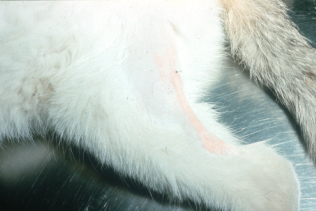
- Eosinophilic granuloma lesions can present as papules, nodules, or plaques that may be linear or oval in configuration. The lesions may be present in various body locations and may become alopecic, eroded or ulcerated. They are typically erythematous or have an orange yellow to salmon color.
- Linear lesions often occur in cats less than 2 years of age and develop on the caudal or medial aspects of the thigh. Occasionally, lesions can be observed on the lateral trunk, neck, thorax, and forelimbs. They can be single or multiple and are usually firm and have a yellowish or pink color.
- Eosinophilic granuloma lesions can present as papules, nodules, or plaques that may be linear or oval in configuration. The lesions may be present in various body locations and may become alopecic, eroded or ulcerated. They are typically erythematous or have an orange yellow to salmon color.
-
-
- Linear granuloma lesions are often discovered by accident because they are frequently asymptomatic. However, in some cases, they can be very pruritic, and an allergic cause should then be pursued.
-
-
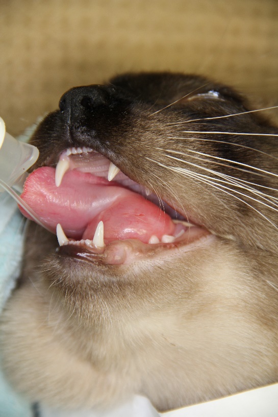
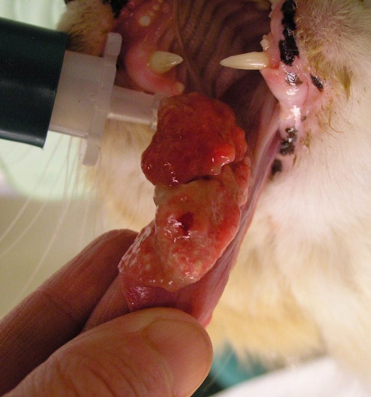
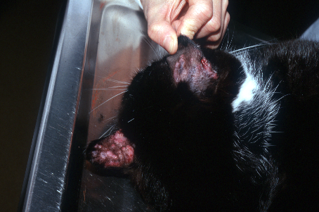
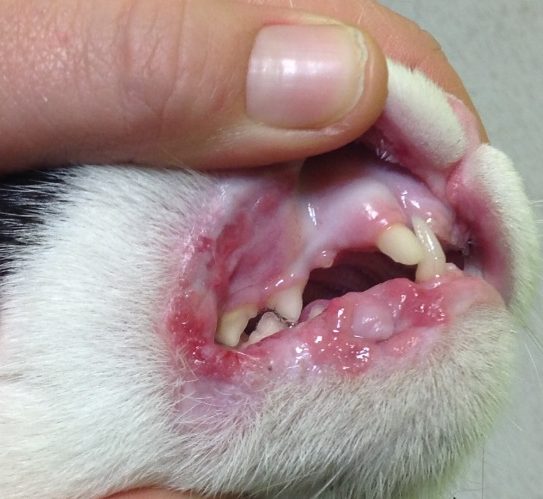
- Individual or grouped nodular lesions can erode or ulcerate and may be located on the trunk, head, ears, hard palate and dorsal or ventral aspect of the tongue.
- Cats with oral lesions may be anorexic, have hypersalivation, problems with apprehension of food, and halitosis.
- Individual or grouped nodular lesions can erode or ulcerate and may be located on the trunk, head, ears, hard palate and dorsal or ventral aspect of the tongue.
-
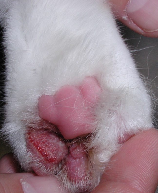
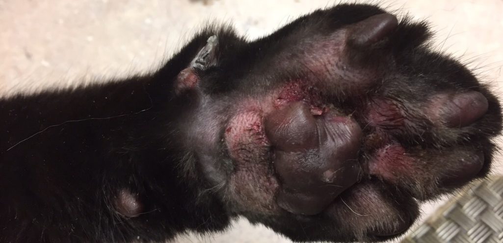
- Lesions can also be present adjacent to the paw pads or interdigitally. These lesions often manifest with crusting and erosions/ulcerations and can cause lameness.
- Areas of erosions/ulcerations are caused by the cat licking at the lesions. These sites will often become secondarily infected.
- Lesions can also be present adjacent to the paw pads or interdigitally. These lesions often manifest with crusting and erosions/ulcerations and can cause lameness.
-
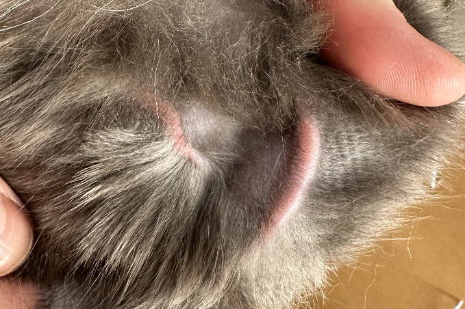
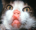
- Eosinophilic granuloma is the most common cause of feline lower lip swelling (pouting) and asymptomatic swollen chin (fat-chinned cats). Lip margin swelling and inflammation may also occur.

-
- Some cats with severe eosinophilic granuloma lesions have eosinophilia and peripheral lymphadenopathy.
- Indolent ulcer and/or eosinophilic plaques may develop concurrently with eosinophilic granuloma lesions.
Important Facts
- The clinical presentation of eosinophilic granuloma lesions varies according to the location of the lesion and can present as papules, nodules, or plaques that may be linear or oval in configuration.
- Linear lesions are more frequently seen on the caudal or medial aspects of the thighs, and they are often asymptomatic and can spontaneously regress.
- Lesions on the paws and oral cavity are typically eroded/ulcerated nodules or plaques, and often cause discomfort for the animals to walk and eat, respectively.
- Eosinophilic granuloma is the most common cause of feline lower lip swelling (pouting) and asymptomatic swollen chin (fat-chinned cats).
- Some cats with severe eosinophilic granuloma lesions have peripheral eosinophilia and peripheral lymphadenopathy.
- A cat may have lesions of eosinophilic granuloma, eosinophilic plaque and indolent ulcer concurrently.
-
Diagnosis
- The diagnosis is based on a detailed history and characteristic clinical signs.
- A thorough history is crucial to help identify an underlying triggering disease.
- Allergic causes, including food allergy, environmental allergy (i.e. feline atopic skin syndrome), and fleabite allergy should be pursued.
- In case of an underlying allergic disease, signs consistent with feline allergic diseases such as head and neck or generalized pruritus and inflammation, miliary dermatitis (fleabite allergy), and symmetrical, self-inflicted, non-inflammatory alopecia should be present.
- Surface impression smears of eroded or ulcerated lesions may reveal eosinophils and their granules, but neutrophils may predominate if secondary infection is present.
- Differential diagnoses include plasma cell pododermatitis (paw pad lesions), sterile or infections inflammatory nodules, foreign body reactions and neoplasia.
- Skin biopsy is recommended if the clinical presentation is unusual and infectious granulomas (e.g. fungal and bacterial) or neoplasia are suspected.
- The key histopathological finding is the infiltration of eosinophils. In some cases, the eosinophils degranulate and form “flame figures,” which are collagen fibers coated with eosinophil granules and degenerated eosinophils. Neutrophils may predominate in lesions with secondary bacterial infection. Histiocytic cells are also common in lesions of eosinophilic granuloma.
- The diagnosis is based on a detailed history and characteristic clinical signs.
Important Facts
- The diagnosis is based on a detailed history and characteristic clinical signs. A thorough history is crucial to help identify an underlying disease.
- Allergic causes, including food allergy, feline atopic skin syndrome and flea allergic dermatitis should be pursued.
- Biopsy of lesions is recommended if the clinical signs are unusual and infectious granulomas and neoplasia are suspected.
-
Treatment
- The underlying triggering disease, if identified, should be treated properly to help prevent recurrence.
- Linear granulomas are usually asymptomatic and do not need to be treated.
- Lesions developing before 1 year of age typically resolve within 3 to 5 months.
- In some cases, lesions will be very pruritic, and an underlying allergic cause should be pursued.
- All aspects of therapy are as described for the indolent ulcer.
Important Facts
- Look for a primary underlying triggering disease (e.g. food allergy, atopic skin syndrome, flea allergic dermatitis) and treat it properly to help prevent recurrence.
- Linear granulomas are usually asymptomatic and do not need to be treated. Most lesions developing before 1 year of age have spontaneous resolution.
- All aspects of therapy are as described for the indolent ulcer.
References
Bloom PB. Canine and feline eosinophilic skin diseases. Vet Clin North Am Small Anim Pract 2006; 36:141-160.
Buckley L and Nuttall T. Feline eosinophilic granuloma complex(ities): Some clinical clarification. J Fel Med Surg 2012; 14: 471-481.
Foster A. Clinical approach to feline eosinophilic granuloma complex. In Pract 2003; 25: 2–10.
McKeever PJ, Nuttall T, Harvey RG. A Color Handbook of Skin Diseases of the Dog and Cat. 2nd edn. London: Manson Publishing Ltd., 2009; 102-105.
Miller WH, Griffin GE, Campbell KL. Muller & Kirk Small Animal Dermatology. 7th edn. St Louis: Elsevier Inc., 2013; 714-718.
Mueller RS, Nuttall T, Prost C et al. Treatment of the feline atopic syndrome – a systematic review. Vet Dermatol 2021; 32: 43-e8.
Porcellato I, Giontella A, Mechelli L et al. Feline eosinophilic dermatoses: a retrospective immunohistochemical and ultrastructural study of extracellular matrix remodeling. Vet Dermatol 2014; 25: 86-226.
Power HT and Ihrke PJ. Selected feline eosinophilic skin diseases. Vet Clin North Am Small Anim Pract 1995; 25: 833–850.
Power HT. Eosinophilic granuloma in a family of specific pathogen-free cats. Proceedings of the American Academy of Veterinary Dermatology/American College of Veterinary Dermatology. Vol 6. San Francisco; 1990, p 45.
Scott, Miller & Griffin. Miscellaneous Skin Diseases. In: Small Animal Dermatology. W.B. Saunders, Philadelphia, 1995, p 902-955.
