17b. Feline Eosinophilic Plaque
-
General Considerations
- Eosinophilic plaque is a common cutaneous lesion of cats.
- It is thought to be a hypersensitivity reaction to fleas, food or environmental allergens, but some cases are idiopathic.
Important Facts
- The etiology of eosinophilic plaque is unknown. However, it is thought to be a hypersensitivity reaction to fleas, food or environmental allergens.
-
Clinical Signs
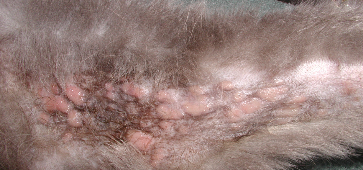
- Eosinophilic plaques are well demarcated, raised but flat-topped, round to oval, erythematous, often eroded and sometimes ulcerated lesions.
- Lesions may be single or multiple and coalescing.
- They most commonly develop on the ventral abdomen and medial thighs but may be present in other areas of the body.
- Pruritus is usually present, and an underlying allergic disorder should be always pursued.
- Cats typically lick at the lesions causing erosions. The damaged skin barrier will likely become infected.
- Eosinophilic plaques are well demarcated, raised but flat-topped, round to oval, erythematous, often eroded and sometimes ulcerated lesions.

-
- Peripheral lymphadenopathy may be present.
Important Facts
- Most eosinophilic plaques occur on the ventral abdomen and medial thighs but may also occur in other skin sites.
- Lesions may be single or multiple and coalescing.
- Cats frequently lick at the plaques causing erosions. The damaged skin barrier will likely become infected.
- Pruritus is usually present, and an underlying allergic cause should be pursued.
-
Diagnosis
- The diagnosis is based on the characteristic clinical signs.
- A thorough history is crucial in helping to identify an underlying triggering disease.
- In the case of an underlying allergic disease, signs consistent with feline allergic diseases such as, head and neck or generalized pruritus and skin inflammation, miliary dermatitis (fleabite allergy), and symmetrical, self-inflicted, non-inflammatory alopecia should be present.
- Surface impression smears of eroded lesions may reveal eosinophils and their granules, but neutrophils may predominate if secondary infection is present.
- Skin biopsy is recommended if the clinical presentation is unusual and infectious granulomas (fungal and bacterial) or neoplasia are suspected.
- The key histopathological finding is the infiltration of various numbers of eosinophils. In some cases, the eosinophils degranulate and form “flame figures” which are collagen fibers coated with eosinophil granules and degenerated eosinophils. Other inflammatory cells are typically present and neutrophils may predominate in cases complicated with a secondary bacterial infection.
- It is necessary to try to identify an underlying triggering condition (e.g. food allergy, feline atopic skin syndrome, fleabite allergy).
- The diagnosis is based on the characteristic clinical signs.
Important Facts
- The diagnosis is based on the pet’s thorough history and characteristic clinical signs.
- Biopsy of lesions is recommended if the clinical signs are unusual and infectious granulomas and neoplasia are suspected.
- It is necessary to try to identify an underlying triggering condition (e.g. food allergy, feline atopic skin syndrome, fleabite allergy).
-
Treatment
- Treatment recommendations are as described previously for the feline indolent ulcer.
- Treat appropriately any identified underlying triggering condition to prevent recurrences.
- Treat secondary bacterial infections since significant improvement or complete resolution of lesions can be noted by treating infections.
- A study showed a significant response compared to placebo when cats with eosinophilic plaques or the eosinophilic ulcer were treated solely with amoxicillin clavulanate based on culture and susceptibility. These findings suggest that bacterial infections play a significant role in lesion development or persistence in some cases
- Keep in mind that some cases can be very difficult to manage, especially if underlying triggering disease cannot be identified and/or controlled properly.
Important Facts
- Treat properly any identified underlying triggering condition to prevent recurrences.
- All aspects of treatment are similar to the ones already described for the indolent ulcer.
References
Bloom PB. Canine and feline eosinophilic skin diseases. Vet Clin North Am Small Anim Pract 2006; 36:141-160.
Buckley L and Nuttall T. Feline eosinophilic granuloma complex(ities): Some clinical clarification. J Fel Med Surg 2012; 14: 471-481.
Foster A. Clinical approach to feline eosinophilic granuloma complex. In Pract 2003; 25: 2–10.
McKeever PJ, Nuttall T, Harvey RG. A Color Handbook of Skin Diseases of the Dog and Cat. 2nd edn. London: Manson Publishing Ltd., 2009; 102-105.
Miller WH, Griffin GE, Campbell KL. Muller & Kirk Small Animal Dermatology. 7th edn. St Louis: Elsevier Inc., 2013; 714-718.
Mueller RS, Nuttall T, Prost C et al. Treatment of the feline atopic syndrome – a systematic review. Vet Dermatol 2021; 32: 43-e8.
Porcellato I, Giontella A, Mechelli L et al. Feline eosinophilic dermatoses: a retrospective immunohistochemical and ultrastructural study of extracellular matrix remodeling. Vet Dermatol 2014; 25: 86-226.
Power HT and Ihrke PJ. Selected feline eosinophilic skin diseases. Vet Clin North Am Small Anim Pract 1995; 25: 833–850.
Scott, Miller & Griffin. Miscellaneous Skin Diseases. In: Small Animal Dermatology. W.B. Saunders, Philadelphia, 1995, p 902-955.
Wildermuth BE, Griffin CE, Rosenkrantz WS. Response of feline eosinophilic plaques and lip ulcers to amoxicillin trihydrate-clavulanate potassium therapy: a randomized, double-blind placebo-controlled prospective study. Vet Dermatol 2011; 22:521-527
