17a. Feline Indolent Ulcer
-
General Considerations
- Indolent ulcer, also called eosinophilic ulcer or rodent ulcer, is a common lesion of cats that develops at the mucocutaneous junctions of the mouth.
Important Facts
- Indolent ulcer, also called eosinophilic ulcer or rodent ulcer, is a common lesion of cats that affects the mucocutaneous junctions of the mouth.
-
Etiology
- There are reports of cats with indolent ulcers having positive intradermal tests and subsequent response to allergen-specific immunotherapy, suggesting an allergic etiology at least in some cases. Moreover, some cats respond to a food elimination trial and indolent ulcer lesions can be present in cats with fleabite allergy.
- Genetic predisposition has been suggested based on a SPF cat colony where there was a high incidence of indolent ulcers and eosinophilic granuloma lesions in related cats. An allergic or parasitic condition could not be identified in these cats. Another evidence to support a possible genetic role in the development of these lesions is a report of 17 related Norwegian forest cats with eosinophilic granuloma and indolent ulcer lesions.
- Some cases are idiopathic.
Important Facts
- Some cases of indolent ulcer are associated with allergies, others with genetic factors and yet others are idiopathic.
-
Clinical Signs
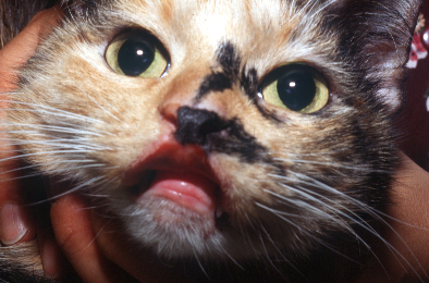
- The lesion is typically well-demarcated with raised margins and a central granular and glistening ulcerated area. Focal to multifocal, whitish necrotic areas are often present. The extent of the lesion is variable; however, most indolent ulcers span the upper lip.
- Pruritus and pain are rare, and peripheral lymphadenopathy may be present.
- The lesion recurs frequently if the underlying primary disease is not identified and if treatment is interrupted. Occasionally, it will be refractory to treatment.

Important Facts
- The ulcer is typically well-demarcated and have raised margins and a central granular and glistening area. Focal to multifocal necrotic areas are often present.
- The extent of the lesion is variable but most indolent ulcers span the upper lip.
- The lesion recurs frequently if the primary disease is not identified or if treatment is interrupted.
- Occasionally, the lesion will be refractory to treatment.
-
Diagnosis
- Differential diagnoses include neoplasia (e.g. squamous cell carcinoma, lymphoma, mast cell tumor) or infectious granulomas (bacterial, fungal, FeLV-associated).
- The characteristic clinical signs are usually diagnostic of indolent ulcers.
- A thorough history is crucial in helping to identify the underlying triggering disease.
- In case of an underlying allergic disease, pruritus and signs consistent with feline allergic diseases such as, head and neck pruritus, miliary dermatitis (fleabite allergy), and symmetrical, self-inflicted, non-inflammatory alopecia should be present.
- It is important to try to identify an underlying primary disease such as food allergy and feline atopic skin syndrome, while also maintaining good flea control.
- Perform cytology from the exudate associated with the lesion. The sample should show a mixed inflammatory infiltrate including eosinophils. Degenerate neutrophils and bacteria are often seen because the skin barrier is damaged opening the door for a secondary infection to establish.
- Skin biopsy is recommended if the clinical presentation is unusual and infectious granulomas (fungal and bacterial) or neoplasia are suspected.
- The key histopathological finding is the presence of variable numbers of eosinophils. In some cases, the eosinophils degranulate and form “flame figures,” which are collagen fibers coated with eosinophil granules and degenerated eosinophils. Neutrophils may predominate if the lesion is infected.
Important Facts
- The history and characteristic clinical signs are usually diagnostic of an indolent ulcer.
- It is important to try to identify any underlying primary disease such as food allergy, feline atopic skin syndrome, and fleabite allergy.
- Biopsy of lesions is recommended if the clinical signs are unusual and infectious granulomas or neoplasia are suspected.
-
Treatment
- Look for an underlying triggering condition and try to resolve it, if feasible, or control it effectively.
- The effective treatment of the underlying disease will positively influence the long-term outcome and will reduce the chances of lesion recurrence.
- Because the primary underlying disease cannot be cured (e.g. allergies) in most cases or is not identified, long-term treatment is often required.
- The medications used to treat the lesions of the eosinophilic granuloma complex have potentially severe side effects. Therefore, monitor the patients carefully during therapy.
- Corticosteroids are often needed to resolve the lesion, and it is the mainstay therapy.
- Oral prednisolone (2-4 mg/kg q 24h) or methylprednisolone (2-3 mg/kg q 24h) may be satisfactory. If long-term maintenance is needed, the lowest dose necessary to maintain the disease under control, administered every-other-day, is recommended.
- Another alternative is oral triamcinolone at the initial dosage of 0.5 to 0.75 mg/kg q 24h then the lowest possible dose that keeps the condition under control should be sought and given q 48h- 72h.
- Methylprednisolone acetate (Depo-Medrol) at the dose of 4 to 5 mg/kg IM (minimum of 20 mg/cat) is usually satisfactory to achieve remission. Two or three injections every 2-3 weeks may be necessary for the lesion to resolve. Avoid administering it more often than every 2 months, otherwise, the risk for the development of diabetes mellitus will likely increase.
- Remember, cats are prone to developing diabetes mellitus due to corticosteroid therapy, especially long-acting injectable glucocorticoids and oral dexamethasone. Therefore, reserve these glucocorticoids for cases refractory to oral prednisolone or that cannot be medicated orally. Monitor the patient closely.
- Cyclosporine is a good alternative to glucocorticoids. Give 5-7 mg/kg q 24h, two hours apart from feeding to improve bioavailability. Keep in mind that a dose of 7-7.5 mg/kg q 24h is recommended for cats and can be more effective than 5 mg/kg q24h in some cases.
- A liquid formulation is approved for use in cats in some countries.
- Cyclosporine has a lag-time of about 4 weeks before its effect can be noted.
- Main side effects are vomiting and/or diarrhea. Other possible side effects are hypersalivation, anorexia, hypertrichosis, gingival hyperplasia, and cutaneous viral papillomatosis.
- Toxoplasmosis has been reported in cats being treated with cyclosporine. However, it appears to be rare at the recommended doses. Preventative measures include avoiding raw meat, keeping cats indoors, and attaching two bells to the collar with the goal of making hunting less successful.
- Interesting, Toxoplasma-naïve cats are considered to be at a higher risk of developing toxoplasmosis when treated with cyclosporine than Toxoplasma-positive cats. Testing cats before treatment is recommended.
- Consider another treatment option if no improvement is noticed after a one-month trial. If a good response is seen, reduce the dose to every-other-day and then twice-weekly therapy, if possible.
- Cats that relapse on alternate-day therapy can be treated daily. However, find the lowest possible dose that maintains the disease controlled.
- Oclacitinib (Apoquel®) has been anecdotally reported to be efficacious in controlling lesions of the eosinophilic granuloma complex in some cats with underlying allergies. Pruritus will resolve within a day; however, it will take longer for the lesions to improve or resolve. The dose is 1 mg/kg q 12h. Try to reduce the dose to once daily after remission is achieved if using oclacitinib long-term.
- Oclacitinib is currently not labeled for cats.
- It is important to perform a CBC every 2 weeks for the first few months to monitor for bone marrow suppression.
- If there are poor responses to corticosteroid, cyclosporine and oclacitinib, chlorambucil (Leukoran®) or gold salts (sodium aurothiomalate; Myochrysine®) is an optionn.
- Chlorambucil dose is 0.1 to 0.2 mg/kg orally q 24-48h. It is available in 2 mg tablets and the tablets should not be crushed or split. There is a lag phase of 3-6 weeks, and an oral glucocorticoid can be given during this lag period. It is important to monitor every 2 weeks for potential bone marrow suppression. After 3-4 months, monitoring can be reduced to every 3 months.
- Sodium aurothiomalate is administered at the dose of 1 mg/kg every 7 days. There is a lag phase of 6 to 12 weeks before clinical response is observed. Monitoring for bone marrow suppression, proteinuria and cutaneous or oral cavity drug eruption is important.
- Treat secondary bacterial infections since significant improvement or complete resolution of lesions can be noted with treatment of infections!
- A study showed a significant response compared to placebo when cats with eosinophilic plaques or eosinophilic ulcers were treated solely with amoxicillin clavulanate based on culture and susceptibility. These findings suggest that bacterial infection plays a significant role in lesion development or persistence in some cases.
- Look for an underlying triggering condition and try to resolve it, if feasible, or control it effectively.
Important Facts
- Look for an underlying condition and try to resolve it, if feasible, or control it effectively.
- The effective treatment of the underlying triggering disease will positively influence the long-term outcome and will reduce the chances of lesion recurrence.
- Oral glucocorticoids, and sometimes injectable, are the mainstay treatment of indolent ulcers.
- Oral cyclosporine has been also shown to be efficacious and is a good alternative to glucocorticoids.
- Oclacitinib (Apoquel®) has been anecdotally reported to be efficacious in controlling lesions of the eosinophilic granuloma complex in some cats. A potential and concerning side effect is bone marrow suppression, therefore, monitor closely with CBCs for the first few months.
- Chlorambucil or gold salts is an option for cases that do not respond to glucocorticoids, cyclosporine and oclacitinib.
- Treat secondary infections because significant improvement or complete resolution of lesions can be noted after infections are resolved.
- Because the primary underlying disease cannot be cured in most cases or is not identified, long-term treatment is often required.
- The medications used to treat lesions of the eosinophilic granuloma complex have potentially severe side effects requiring long-term monitoring of the patients during therapy.
References
Bajwa J. Feline indolent ulcers and their significance. Dermatologie Veterinaire 2019; 60: 1009-1011.
Bloom PB. Canine and feline eosinophilic skin diseases. Vet Clin North Am Small Anim Pract 2006; 36:141-160.
Buckley L and Nuttall T. Feline eosinophilic granuloma complex(ities): Some clinical clarification. J Fel Med Surg 2012; 14: 471-481.
Colombini S and Hodgin EC. Induction of feline flea allergy dermatitis and the incidence and histopathological characteristics of concurrent indolent lip ulcers. Vet Dermatol 2001; 12: 155-161.
Foster A. Clinical approach to feline eosinophilic granuloma complex. In Pract 2003; 25: 2–10.
McKeever PJ, Nuttall T, Harvey RG. A Color Handbook of Skin Diseases of the Dog and Cat. 2nd edn. London: Manson Publishing Ltd., 2009; 102-105.
Miller WH, Griffin GE, Campbell KL. Muller & Kirk Small Animal Dermatology. 7th edn. St Louis: Elsevier Inc., 2013; 714-718.
Mueller RS, Nuttall T, Prost C et al. Treatment of the feline atopic syndrome – a systematic review. Vet Dermatol 2021; 32: 43-e8.
Porcellato I, Giontella A, Mechelli L et al. Feline eosinophilic dermatoses: a retrospective immunohistochemical and ultrastructural study of extracellular matrix remodeling. Vet Dermatol 2014; 25: 86-226.
Power HT and Ihrke PJ. Selected feline eosinophilic skin diseases. Vet Clin North Am Small Anim Pract 1995; 25: 833–850.
Scott, Miller & Griffin. Miscellaneous Skin Diseases. In: Small Animal Dermatology. W.B. Saunders, Philadelphia, 1995, p 902-955.
Wildermuth BE, Griffin CE, Rosenkrantz WS. Response of feline eosinophilic plaques and lip ulcers to amoxicillin trihydrate-clavulanate potassium therapy: a randomized, double-blind placebo-controlled prospective study. Vet Dermatol 2011; 22:521-527.
