15. Cutaneous Asthenia – Canine and Feline
Learning Objectives
- Know! Cutaneous asthenia, also known as Ehlers-Danlos syndrome and dermatoparaxis, is a rare hereditary group of conditions characterized by abnormal collagen development.
- Know! A dominant form of the disease has been documented in dogs and cats and is associated with abnormal collagen packing resulting in decreased skin tensile strength. In cats, a recessive form has been also reported. The recessive form is associated with a deficiency of N-procollagen peptidase enzyme.
- Know! Dogs and cats are born with this condition, but the owner may only notice the problem when the pet develops skin lesions, which may only occur weeks or even months after birth. The skin may show multiple scars and will lacerate easily with minor trauma. Minimum blood is usually associated with skin lacerations. Some animals will also have hyperextensibility of the skin and/or joint laxity. Hematomas, elbow hygromas and umbilical and inguinal hernias may be present in some cases. Ocular problems may also be part of clinical signs.
- Remember! Cats may develop fragile skin due to naturally occurring or iatrogenic hyperadrenocorticism, diabetes mellitus, liver disease or with excessive use of megestrol acetate. However, this acquired skin fragility will only develop in middle to old age, which is a relevant differentiating factor.
- Know! Diagnosis can usually be done based on history and clinical signs. Remember, there are other conditions that can cause skin fragility in cats, but these cases are related to systemic diseases and certain medications and only develop later in life. Skin biopsy and collagen stain (i.e. Masson’s trichrome) may support the diagnosis. However, whole genome sequencing studies, electron microscopy to identify structural collagen abnormalities and biochemical analysis of the dermal collagen are needed to determine the collage defect. Currently, most studies are using whole genome sequencing analyses to identify the genetic mutation because is more effective.
- Know! Owners must be informed of the chronic and non-curable nature of the disease. Animals should not be used for breeding. Skin lacerations should be sutured and the animal’s lifestyle adjusted to minimize trauma.
-
General Considerations
- Cutaneous asthenia, also known as Ehlers-Danlos syndrome and dermatoparaxis, is a rare hereditary group of disorders characterized by abnormal collagen development.
- Several forms of cutaneous asthenia have been recognized in man, dogs, cats, cattle, horses, mink, and sheep based on clinical, genetic, and biochemical variations.
- The common basis for all these forms is that they are accompanied by connective tissue weakness due to abnormalities in biosynthesis or post-translational modifications of collagen.
Important Facts
- Cutaneous asthenia, also known as Ehlers-Danlos syndrome and dermatoparaxis, is a rare hereditary group of disorders characterized by abnormal collagen development.
- Several forms of cutaneous asthenia have been recognized in man, dogs, cats, cattle, horses, mink, and sheep based on clinical, genetic, and biochemical differentiation.
-
Pathogenesis
- In dogs, cutaneous asthenia is a simple autosomal dominant condition associated with collagen packing defect characterized by focal or diffuse areas of severely disorganized fibers with many abnormally large fibrils. The disease is called “classical Ehlers-Danlos syndrome” and is associated with variants in COL5A1 and COL5A2 genes.
- Cats have two forms of cutaneous asthenia, a dominant form similar to the dog’s disease and a recessive form.
- The recessive form is characterized by decreased activity of N-procollagen peptidase enzyme, which results in the affected collagen forming twisted ribbons instead of cylindrical fibrils and fibers.
- The dominant form is similar to the dog’s condition and is associated with collagen packing defect. A COL5A1 variant was reported in a cat.
- The loss of rigidity and tensile strength associated with the collagen defect leads to skin fragility and ultimately tearing.
Important Facts
- In dogs, cutaneous asthenia is a simple autosomal dominant condition associated with collagen packing defect.
- Cats have two forms of cutaneous asthenia: a dominant form similar to the dog’s condition and a recessive form characterized by deficiency of N-procollagen peptidase enzyme, which results in the affected collagen forming twisted ribbons instead of cylindrical fibrils and fibers.
-
Clinical Signs
- Cutaneous asthenia has been reported in the beagle, boxer, dachshund, English setter, Irish setter, keeshond, toy poodle, English springer spaniel, German shepherd dog, Garafian shepherd, greyhound, Yorkshire terrier, Irish setter, Alaskan malamute, Welsh corgi, soft-coated wheaten terrier, red kelpie, Keeshond, Saint Bernard, Fila Brasileiro, and mixed breed dogs.
- In cats, it has been documented in the domestic shorthair or longhair, Himalayan, Persian and Burmese cats.
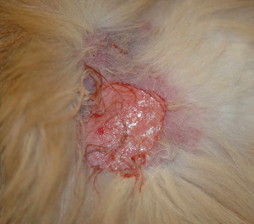
- Fragility of the skin since birth is the most characteristic clinical sign. However, it may not be recognized by the owner until lesions develop, which may only occur few weeks to months after birth.
- Clinically, animals will have multiple skin tears and scars. The skin lacerations are associated with minimal or no bleeding and healing occurs rapidly by the formation of a thin and irregular scar tissue. The skin tensile strength of affected dogs is reduced 40-fold and that of cats 10-fold.
-
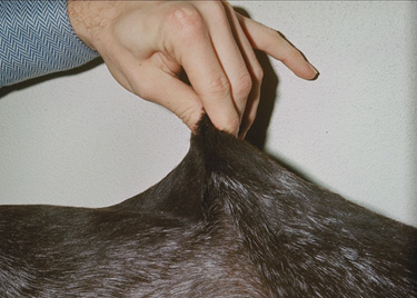
- Some animals will also have hyperextensibility of the skin and/or joint laxity. The skin hyperextensibility may be the only presentation in some cases.
- Extensibility is determined by extending a dorsolumbar skin fold to the maximal distance above the spine. This distance and the body length from the occipital crest to the tail base are measured. These parameters are used in the formula below:
- Extensibility index = Vertical height of skin fold/body length X 100.
- In affected dogs the skin extensibility is >14.5% and in cats >19%.
- Extensibility is determined by extending a dorsolumbar skin fold to the maximal distance above the spine. This distance and the body length from the occipital crest to the tail base are measured. These parameters are used in the formula below:
- Some animals will also have hyperextensibility of the skin and/or joint laxity. The skin hyperextensibility may be the only presentation in some cases.
-
- Hematomas, elbow hygromas, join laxity and umbilical or inguinal hernias may occur in some animals.
- Ocular problems such as microcornea, lens luxation, cataracts, and scleroderma may be part of the clinical signs in some cases.
- Cats may also develop fragile skin due to spontaneous or iatrogenic hyperadrenocorticism, diabetes mellitus, liver disease, or with excessive use of megestrol acetate. However, skin fragility is acquired in these cases and will only develop in middle age to old cats, which is an important differentiating factor.
Important Facts
- Fragility of the skin since birth is the most characteristic clinical sign. However, it may not be recognized by the owner until lesions develop, which may only happen few weeks to months after birth.
- Clinically, animals will have multiple skin tears and scars with minimum hemorrhage.
- Some animals will also have hyperextensibility of the skin and/or joint laxity and rarely, animals may present solely with skin hyperextensibility.
- Cats may also develop fragile skin due to spontaneous or iatrogenic hyperadrenocorticism, diabetes mellitus, or with excessive use of megestrol acetate. However, skin fragility associated with these diseases or circumstances only develops late in life, which is an important differentiating factor.
-
Diagnosis
- The patient’s history (especially age of disease onset) and characteristic clinical signs are usually sufficient to make a diagnosis of cutaneous asthenia.
- Histopathological findings are variable. Using hematoxylin and eosin stain, collagen fibers in the affected skin are small and sparse compared with control sections. Some fibers can appear fragmented, shortened, disorganized and bundle sizes may be irregular. However, in some cases (mostly cats) dermal collagen appears normal. Masson’s trichrome stain is useful in identifying collagen abnormalities.
- Currently, whole genome sequencing analyses is done to identify the gene mutation associated with the collagen defect.
- Electron microscopic is sensitive to identify structural collagen abnormalities and biochemical analysis of the dermal collagen can be done to determine the specific abnormality in collagen synthesis.
Important Facts
- History and characteristic clinical signs are usually sufficient to make a diagnosis of cutaneous asthenia.
- Whole genome sequences and electron microscopic and biochemical analyses of dermal collagen are needed to characterize the specific collagen synthesis defects.
-
Treatment
- There is no specific treatment for cutaneous asthenia.
- Lacerations can be sutured using tension patterns, but they often heal spontaneously and uneventfully.
- The animal should be protected from any minor or major trauma.
- Cats should be declawed to prevent wounding during grooming or scratching.
- Affected animals cannot be used for breeding and owners need to know that the disease cannot be cured.
Important Facts
- There is no specific treatment for cutaneous asthenia.
- Lacerations can be sutured using tension patterns, but they often heal spontaneously and uneventfully.
- The animal should be protected from any minor or major trauma.
- Affected animals should not be used for breeding.
References
Barnett KC, Cottrell BD. Ehlers-Danlos syndrome in a dog: Ocular, cutaneous and articular abnormalities. J Small Anim Pract 1987; 28:941.
Burton G, Stenzel D, Mason KV. Cutaneous asthenia in Burmese cats: a vasculopathy? Vet Dermatol 2000; 11:31.
Fernandez CJ, et al. Staining abnormalities of dermal collagen in cats with cutaneous asthenia or acquired skin fragility as demonstrated with Masson’s trichrome stain. Vet Dermatol 1998; 9:49-54.
McKeever PJ, Nuttall T, Harvey RG. A Color Handbook of Skin Diseases of the Dog and Cat. 2nd edn. London: Manson Publishing Ltd., 2009; 142-143.
Miller WH, Griffin GE, Campbell KL. Muller & Kirk Small Animal Dermatology. 7th edn. St Louis: Elsevier Inc., 2013; 602-604.
Paciello O, Lamagna F, Lamagna B, et al. Ehlers-Danlos-like syndrome in 2 dogs: clinical, histologic, and ultrastructural findings. Vet Clin Pathol 2003; 32:13-18.
Patterson DF, Minor RR. Hereditary fragility and hyperextensibility of the skin of cats. A defect in collagen fibrillogenesis. Lab Invest 1977; 37:170-179.
Spycher M, Bauer A, Jagannathan V et al. A frameshift variant in the COL5A1 gene in a cat with Ehlers-Danlos syndrome. Anim Genet doi: 10.1111/age.12727.
