12. Perianal Fistulas – Canine
Learning Objectives
- Remember! Perianal fistulas or sinuses is a very difficult disease to manage.
- Know! The disease etiopathogenesis is unknown. However, the response to cyclosporine suggests that an immune mediated process plays a role. Moreover, the strong breed predilection indicates that genetics also plays a role.
- Know! The disease occurs much more frequently in German shepherd dogs, which account for more than 80% of the cases. However, many other breeds have been diagnosed with perianal sinuses. Most affected dogs are 3 to 6 years old at the time of diagnosis. Clinical signs are characterized by multiple sinus tracts and ulcers localized on the tissue surrounding the anus, but the disease can extend to affect the anal ring. Lesions are very painful, and the dogs present with tenesmus, dyschezia, hematochezia and constipation. Diarrhea may occur in some dogs. Owners often observe the pet licking the perianal region and a foul odor.
- Know! The diagnosis is based on the patient’s history and characteristic clinical signs. Skin biopsy is only required in the presence of an unusual clinical presentation.
- Keep in mind that antibiotic therapy has very little impact on the clinical outcome! Be sure to perform bacterial culture and sensitivity tests if you are considering systemic antibiotic therapy. The authors do not typically use systemic antibiotics as part of the treatment regimen of perianal fistulas.
- Remember! Evaluate every case for concurrent inflammatory bowel disease (i.e. colitis and/or proctitis).
- Know! The mainstay therapy for perianal fistulas is oral cyclosporine; which can be used in conjunction with ketoconazole to reduce the dose in half and, therefore, decrease the treatment cost. Oral glucocorticoid with or without sulfasalazine is an option for owners who cannot afford cyclosporine, but it typically does not have a notable impact in improving the lesions but reduce the associated discomfort that the pet experiences. Azathioprine can be used for severe and refractory cases. Topical tacrolimus 0.1% can be used combined with other treatments but it does not work well as sole therapy in severe cases. However, it can be used as sole therapy to maintain remission. Other treatment modalities that need further investigation include fluorescent light energy, oclacitinib and mesenchymal stem cell therapy. Surgery is rarely performed nowadays but can be considered as part of the treatment regimen in some cases.
-
General Considerations
- Perianal fistulas are also called perianal sinuses or anal furunculosis.
- Perianal fistula is a misnomer because the tissue tracts do not communicate with the rectal lumen. The proper name that should be widely adopted is “perianal sinuses”.
- It is a chronic and debilitating disease of the perianal tissue.
- The etiopathogenesis is unknown.
- Research has explored many possible etiologies including overproduction by local secretory glands, poor ventilation associated with low tail carriage, anal sac disease, and hip dysplasia.
- The very good response to cyclosporine therapy supports the role of an immune mediated process involving primarily T-lymphocytes; however, the pathomechanism is not completely known.
- Some people have suggested that canine perianal fistulas is similar to fistulizing Crohn’s disease in people.
- Breed predisposition suggests that genetics plays a role.
- Studies have been conducted in search for genes or gene regions associated with perianal fistulas in German shepherd dogs but results have not been definitive yet.
- Perianal fistulas are also called perianal sinuses or anal furunculosis.
Important Facts
- The etiopathogenesis of perianal sinuses is currently unknown.
- The good response to cyclosporine therapy supports the role of an immune mediated process.
- Breed predisposition suggests that genetics also plays a role.
-
Signalment
- German shepherd dogs are predisposed and reported to account for more than 80% of the cases. However, perianal fistulas can develop in any other breed or mixed-breed dogs.
- Most affected animals are 3 to 6 years of age at the time of diagnosis.
-
Clinical Signs
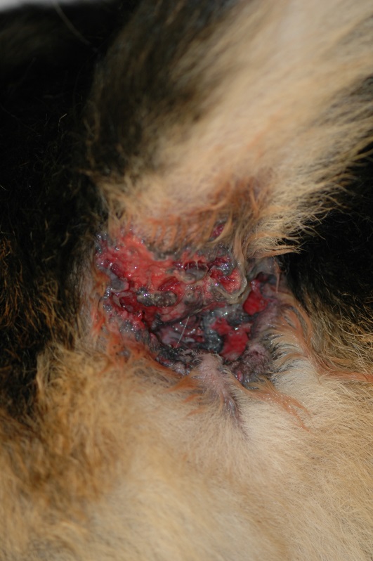
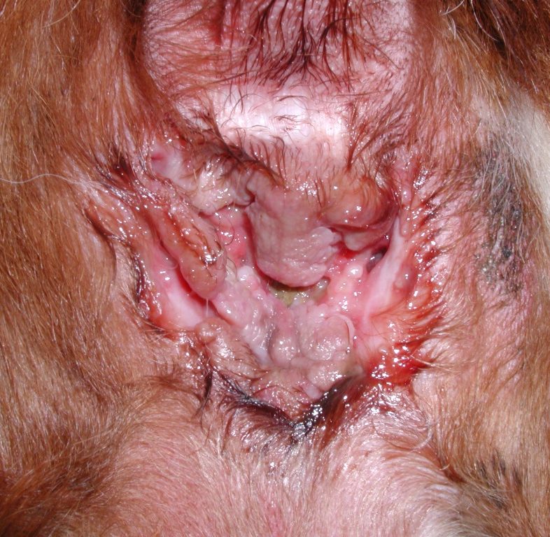
- Clinical signs are characterized by single and more commonly multiple sinus tracts and ulcers affecting the tissue surrounding the anus. The ulcers can be large and deep or crateriform. The disease can progress to involve the anal ring.
- Tissue fibrosis is often seen as the affected skin attempts to heal.
- The affected areas are usually very painful.
- Clinical signs are characterized by single and more commonly multiple sinus tracts and ulcers affecting the tissue surrounding the anus. The ulcers can be large and deep or crateriform. The disease can progress to involve the anal ring.
-
- Affected dogs typically have tenesmus (i.e. straining to defecate), dyschezia (i.e. painful defecation), hematochezia (i.e. fresh blood in the stool) and constipation. Owners may also observe the pet licking at the affected area and a foul odor. Diarrhea may occur in some cases.
- Many dogs with perianal fistulas have concurrent colitis and proctitis. However, signs of colitis such as increase frequency of defecation, diarrhea, and mucus in the feces may not be present or can be present in dogs without colitis. Colonoscopy and biopsy are needed to confirm a presumptive clinical diagnosis.
- Many patients with perianal fistulas will have anal sacculitis, anal sac impaction, or anal sac rupture secondary to inflammation and fibrosis of the surrounding tissue.
- Affected dogs typically have tenesmus (i.e. straining to defecate), dyschezia (i.e. painful defecation), hematochezia (i.e. fresh blood in the stool) and constipation. Owners may also observe the pet licking at the affected area and a foul odor. Diarrhea may occur in some cases.
Important Facts
- Perianal fistulas is a disease much more common in German shepherd dogs, but other breeds or mixed-breed dogs can also be affected.
- Most affected animals are middle age at the time the disease develops.
- Clinical signs are characterized by multiple sinus tracts and ulcers surrounding the anus.
- The affected area is painful and the animals often have tenesmus (i.e. straining to defecate), dyschezia (i.e. painful defecation), hematochezia (i.e. blood in the stools) and constipation. Owners may also observe the pet licking at the perianal area and a foul odor. Diarrhea may occur in some cases.
- Many dogs with perianal fistulas will have concurrent colitis and proctitis but the classical clinical signs of colitis may not be present. Colonoscopy and biopsy are needed to confirm a presumptive clinical diagnosis.
-
Diagnosis
- The diagnosis is based on the patient’s history and characteristic clinical signs.
- Differential diagnoses include (i) mucocutaneous lupus erythematosus (ulcerative lesions are formed on the anal ring but sinus tracts are not typically formed), (ii) perianal neoplasms (present with dermal nodules that may ulcerate but do not typically form sinuses), and (iii) glandular tissue incompletely removed after anal sacculectomy, which may result in chronic perianal tracts that mimic perianal fistulas.
- Histopathology is not required to confirm a clinical diagnosis but should be considered in unusual clinical presentations.
- Many patients with perianal fistulas will have concurrent anal sac disease; therefore, rectal palpation and careful examination of the anal sacs and their content should be part of the physical examination.
- Culture and sensitivity are recommended if systemic antibiotic therapy is considered as part of the treatment regimen.
- Secondary bacterial infection and bacterial overgrowth are inevitable in these cases. The author does not typically treat the secondary infection/overgrowth with systemic antibiotics because, in most cases, it does not have a significant impact on the clinical outcome and the bacterial overgrowth/infection will certainly recur.
- Evaluate every case for concurrent colitis and proctitis.
Important Facts
- Diagnosis is based on the patient’s history and characteristic clinical signs.
- Culture and sensitivity are recommended if systemic antibiotic therapy is considered as part of the treatment regimen. However, the author discourages the use of systemic antibiotic in these cases as it has little to no impact in clinical outcome.
- Evaluate every case for concurrent colitis and proctitis.
-
Treatment
- Oral cyclosporine is currently the mainstay therapy for perianal fistulas.
- Initial doses range from 1.5 mg/kg q 12-24h to 5 mg /kg q 12-24h.
- Initial doses of cyclosporine at 2.5 – 5.0 mg/kg q 24h combined with ketoconazole at 5 mg/kg q 24h can be used when the cost of cyclosporine is a concern. Ketoconazole inhibits a hepatic P450 microsomal enzyme involved in the metabolism of cyclosporine and inhibits intestinal P-glycoprotein allowing for the reduction of its dose in half.
- When combining cyclosporine with ketoconazole, the authors prefer to use 2.5 mg/kg q 24h to avoid significant immunosuppression because the concentration of cyclosporine can reach very high levels if doses above 2.0-2.5 mg/kg q 24h are used.
- Make sure to use the modified microemulsified formulation of cyclosporine and to give it without food to increase bioavailability.
- Improvement should be expected in 4 weeks; however, it may take up to 16 weeks (range 8 – 16 weeks) for remission to occur.
- After remission is achieved, find the lowest dose that maintains the disease controlled.
- Tacrolimus ointment (0.1% Protopic®) applied twice daily can be combined with oral cyclosporine or other immunosuppressant drugs to maintain the disease under control. However, it has not been effective as initial and sole therapy in chronic and severe cases. Nonetheless, tacrolimus ointment can be tried as sole therapy in mild cases. In some cases it may be effective as sole therapy to prevent recurrences.
- Prednisolone at 1.1 mg/kg q 12h (or 2.2 mg/kg q 24h) for 14 days may be an option for owners that cannot afford cyclosporine therapy. After 14 days find the lowest possible dose that controls the disease and administer it every-other-day.
- Glucocorticoids do not have a significant impact in improving skin lesions in most cases. However, they often reduce the discomfort in defecating.
- Anecdotal reports suggest that using sulfasalazine at 1g orally q 8h in combination with prednisolone therapy at 1 mg/kg q 24h may result in better outcome than using prednisolone as sole therapy.
- Azathioprine at 2.2 mg/kg q 24h can be used in cases that do not respond well to other treatments. It has a lag phase of 4 to 6 weeks. Monitor closely for potential side effects, mainly bone marrow suppression. It may take 4 months for clinical improvement or resolution to occur, if it ever occurs.
- A long-term therapeutic regimen that controls the disease with minimal side effects should be pursued in all cases. Treatment discontinuation will likely lead to recurrences.
- Oclacitinib (Apoquel®)
- Oclacitinib was successfully used to treat two German shepherd dogs with perianal fistulas. One of the dogs had not responded to cyclosporine and had an inconsistent response to mycophenolate mofetil.
- The dosage ranged from 0.88 to 1.125 mg/kg q 12h. Improvement in clinical signs was noted as early as 4 days in one dog (improvement in demeanor and dyschezia) and after three weeks in the other dog. Remission occurred in 10 and 12 weeks. Dosages were reduced thereafter, but were still higher than the label dose.
- The good outcome of these two cases suggests that oclacitinib (or other JAK inhibitors) may be a treatment option for perianal fistulas in dogs. However, more studies are needed to determine the ideal induction and maintenance dosages and the long-term safety profile.
- For more detail about the case reports, refer to Harvey and Horton 2023.
- Fluorescence light energy (FLE):
- Fluorescence light energy decreases inflammation by downregulating inflammatory cytokines such as TNFα and IL-6. It also promotes wound healing.
- Four German shepherd dogs were treated with FLE applied to affected areas once weekly. Two consecutive applications of 2 minutes each were done in the same session. The dogs were followed up weekly by the investigators.
- All dogs showed significant decrease in straining to defecate, vocalization, and licking at the perianal area after 2 weeks of therapy. At 5 weeks post-treatment, lesion scores decreased significantly compared to pretreatment. Only one dog needed more than seven applications (range 4-13).
- No recurrence was noted 6 months after treatment discontinuation.
- For more information about the study, refer to Marchegiani et al, 2020.
- Mesenchymal stem cell injections:
- Mesenchymal stem cells have immunomodulatory properties including the decrease in proliferation and activation of T lymphocytes and dendritic cells and the increase in production of T-regulatory cells.
- A single dose of human embryonic stem cell-derived mesenchymal stem cells was injected within the lesions of six dogs with perianal fistulas that had failed treatment with cyclosporine for at least 6 months. Resolution of clinical signs occurred in all six dogs by 3 months after the injection of stem cells. The recurrence of lesions was observed in two dogs by 6 months post-injections.
- Controlled randomized clinical trials including large numbers of dogs are needed before any conclusion can be made about the safety and efficacy of this treatment modality in the management of perianal fistulas. For more information about the study, refer to Ferrer et al, 2016.
- Hydrocortisone acetate suppository (25 mg) can alleviate the pain to defecate in dogs with severe dyschezia. Administer one suppository q 12h for 2 weeks. Check the feces closely after the dog defecates and discard any excreted suppository to avoid accidental ingestion by wildlife or other animals.
- Surgery is currently only considered an option for early cases and for those animals that fail to respond satisfactorily to medical therapy.
- Various surgery modalities have been used including en bloc surgical excision of affected tissue and anal sacs, laser excision, cryosurgery, and tail amputation at the base.
- A complete to near complete response was reported in more than 88% of the dogs treated with en bloc surgical resection, bilateral sacculectomy and diet change. Response was maintained during 1 year post-surgery.
- Post-operative complications include fistula recurrence (>50%), anal strictures, fecal incontinence, tenesmus, and dyschezia.
- Various surgery modalities have been used including en bloc surgical excision of affected tissue and anal sacs, laser excision, cryosurgery, and tail amputation at the base.
- Oral cyclosporine is currently the mainstay therapy for perianal fistulas.
-
Prognosis
- Early diagnosis and aggressive medical treatment are crucial to achieving a desirable outcome.
Important Facts
- Perianal fistulas is a challenging condition to manage.
- Many treatment modalities have been used to manage this disease including oral cyclosporine, immunosuppressive doses of corticosteroids, azathioprine, topical tacrolimus, oclacitinib, fluorescence light energy, mesenchymal stem cell therapy, and surgery.
- Clinical improvement may be seen as early as 2 weeks depending on the case and treatment used. However, it may take 12 weeks or more for resolution of lesions, if it will ever occur.
- Recurrence rates can be as high as 50% if treatment is discontinued, therefore, try to find a maintenance treatment regimen that controls the disease with minimal side effects.
- Early diagnosis and aggressive medical treatment are crucial to achieving a desirable outcome.
References
Cain CL. Canine perianal fistulas: Clinical presentation, pathogenesis and management. Vet Clin Small Anim Pract 2018; 49: 53-65.
Day MJ. Immunopathology of anal furunculosis in the dog. J Small Anim Pract 1993; 34:381-389.
Doust R, Griffiths LG, Sullivan M. Evaluation of once daily treatment with cyclosporine for anal furunculosis in dogs. Vet Rec 2003; 152:225-229.
Ferrer L, Kimbrel EA, Lam A et al. Treatment of perianal fistulas with human embryonic stem cell-derived mesenchymal stem cells: a canine model of human fistulizing Crohn’s disease. Regen Med 2016; 11: 33-43.
Harkin KR, Walshaw R, Mullaney TP. Association of perianal fistula and colitis in the German shepherd dog: response to high-dose prednisone and dietary therapy. J Am Anim Hosp Assoc 1996; 32: 515-520.
Harvey R and Horton H. Successful treatment of perianal fistulas in two dogs with oclacitinib. Vet Dermatol 2023; DOI: 10.1111/vde.13171.
Jamieson PM, Simpson JW, Kirby BM, et al. Association between anal furunculosis and colitis in the dog: preliminary observations. J Small Anim Pract 2002; 43: 109-114.
Lombardi RL and Marino DJ. Long-term evaluation of canine perianal fistula disease treated with exclusive fish and potato diet and surgical excision. J Am Anim Hosp Assoc 2008; 44: 302-307.
Marchegiani A, Tambella AM, Fruganti A et al. Management of canine perianal fistula with fluorescence light energy: preliminary findings. Vet Dermatol 2020; DOI: 10.1111/vde.12890
McKeever PJ, Nuttall T, Harvey RG. A Color Handbook of Skin Diseases of the Dog and Cat. 2nd edn. London: Manson Publishing Ltd., 2009; 174-175.
Mathews KA et al. Cyclosporine treatment of perianal fistulas in dogs. Canadian Vet Journal 1997. 38:39-41.
Mathews KA, Sukhiani HR. Randomized controlled trial of cyclosporine for treatment of perianal fistulas in dogs. J Am Vet Med Assoc 1997; 211:1249-1253.
Misseghers BS, Binnington AG, Mathews KA. Clinical observations of the treatment of canine perianal fistulas with topical tacrolimus in 10 dogs. Can Vet J 2000; 41:623-627.
