9. Steven Johnson Syndrome and Toxic Epidermal Necrolysis
Learning Objectives
- Know! Steven Johnson syndrome (SJS), toxic epidermal necrolysis (TEN) and their overlaps are rare and life-threatening diseases of dogs and cats involving the mucosal and skin surfaces.
- Remember! Most cases are triggered by drugs but other triggering causes such as infection, vaccine reaction, or neoplasia may also play a role. A fair number of cases are idiopathic.
- Know! SJS, TEN and their overlaps are characterized by the rapid formation of multiple or generalized erythematous macules and patches localized to various parts of the body including oral and/or other mucosal sites. Lesions progress to form ulceration that can extend from < 10% of the body surface area (SJS) to >30% (TEN). Erythroderma affects > 50% of the body surface. Fever, anorexia, depression, and lethargy accompany the lesions.
- Know! The diagnosis is based on characteristic history, clinical signs and histopathological findings. It is important to take into consideration the extent of the epithelial detachment and erythroderma because the histopathological findings support a clinical diagnosis but do not distinguish EM, SJS, TEN and their overlaps.
- Remember! The triggering cause needs to be identified and removed to positively affect the outcome.
- Know! Supportive care is an important part of the treatment regimen to prevent dehydration and sepsis. The use of anti-inflammatory to immunosuppressive doses of glucocorticoids as sole therapy or combined with cyclosporine, or other immunosuppressive drugs, can be tried. Intravenous human immunoglobulin (IVIG) should be considered for life-threatening cases or cases refractory to glucocorticoids, cyclosporine or other immunosuppressive therapies.
-
General Considerations
- Steven Johnson syndrome (SJS), toxic epidermal necrolysis (TEN) and their overlaps are rare and life-threatening diseases of dogs and cats involving the mucosal and skin surfaces.
- In dogs and cats, like people, SJS and TEN are considered variants of the same disease spectrum, with SJS being less severe. However, in contrast to humans, erythema multiforme (EM) is not considered a separate disease in dogs and cats and rather a milder manifestation of the spectrum EM — SJS — TEN. It is important to keep in mind that EM minor, EM major and hyperkeratotic EM variants, differ from SJS and TEN in characteristic clinical presentation and most common triggers (refer to Erythema Multiforme chapter).
-
Cause and Pathogenesis
- In dogs and cats, like humans, the main reported triggering cause of SJS and TEN is drugs. The main drugs implicated include antibiotics (e.g. cephalosporins, penicillins, trimethoprim sulfonamides), levamisole, diethylcarbamazine, cannabidiol-containing hemp oil and phenobarbital. Flea dips containing D-limonene have been also implicated in the development of TEN in dogs and cats. Moreover, infection and neoplasia may also trigger SJS/TEN. Some cases are idiopathic.
- The pathogenesis is currently unknown but similar to EM, the panepidermal keratinocyte death (apoptosis) is mediated by soluble mediators, such as TNF-alpha and soluble Fas-ligand, released by activated T-lymphocytes. Granulysin of cytotoxic T-lymphocytes and natural killer cells also appear to be an important player in keratinocytes damage.
Important Facts
- SJS and TEN are rare and life-threatening diseases involving the mucosal and skin surfaces.
- It is often triggered by drugs but other triggering factors such as infection, vaccine reaction, or neoplasia may play a role in disease development.
- The pathomechanism is not known, but TNF- alfa, Fas-ligand and proteases of cytotoxic T-lymphocytes and natural killer cells appear to play a role in causing the panepidermal keratinocyte death characteristic of SJS/TEN.
-
Clinical Signs
- There are no age, sex or breed predilection.
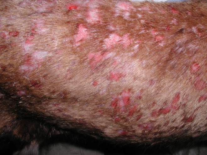
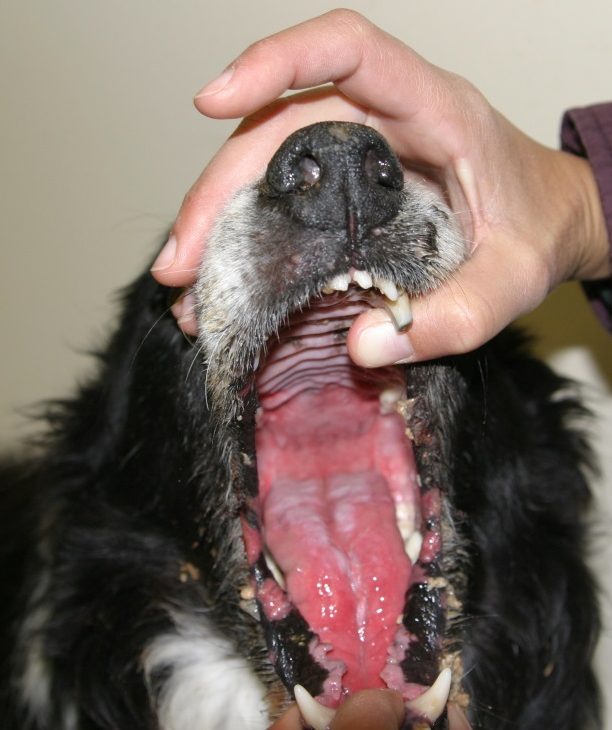
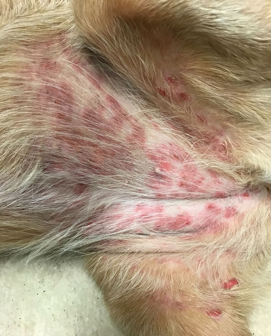
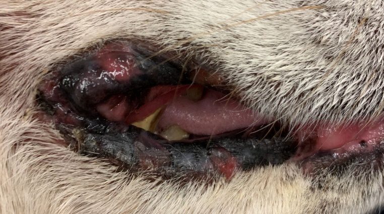
- SJS, TEN and the overlap syndrome (OVS) are characterized by an acute onset of painful, multifocal to generalized erythematous macules, patches, and/or diffuse areas of erythroderma. These lesions progress to deep erosions and ulcerations.
- The degree of epithelial detachment helps differentiate SJS, TEN and OVS :
- The epithelial detachment in SJS is < 10%, in the OVS it ranges from 10% to 30% and in TEN it is > 30%.
- Erythroderma involves >50% of the body surface area in SJS, TEN and OVS.
- A positive Nikolsky sign may be present. This means that lesions can be induced by applying lateral pressure to normal appearing skin adjacent to affected areas indicating poor cellular cohesion.
- Pseudo-Nikolsky sign is seen more often in animals and is characterized by inducing the detachment of the epidermis from the dermis after lateral pressure is applied to erythematous skin not normal appearing skin.
- Lesions can localize on any part of the body, but particularly on the head, trunk, mucocutaneous junctions (especially periocular and perilabial areas) and oral or other mucosal site. Footpads can also be affected.
-
- Lesions are typically painful, but pruritus is uncommon.
- SJS/TEN should be considered a true dermatological emergency due to the fast disease progression and severe systemic signs associated with the extensive epidermal detachment, which can be > 30% of the body surface area.
- Systemic signs may include fever, anorexia, lethargy, hypothermia, dehydration, hypoalbuminemia, hypoproteinemia, and sepsis.
- Prodromal signs such as fever, lethargy, and pruritus may be present before lesion development.
Important Facts
- SJS/TEN is characterized by an acute onset of multiple to generalized erythematous macules and patches localized on any part of the body, but particularly on the trunk, mucocutaneous junctions and oral (or other) mucosa.
- Lesions evolve to develop deep erosions and ulcerations.
- Systemic signs accompany skin lesions and may include fever, anorexia, lethargy, hypothermia, dehydration, hypoalbuminemia, hypoproteinemia, and sepsis.
-
Diagnosis
- Differential diagnoses include severe burn, EM major, paraneoplastic pemphigus, pemphigus vulgaris, autoimmune subepidermal blistering diseases, toxic shock syndrome, canine Sweet’s-like syndrome and sterile pustular erythroderma of miniature schnauzers.
- The diagnosis is based on a characteristic history, clinical signs and histopathological findings. It is important to take into consideration the extent of epithelial detachment and erythroderma because the histopathological findings support clinical diagnosis but do not distinguish among EM, SJS, TEN and OVS. Systemic signs are present in SJS, TEN and OVS but only in severe cases of EM (i.e. EM major).
- Biopsy erythematous non-ulcerated lesions because the epidermis contains the diagnostic features. The histopathological findings of EM, SJS, TEN and OVS are characterized by individual to confluent keratinocyte apoptosis and satellitosis at multiple levels of the epidermis.
- In contrast to humans, full-thickness coagulation necrosis is uncommon in SJS, TEN and OVS.
- Lymphocytic interface dermatitis with hydropic degeneration of basal keratinocytes is often present.
- An additional database to uncover internal manifestations of the disease should include a CBC, serum chemistry profile and urinalysis.
- Possible abnormalities include leukocytosis, electrolyte abnormalities, hypoproteinemia, and altered renal function.
Important Facts
- The diagnosis is based on a characteristic history, clinical signs and skin biopsy findings.
- The extent of epithelial detachment and erythroderma are important clinical features to help distinguish among EM, SJS, TEN and OVS as the histopathological findings overlap and cannot differentiate the disease forms.
- It is important to biopsy non-ulcerated lesions because the epidermis has diagnostic features.
- In all disease forms, the histopathological feature is keratinocyte apoptosis at multiple epidermal layers. Moreover, lymphocyte satellitosis, hydropic degeneration of basal keratinocytes as well as lymphocytic interface dermatitis are often present.
- An additional database to uncover internal manifestations of the disease should include a CBC, serum chemistry profile and urinalysis.
-
Treatment
- Animals with extensive epithelial detachment and erythroderma should be referred to a hospital intensive care unit as soon as possible. These cases lose a significant amount of fluid, protein, electrolytes and can become hypothermic. Moreover, because the skin barrier is lost in areas of epithelial detachment, the patients are susceptible to infection, which can evolve to sepsis. Pain therapy is also an important part of the treatment regimen.
- Identification and elimination of the trigger is crucial to positively influence the prognosis.
- No single therapy has been shown to be effective for all cases.
- Anti-inflammatory to immunosuppressive doses of glucocorticoid (1.1 – 2.0mg/kg/day for dogs and 2.0- 4.0 mg/kg/day for cats) can be tried in some cases. Decrease the initial high dose as soon as possible to the lowest possible dose that controls the disease or stop therapy if not helping.
- Cyclosporine at the dose of 5-7 mg/kg/day has been used as the initial immunosuppressive therapy in conjunction with glucocorticoids in dogs and cats.
- Intravenous human immunoglobulin (IVIG) has been used with success in a dog and can be an option for dogs and cats with this life-threatening disease or cases refractory to glucocorticoids, cyclosporine or other immunosuppressive therapies.
-
Prognosis
- Prognosis for SJS, TEN and OVS is guarded to poor.
- The mortality rate is high, especially in cases of OVS and TEN.
Important Facts
- Animals with extensive epithelial detachment and erythroderma should be referred to a hospital intensive care unit as soon as possible for supportive therapy.
- Anti-inflammatory to immunosuppressive doses of glucocorticoid can be tried as sole therapy or combined with cyclosporine or another immunosuppressive drug.
- Intravenous human immunoglobulin is an option for life threatening cases or cases refractory to glucocorticoids, cyclosporine or other immunosuppressive therapies.
- The triggering cause needs to be identified and removed to positively affect the outcome.
References
Banovic F, Dunston S, Linder KE et al. Apoptosis as a mechanism for keratinocyte death in canine toxic epidermal necrolysis. Vet Pathol 2017; 54: 249-253.
Banovic F, Olivry T, Bazzle L, et al. Clinical and microscopic characteristics of canine toxic epidermal necrolysis. Vet Pathol 2015; 52: 321-330.
Bizikova P, Linder KE, Anderson JG. Erosive and ulcerative stomatitis in dogs and cats: which immune-mediated diseases to consider? J Am Vet Med Assoc 2023; doi.org/10.2460/javma.22.12.0573.
Hinn AC, Olivry T, Luther PB, et al. Erythema multiforme, Stevens-Johnson syndrome, and toxic epidermal necrolysis in the dog: classification, drug exposure and histopathological correlations. J Vet Allergy Clin Immunol 1998; 6:13-20.
Frank AA, Ross JL, Sawvell BK. Toxic epidermal necrolysis associated with flea dips. Vet Hum Toxicol 1992; 34:57-61.
McKeever PJ, Nuttall T, Harvey RG. A Color Handbook of Skin Diseases of the Dog and Cat. 2nd edn. London: Manson Publishing Ltd., 2009; 194-196.
Miller WH, Griffin GE, Campbell KL. Muller & Kirk Small Animal Dermatology. 7th edn. St Louis: Elsevier Inc., 2013; 477-479.
Nuttall TJ, Mallam T. Successful intravenous human immunoglobulin treatment of drug induced Stevens-Johnson-Syndrome in a dog. J Small Anim Pract 2004; 45:357-361.
Scott DW et al. Toxic epidermal necrolysis in two dogs and a cat. J Am Anim Hosp Assoc 1979; 15:271.
Yager JA. Erythema multiforme, Stevens-Johnson syndrome and toxic epidermal necrolysis: a comparative review. Vet Dermatol 2014; 25: doi:10.1111/vde.12142.
