8. Erthema Multiforme
Learning Objectives
- Know! Erythema multiforme is an uncommon disease in dogs and rare in cats and believed to be mediated by T-cells damage of keratinocytes leading to panepidermal apoptosis. Reported triggering factors include infectious diseases, drugs, food antigens and neoplasia. Always look for the triggering factors when managing erythema multiforme, however, EM is often idiopathic!
- Remember! There are three forms of EM currently recognized in dogs: (i) EM minor, EM major, and (iii) hyperkeratotic EM. Mucosal involvement is used to differentiate EM minor and EM major. No or one mucosal site is affected in EM minor and more than one mucosal site is affected in EM major. In both forms, skin detachment occurs in less than 10% of the body surface area. Erythroderma (severe and diffuse erythema), if present, affects less than 50% of the body surface in EM minor and major. The “hyperkeratotic EM” is a separate variant and mucosal involvement and extent of skin lesions do not apply. This EM form has a protracted course and is characterized by marked hyperkeratotic lesions.
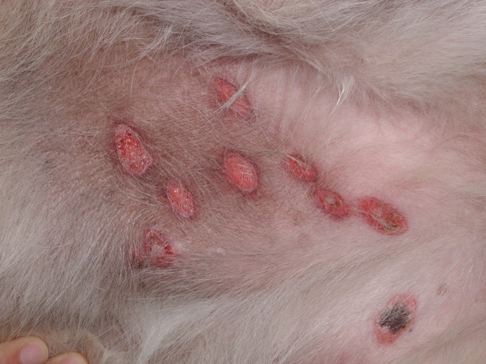
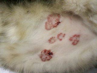
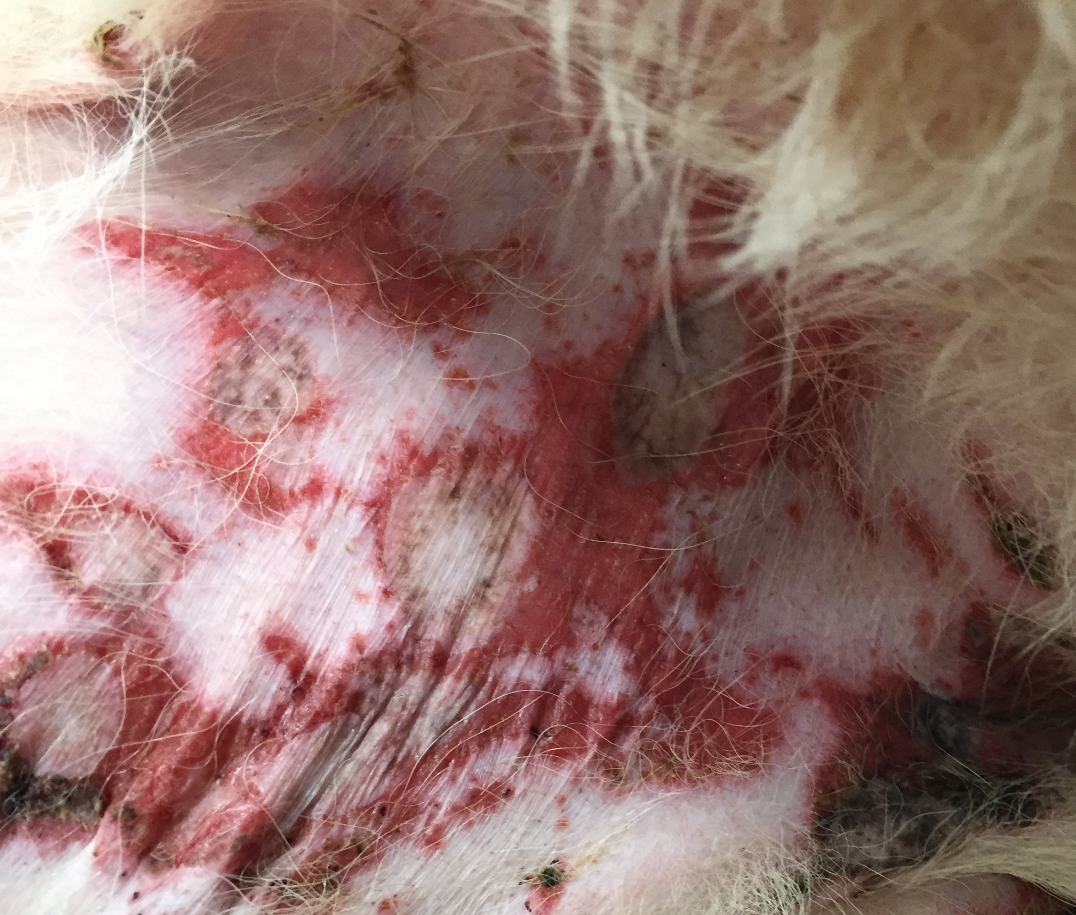
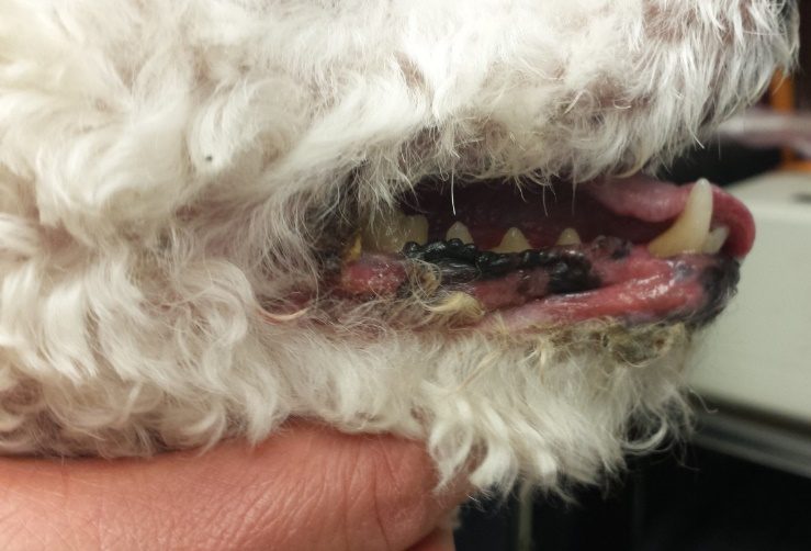
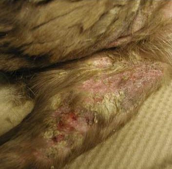
- Know! The most common lesion in EM minor and major is an erythematous maculopapular lesion that can become eroded and ulcerated. Lesions can acquire a round, oval, arciform and/or serpiginous pattern. In contrast with human EM, target lesions are not often present in dogs and cats. Lesions are more often seen on the limbs, ventrum, axillae, and ears. Vesicles, bullae and ulcers, which typically affect mucocutaneous junctions and oral mucosa are generally present in EM major. Lesions of hyperkeratotic EM are characterized by erythematous macules and plaques associated with thick and tightly adhered keratotic crusts. Lesions are typically present on the trunk, ventrum, head, pinnae, axillae and extremities. Mucosal sites are not affected, but lesions can be present on or adjacent to mucocutaneous sites.
- Know! Very few cases have been described in cats, but lesions are typically ulcerative and crusted and localized to the trunk and mucocutaneous junctions. Vesicles and bullae have also been described.
- Know! The diagnosis is based on the pet’s history, clinical signs and histopathological findings. Consistent histopathological findings are single cell necrosis of keratinocytes (apoptosis) at all epidermal layers and satellitosis (i.e. lymphocytes close or touching the apoptotic keratinocyte) and various degrees of vacuolar degeneration of basal keratinocytes and lymphocytic interface dermatitis.
- Know! If the triggering cause is identified and removed or successfully treated, the disease will resolve so try hard to identify possible triggers. Glucocorticoids is the first line of therapy. Azathioprine, cyclosporine, mycophenolate mofetil, oclacitinib or intravenous human immunoglobulin have been used for severe cases or cases refractory to glucocorticoids or that cannot tolerate it. Pentoxifylline can be added to the treatment regimen as adjunctive therapy.
-
General Considerations
- Erythema multiforme (EM) is an uncommon disease in dogs and rare in cats.
- In veterinary medicine, EM is considered the mildest manifestation of a progressive disease spectrum as follows: EM minor – EM major – Hyperkeratotic EM variant – Stevens-Johnson syndrome (SJS), and toxic epidermal necrolysis (TEN), which is the most severe form. In human medicine, EM is currently considered a separate disease entity from SJS and TEN.
-
Cause and Pathogenesis
- The putative triggering factors include infection (e.g. bacterial, parvovirus, herpesvirus, distemper virus), cutaneous drug reaction (e.g. cephalosporins, trimethoprim potentiated sulfonamides, penicillins, and others), food antigens, or internal disorders (e.g. connective tissue diseases, neoplasia).
- Cutaneous drug reaction is considered a rare cause of EM in dogs and but it is the main culprit of SJS/TEN.
- A fair number of cases are idiopathic.
- The pathomechanism is not well-understood. It is postulated that cytotoxic T-lymphocytes (CD8+), stimulated by antigen-presenting Langerhans’ cells, become sensitized against keratinocyte antigens causing damage by releasing lymphokines such as, interferon-gamma (IFN- gamma) and tumor necrosis factor- alpha (TNF alpha), resulting in keratinocyte death (i.e. apoptosis) that occurs at all epidermal levels.
- It is also possible that the activation of T-helper cells (CD4+) by antigen-presenting Langerhans cells sets the stage for the cytotoxic T-cell attack. Natural killer cells may also play a role. These cells carry in their cytoplasmic granules, proteases with cytolytic effect.
- The putative triggering factors include infection (e.g. bacterial, parvovirus, herpesvirus, distemper virus), cutaneous drug reaction (e.g. cephalosporins, trimethoprim potentiated sulfonamides, penicillins, and others), food antigens, or internal disorders (e.g. connective tissue diseases, neoplasia).
Important Facts
- Erythema multiforme is an uncommon disease in dogs and rare in cats.
- The pathogenesis is unknown, but it is speculated that T-cell damage to keratinocytes, mediated by IFN- alpha and TNF- alpha, result in keratinocyte cell death (i.e. apoptosis) at all epidermal levels. Cytotoxic T-lymphocytes (CD8+) and T-helper lymphocytes (CD4+) are believed to be the main players.
- The reported triggering factors include infection, cutaneous drug reaction, food antigens, or internal disorders such as neoplasia. Many cases are idiopathic.
-
Clinical Signs
- Humans with EM have very well-defined and characterized target lesions. In veterinary medicine, a strict classification of EM based on clinical signs is not appropriate because of the various degrees of overlap that exist between EM, SJS and TEN.
- The eruption of lesions is typically symmetrical and often involves the limbs, ventrum, axillae, groin, and ears. Mucosae and mucocutaneous junctions are also affected.
- If none or one mucous membrane is affected, the disease is classified as EM minor and if more than one mucosa is affected the disease is classified as EM major.
- Epithelial detachment, leaving areas of erosion and ulceration, involves <10% of the body surface area in both EM minor and EM major.
- Erythroderma (i.e. diffuse and severe erythema), if present, affects <50% of the body surface area.
- Lesions in dogs are variable and include, erythematous macules and papules (most common), urticarial plaques, and vesicles and bullae that evolve to erosions, ulcerations and crusting/scaling when they rupture.
- The erythematous maculopapular lesions may present with central erosions and shallow ulcers.
- Target lesions, characterized by round to oval lesions with a palpable or non-palpable erythematous border and a clear center, are not commonly seen.
- Lesions may also assume a serpiginous to arciform pattern.
-
- Patients with EM major may have systemic signs such as fever, anorexia, and depression. Moreover, vesicles, bullae and ulcerations involving the oral mucosa and/or mucocutaneous junctions are more often seen with EM major than EM minor.
-
- Hyperkeratotic EM variant:
- This form of EM has a protracted course. Because of its different clinical presentation and chronic course, some people question if this disease is indeed EM, but it is currently considered a variant of EM.
- A report on 17 cases showed that most dogs started developing lesions in mid-adulthood (i.e. 5 years of age or older) but the disease can develop in dogs as young as 1 year of age. Male dogs were overrepresented in this case series.
- Lesions are characterized by multifocal to coalescing, linear to annular macules and plaques associated with erythema and tightly adhered and thick crusts of dark brown to black to yellowish color.
- In the case series, lesions were present on the trunk and ventral abdomen (100% of cases), pinnae and head (94%), distal limbs (82%) and axillae (76% of cases).
- Lesions were also present on or around the genitalia, lips, oral cavity and anal area in 16 of the 17 dogs (94%).
- Other than lethargy in 47% of the cases, no systemic signs were noted.
- No triggering cause was identified in any of the cases.
- Main differential diagnoses for hyperkeratotic EM include generalized discoid lupus erythematosus and superficial necrolytic dermatitis.
- Hyperkeratotic EM variant:
-
- Pain and pruritus may be present in all forms of EM.
- Very few cases have been described in cats, but lesions are typically ulcerative and crusted and localized to the trunk and mucocutaneous junctions. Vesicles and bullae have also been described.
Important Facts
- The eruption of EM lesions is usually acute and symmetrical, and often involves the limbs, ventrum, axillae, groin, and ears.
- Lesions of EM minor and major in dogs are variable and include, erythematous macules and papules (most common), urticarial plaques, and vesicles and bullae that evolve to form erosions, ulcerations and crusting/scaling when they rupture.
- In contrast to humans, target lesions are not commonly seen in dogs with EM.
- EM minor is characterized by none or one mucosal involvement and EM major by more than one mucosal site affected. In EM minor and major, epidermal detachment affects less than 10% of the body surface area and erythroderma, if present, involves less than 50% of the body.
- Patients with EM major can also develop fever, anorexia and depression.
- A canine EM variant named “hyperkeratotic EM” is more often seen in mid-adulthood (>5 years old) and it has a chronic course. Lesions are characterized by erythematous macules and plaques associated with tightly adhered and thick crusts. They are typically present on the trunk, ventral abdomen, pinnae, head, axillae and extremities.
- Pruritus and pain may be present in all forms of EM.
- In cats, lesions are typically ulcerative and crusted and localized to the trunk and mucocutaneous junctions. Vesicles and bullae have also been described in cats.
-
Diagnosis
- The diagnosis of EM is based on the patient’s history, clinical signs and histopathological findings.
- Detailed history is very important to help identify the triggering cause.
- Preferred biopsy sites are erythematous macules/papules or plaques. Avoid sampling eroded or ulcerated lesions because the sample will not be diagnostic without the epidermis.
- Characteristic histopathological findings include apoptotic keratinocytes (i.e. eosinophilic keratinocytes) with satellitosis (i.e. lymphocytes close or touching apoptotic keratinocytes) at all epidermal levels. Vacuolar degeneration of the basal cell layer and lymphocytic interface reaction are typically present. Parakeratotic hyperkeratosis, often intermixed with orthokeratosis, is typical of hyperkeratotic EM.
- Differential diagnoses for cases that present with vesicles, bullae and ulcerations include the various subepidermal bullous diseases (e.g. mucous membrane pemphigoid, bullous pemphigoid, epidermolysis bullosa acquisita etc), pemphigus vulgaris, paraneoplastic pemphigus, Sweet’s-like syndrome, and sterile pustular erythroderma of miniature schnauzers.
- Differential diagnoses for the hyperkeratotic variant include generalized lupus erythematosus and superficial necrolytic dermatitis.
- The diagnosis of EM is based on the patient’s history, clinical signs and histopathological findings.
Important Facts
- Diagnosis is based on a detailed history, clinical signs and histopathological findings.
- Try to identify the triggering factors such as, infections agents, drugs and neoplasia.
-
Treatment
- If the causative insult can be identified and treated or removed, the condition can be expected to regress spontaneously.
- Oral prednisolone/sone (1.5 to 2.5 mg/kg/day) have been reported to induce 75% improvement to complete remission in dogs with hyperkeratotic EM, which can be a challenge to treat. Oral dexamethasone at 0.05 mg/kg/day can be tried if prednisolone/sone are not effective.
- Oral glucocorticoid can be used in conjunction with immunosuppressive drugs during their lag phase (i.e. cyclosporine, azathioprine, mycophenolate mofetil) and then discontinued or maintained at the lowest possible dose that maintains the disease controlled.
- The following drugs can be tried in severe or cases that do not respond solely to oral glucocorticoids or cannot tolerate glucocorticoids:
- Azathioprine: 2.5 mg/kg/day
- Cyclosporine: 5-10 mg/kg/day
- Intravenous human immunoglobulin (IVIG): 0.5-1.5 g/kg/IV
- Mycophenolate mofetil: 20-40 mg/kg q 24h divided into 2 to 3 dosages. A full clinical response typically occurs in 2-6 weeks
- Oclacitinib (Apoquel®): 0.6 to 1.0 mg/kg q 12h has been reported to be effective in treating hyperkeratotic EM and can be an option for EM minor and major. Improvement can be seen as early as 7 days and complete remission in 6 to 12 weeks.
- Pentoxifylline has been used as adjunctive therapy at 10-30 mg/kg 2-3 times a day in recurrent and non-drug associated cases.
-
Prognosis
- Prognosis is excellent if the triggering cause can be identified and removed or successfully treated.
- The prognosis is reserved to good if the triggering cause cannot be identified and removed.
Important Facts
- Look for the triggering factor! If the causative insult can be treated or removed, EM is expected to regress spontaneously.
- Anti-inflammatory doses of prednisolone/sone or dexamethasone will often decrease the inflammation and pruritus when present and are often used as the first treatment option.
- Azathioprine, cyclosporine, mycophenolate mofetil, oclacitinib and intravenous human immunoglobulin have been used in severe cases or cases refractory to glucocorticoid therapy.
- Pentoxifylline combined with other immunomodulatory drugs can be an option in recurrent and non-drug associated cases.
References
Banovic F, Olivry T, Bazzle L, et al. Clinical and microscopic characteristics of canine toxic epidermal necrolysis. Vet Pathol 2015; 52: 321-330.
Banovic F, Olivry T, Artlet B et al. Hyperkeratoric erythema multiforme variant in 17 dogs. Vet Dermatol 2023; 34: 125-133. Doi: 10.1111/vde.13141.
Bizikova P, Linder KE, Anderson JG. Erosive and ulcerative stomatitis in dogs and cats: which immune-mediated diseases to consider? J Am Vet Med Assoc 2023; doi.org/10.2460/javma.22.12.0573.
Byrne KP, Giger U. Use of human immunoglobulin for treatment of severe erythema multiforme in a cat. J Am Vet Med Assoc 2002; 220:197-201.
Favrot COT, Dunston SM, Degorce-Rubiales F et al. Parvovirus infection of keratinocytes as a cause of canine erythema multiforme. Vet Pathol 2000; 37: 647-649.
High EJ, Linder KE, Mamo LB et al. Rapid response of hyperkeratotic erythema multiforme to oclacitinib in two dogs. Vet Dermatol 2020; 31: 330-e86.
Hinn AC, Olivry T, Luther PB, et al. Erythema multiforme, Stevens-Johnson syndrome, and toxic epidermal necrolysis in the dog: classification, drug exposure and histopathological correlations. J Vet Allergy Clin Immunol 1998; 6:13-20.
Itoh T, Nibe K, Kojimoto A et al. Erythema multiforme possibly triggered by food substances in a dog. J Vet Med Sci 2006; 68: 869-871.
McKeever PJ, Nuttall T, Harvey RG. A Color Handbook of Skin Diseases of the Dog and Cat. 2nd edn. London: Manson Publishing Ltd., 2009; 194-196.
Miller WH, Griffin GE, Campbell KL. Muller & Kirk Small Animal Dermatology. 7th edn. St Louis: Elsevier Inc., 2013; 472-477.
Ramos SJ, Beale VM, Langohr IM et al. Erythema multiforme major in a dog treated with intravenous human immunoglobulin and immunosuppressive therapy. J Am Anim Hosp Assoc 2020; DOI 10.5326/JAAHA-MS-6896.
Scott DW, Miller WH. Erythema multiforme in dogs and cats: literature review and case material from the Cornell University College of Veterinary Medicine (1988-96). Vet Dermatol 1999; 10:297-309.
Yager JA. Erythema multiforme, Stevens-Johnson syndrome and toxic epidermal necrolysis: a comparative review. Vet Dermatol 2014; 25: doi:10.1111/vde.12142.
