7. Histiocytic Proliferative Disorders
Learning Objectives
- Know! Histiocytic proliferative diseases are more common in dogs than cats.
- Know! In dogs, the histiocytic disorders that affect the skin are classified as reactive (cutaneous histiocytosis and systemic histiocytosis) and neoplastic (cutaneous histiocytoma and localized or disseminated histiocytic sarcoma).
- Know! Rarely, histiocytomas will present as multiple nodules which will be much slower to regress and can rarely extend to lymph nodes or other internal organs. This condition is called, “Cutaneous Langerhans Cells Histiocytosis”, and shar pei dogs appear to be predisposed but it can affect any breeds
- Know! Cats also have histiocytic sarcomas, which resembles the dog’s disease. They also have a unique disease called progressive histiocytosis, which is considered low-grade histiocytic sarcoma.
- Know the clinical presentations and breed predispositions of the histiocytic proliferative disorders of dogs and cats discussed here.
- Know that the diagnosis is based on the history, clinical signs, signalment and histopathological findings. Immunophenotypic markers may be required to confirm a diagnosis and to differentiate some of these disorders from sterile pyogranuloma/granuloma syndrome.
- Know how to manage the histiocytic proliferative disorders discussed here and their prognosis.
-
General Considerations
- Histiocytic proliferative disorders are common in dogs and rare in cats.
- In dogs, the histiocytic disorders that affect the skin are classified as reactive (cutaneous histiocytosis and systemic histiocytosis) and neoplastic (cutaneous histocytoma and localized or disseminated histiocytic sarcoma).
- Cutaneous and systemic reactive histiocytosis originate from activated interstitial dendritic cells (DCs) and lymphocytes. Cutaneous histiocytoma originates from Langerhans cells (LCs) and histiocytic sarcoma originates from interstitial DCs.
- In cats, LCs disorders have not been identified in the skin, only lungs.
- Histiocytic sarcoma also affects cats. Like in dogs, it originates from interstitial DCs.
- A unique histiocytic disorder of cats that affects the skin is progressive histiocytosis, which is considered low-grade histiocytic sarcoma with an indolent behavior initially.
Important Facts
- Histiocytic proliferative disorders are common in dogs and rare in cats.
- The canine histiocytic disorders that affect the skin include (i) cutaneous reactive histiocytosis, (ii) systemic reactive histiocytosis, (iii) histiocytoma and (iv) histiocytic sarcoma.
- Except for cutaneous histiocytoma that originates from Langerhans cells, the others diseases originate from interstitial dendritic cells.
- No histiocytic disorder of LCs origin has been recognized in cats. However, cats can also have histocytic sarcoma and a unique histiocytic disease named progressive histiocytosis.
-
Cause and Pathogenesis
- The cause of these diseases is currently unknown, but it can be speculated that the reactive disorders result from an aberrant immune response to a yet unidentified antigen. The positive response of the reactive diseases to immunosuppressive drugs supports this theory.
-
Clinical Signs
- Dogs:
- Cutaneous reactive histiocytosis:
- Cutaneous reactive histiocytosis affects primarily the skin and subcutaneous tissue but can extend to draining lymph nodes.
- There is no breed, sex, or age (typically 3 to 9 years) predilection.
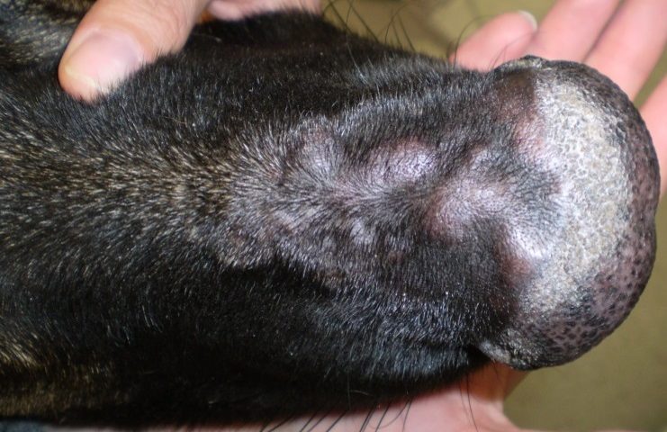
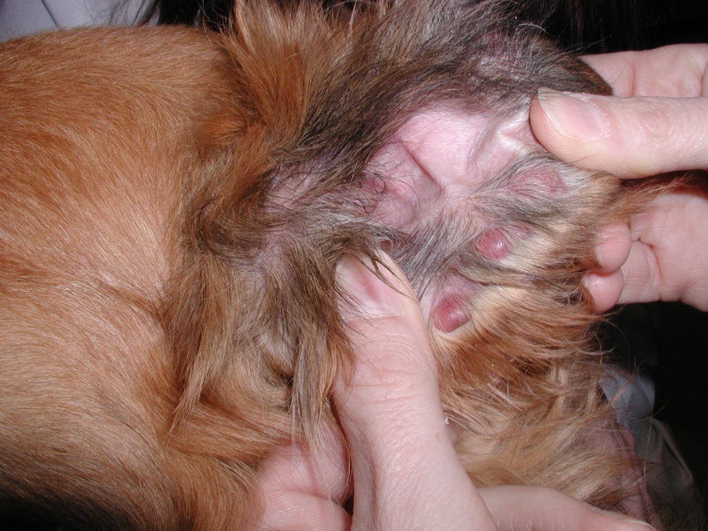
- It is generally associated with one (rarely) or multiple dermal to subcutaneous nodules and/or plaques often localized to the head, nasal planum, muzzle, neck, perineum, scrotum and extremities (including footpads).
- Cutaneous reactive histiocytosis:
- Dogs:
-
-
-
- Lesions may be alopecic or hairy, are typically erythematous and non-pruritic or painful.
-
-
-
-
-
- The nodules and plaques may eventually become eroded or ulcerated.
-
-
-
-
-
- Lesions wax and wane (i.e. resolve or regress in one area and appear in others) and rarely spontaneously resolve, which makes assessment of response to therapy difficult.
- Extension to local cutaneous lymph nodes may occur.
- Systemic histiocytosis:
- It occurs less frequently than cutaneous histiocytosis.
- It affects the skin and extracutaneous sites such as lungs, spleen, liver, and bone marrow. In some cases, only extracutaneous sites are involved.
- Bernese mountain dogs are more frequently affected but the disease has also been described in other breeds such as the golden retriever, Labrador retriever, boxer, basset hound, Irish wolfhound, and Rottweiler. There is evidence for familial occurrence of the disease in Bernese mountain dogs and Irish wolfhounds.
- It typically affects young to middle age dogs (2 to 8 years) and there is no sex predilection.
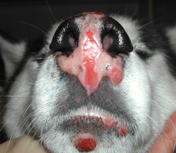
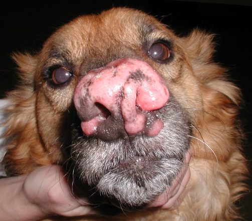
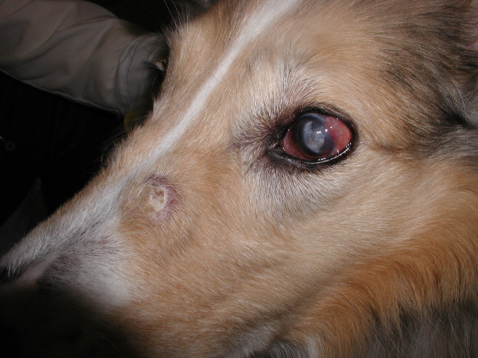
- Lesions have a marked tendency to involve not only the skin but also the ocular and nasal mucosae and peripheral lymph nodes. Skin lesions are similar to those of cutaneous histiocytosis. Lesions may extent to other eye structures than the conjunctival mucosa such as, the sclera, ciliary body, extraocular muscles, and retrobulbar tissues.
-
-
-
-
-
- The severity of clinical signs correlates with the extent of the disease and may include anorexia, weight loss, stertorous respiration and conjunctivitis.
- Like cutaneous reactive histiocytosis, lesions may wax and wane (i.e. resolve or regress in one area and appear in others) and rarely spontaneously resolve, which makes assessment of response to therapy difficult.
- Commonly seen clinicopathologic findings include anemia, monocytosis and lymphopenia.
- Cutaneous histiocytoma:
- Histiocytoma is a common canine tumor of benign behavior in most cases.
- It is considered by many a reactive disease of Langerhans cell origin other than a neoplasm because of its benign behavior characterized by spontaneous regression in most cases. However, metastasis of a solitary histiocytoma to local lymph node has been reported.
- It typically occurs in dogs less than 3 years of age, but it can also develop in older dogs.
- Histiocytoma can occur in any breed but boxers, dachshunds, cocker spaniels, Great Danes, bull terriers, and Shetland sheepdogs may be overrepresented.
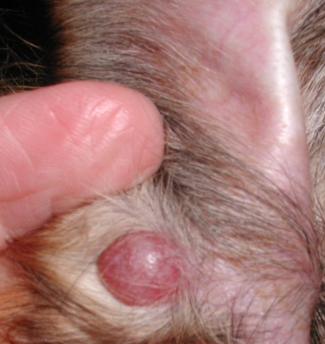
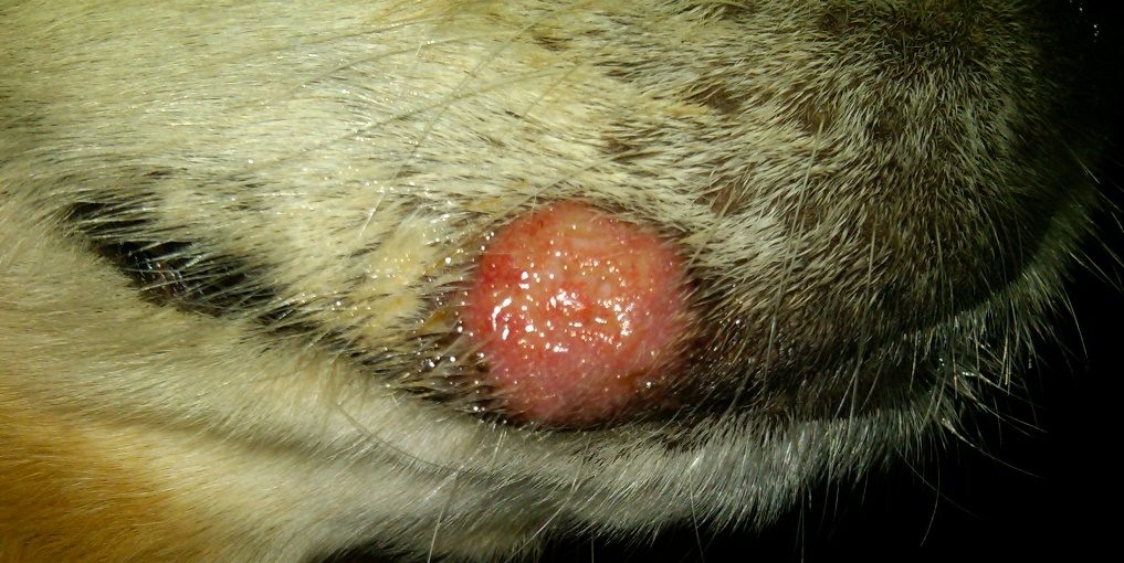
- Nodules are typically single, small, hairless, erythematous, and localized to the head, pinna, neck, or limbs.
-
-
-
-
-
- Single nodules often spontaneous resolve within 1 to 3 months and they typically ulcerate before resolving.
-
-
-
-
-
- Histiocytomas may rarely present (<1%) as multiple (hundreds) skin and mucosal (eye and oral) nodules. Peripheral lymph nodes and rarely systemic involvement may occur. This disease is named “Cutaneous Langerhans Cell Histiocytosis”. The nodules will eventually spontaneously resolve but it may take 10 months or more. Prognosis is significantly worse with lymph nodes and internal organs involvement.
- Shar pei dogs appear to be more prone to develop “Cutaneous Langerhans Cell Histiocytosis” but this disease can affect any breed.
- Histiocytomas may rarely present (<1%) as multiple (hundreds) skin and mucosal (eye and oral) nodules. Peripheral lymph nodes and rarely systemic involvement may occur. This disease is named “Cutaneous Langerhans Cell Histiocytosis”. The nodules will eventually spontaneously resolve but it may take 10 months or more. Prognosis is significantly worse with lymph nodes and internal organs involvement.
- Histiocytic sarcoma:
- Histiocytic sarcoma is classified into localized when it originates at a single tissue site or single organ (with solitary or multiple foci) or disseminated disease when it extends beyond the local draining lymph node to involve distant sites. Disseminated histiocytic sarcoma was previously called malignant histiocytosis.
- Bernese mountain dogs are predisposed, and pedigree analysis suggests a polygenic mode of inheritance in this breed. The disease also occurs frequently in flat-coated retrievers, Rottweilers, and golden retrievers and rarely in other breeds.
- Histiocytic sarcoma affects more often middle age to older dogs and there is no sex predilection.
- Dogs with localized disease typically present with a soft-tissue mass that grows rapidly. It often develops on the limbs close to a joint where it can extend to the joint capsule, ligaments, tendons and muscles. Primary lesions can also develop in the liver, spleen, lung, pancreas, and central nervous system. Localized histiocytic sarcoma has a reported metastatic rate of 91%.
- Disseminated histiocytic sarcoma does not often affect the skin and typically occurs in the spleen, liver, bone marrow, lungs, and lymph nodes. Most affected dogs have a history of anorexia, depression, anemia, weight loss and other signs depending on the organs affected.
- The hemophagocytic histiocytic sarcoma carries the worse prognosis of all histiocytic sarcoma forms with a reported median survival time of only 4 weeks. Patients have severe regenerative anemia, thrombocytopenia, hypoalbuminemia, and hypocholesterolemia. This form originates from macrophages and not Langerhans cells and interstitial dendritic cells, which are involved in the other histiocytic diseases of dogs and cats.
-
- Cats:
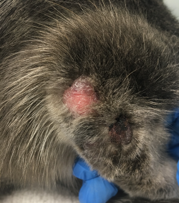
- Feline progressive histiocytosis:
- It typically affects cats between 7 and 17 years of age.
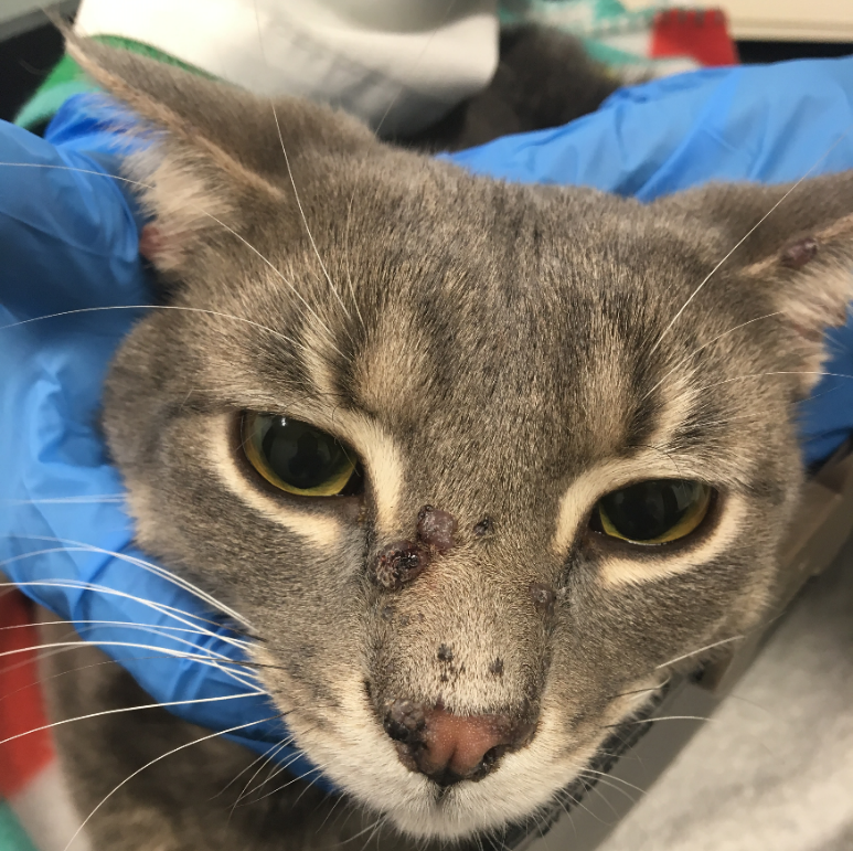
- Most cats develop multiple papules, nodules and/or plaques that are generally non-painful and non-pruritic.
- Lesions are typically alopecic and may ulcerate. They are commonly located at the head, limbs and trunk.
- Some cats may have invasive disease affecting lymph nodes and internal organs including the lungs, kidneys, spleen, and liver.
- Lesions do not spontaneously resolve but they may wax and wane.
- Feline progressive histiocytosis is considered a low-grade histiocytic sarcoma where morphological appearance of histiocytes early in the disease has minimal atypia but they cannot be differentiated from histiocytes seen with histiocytic sarcoma in late-stage lesions.
- The prognosis is poor because cats typically do not respond to therapy.
- Feline progressive histiocytosis:
-
-
-
- Histiocytic sarcoma
- Histiocytic sarcoma can also develop in cats and the disease resembles that of dogs according to the location of lesions and disease progression. However, the disseminated form is much less common in cats than dogs.
- Histiocytic sarcoma
-
Important Facts
- Canine cutaneous reactive histiocytosis can occur in any breed, sex or age.
- Lesions are typically multiple and characterized by nodules or plaques that more often affect the head, nose, neck, perineum, scrotum and extremities (including footpads).
- Lesions of cutaneous reactive histiocytosis wax and wane and seldom spontaneously resolve. They can rarely extend to local skin lymph nodes.
- Systemic reactive histiocytosis is more frequently diagnosed in Bernese mountain dogs but it has been also reported in various other breeds.
- It typically affects young to middle-aged dogs (2 to 8 years) and there is no sex predilection.
- Lesions develop on the skin, ocular and nasal mucosae and peripheral lymph nodes, but it may progress to affect the lungs, spleen, liver and bone marrow.
- Cutaneous histiocytoma is a common canine tumor that spontaneously resolves, many cases within about 3 months. It generally affects dogs younger than 3 years of age. Histiocytoma usually manifests as a single, hairless, erythematous nodule localized on the head, ear pinna, neck or limbs.
- Rarely, histiocytomas will present as multiple nodules that are much slower to regress and can rarely extend to lymph nodes or other internal organs. This condition is called, “Cutaneous Langerhans Cells Histiocytosis”, and Shar pei dogs appear to be predisposed but it can affect any breed.
- Histiocytic sarcoma can be localized or disseminated. Bernese mountain dogs are predisposed but the disease also occurs frequently in flat-coated retrievers, Rottweilers, and golden retrievers and rarely in other breeds.
- Dogs with localized histiocytic sarcomas typically present with a soft-tissue mass that grows rapidly. It often develops on the limbs close to a joint where it can extent to the joint capsule, ligaments, tendons and muscles. Primary lesions can also develop in the liver, spleen, lung, pancreas, and central nervous system.
- Disseminated histiocytic sarcoma does not often affect the skin and typically occurs in the spleen, liver, bone marrow, lungs, and lymph nodes.
- Histiocytic proliferative disorders of cats include histiocytic sarcoma, which has the same lesion distribution and disease progression as the dog’s disorder, and progressive histiocytosis.
- Progressive histiocytosis is considered a low-grade histiocytic sarcoma that typically develops in middle age to older cats. Nodules and/or plaques typically develop on the head, limbs and trunk.
- Prognosis of progressive histiocytosis is poor because cats typically do not respond to many treatment modalities.
-
Diagnosis:
- The history, clinical signs and patient’s signalment will help with the diagnosis.
- Definitive diagnosis can only be made based on histopathological findings. Identification of immunophenotypic markers is required to different some of these disorders from sterile pyogranuloma/granuloma syndrome and to confirm their diagnosis. Markers for cutaneous and systemic histiocytosis are identical but different from markers of histiocytoma and histiocytic sarcoma, which also differ from each other.
- Histopathological features that can be helpful in differentiating the histiocytic disorders include:
- Cutaneous histiocytomas – some degree of epitheliotropism and, in late regressing lesions, prominent lymphocytic infiltrate; the mitotic index of histiocytic cells may be high. The tumor cells have tropism for the superficial dermis creating a “top heavy” distribution.
- Cutaneous and systemic histiocytosis – histiocytes and lymphocytes aggregate around mid-dermis vessels and adnexa and often infiltrate the vessel wall; the infiltrate coalesces and extends to the deep dermis and subcutaneous tissue (“bottom heavy”).
- Histiocytic sarcoma and late stage progressive histiocytosis – marked anisocytosis and anisokaryosis, multinucleate giant cells with marked atypia, and numerous mitotic figures.
Important Facts
- The history, clinical signs and patient’s signalment will help with the diagnosis.
- Definitive diagnosis can only be made based on histopathological findings and, in some cases, identification of immunophenotypic markers.
-
Treatment:
- Cutaneous histiocytoma associated with a single lesion typically resolves spontaneously. Surgical removal can be done for lesions that do not regress within 6 months or become infected. Immunosuppressive drugs (e.g. glucocorticoids or cyclosporine) are not indicated because they will interfere with the natural immune response (CD8+ cytotoxic T-lymphocytes) to the tumor cells.
- Cutaneous reactive histiocytosis cases typically respond well to immunosuppressive/immunomodulatory drugs such as, glucocorticoids, cyclosporine or azathioprine. The combination of tetracycline and niacinamide is an option either as sole therapy for mild cases or as adjunctive therapy for more generalized cases. However, practice antibiotic stewardship and avoid the use of an antibiotic with immunomodulatory properties to treat inflammatory diseases.
- Most dogs will need long-term therapy to prevent recurrences.
- Systemic histiocytosis generally does not respond to high doses of glucocorticoids. The drugs currently recommended for this disease include azathioprine, leflunomide and cyclosporine but none works for all cases. The prognosis is guarded to poor and many dogs are euthanized due to poor response to therapy.
- Cutaneous Langerhans Cell Histiocytosis are typically refractory to treatment. Glucocorticoids and cyclosporine are not efficacious and may impede spontaneous remission, which can occur in about 50% of the cases if lymph node involvement is not present.
- C-kit inhibitors (toceranib and masitinib) prolonged the life of a dog with internal disease after various chemotherapeutic agents had failed.
- The prognosis of histiocytic sarcomas is very poor. Localized disease may be more amenable to treatment and radical surgical excision with or without radiotherapy may be an option for some cases. Very few cases have been treated with chemotherapy to allow assessment of efficacy. An oncologist should be ideally consulted to discuss treatment options.
- Feline progressive histiocytosis has a poor prognosis because most cats do not respond to various treatment modalities.
Important Facts
- Most cutaneous histiocytoma, when single, spontaneously resolve within 1 to 3 months. Immunosuppressive drugs (e.g. glucocorticoids or cyclosporine) are not indicated because they will interfere with the natural immune response to the tumor cells.
- Cutaneous reactive histiocytosis typically responds well to glucocorticoids, cyclosporine, azathioprine and/or the combination of tetracycline and niacinamide. Most dogs will need long-term therapy to prevent recurrences.
- Systemic histiocytosis typically does not respond to high doses of glucocorticoids and the recommended drugs include azathioprine, cyclosporine or leflunomide. The prognosis is guarded to poor and many dogs are euthanized due to poor treatment response.
- Cutaneous Langerhans Cell Histiocytosis are typically refractory to treatment. Glucocorticoids and cyclosporine are not efficacious and may impede spontaneous remission, which can occur in about 50% of the cases if lymph node involvement is not present.
- The prognosis of histiocytic sarcomas is poor and an oncologist should be consulted to discuss treatment options.
- Most cats with progressive histiocytosis do not respond to therapy.
References
Affolter VK, Moore PF. Canine cutaneous and systemic histiocytosis: reactive histiocytosis of dermal dendritic cells. Am J Dermatohistopathol 2000; 22:40-48.
Affolter VK, Moore PF. Localized and disseminated histiocytic sarcoma of dendritic cell origin in dogs. Vet Pathol 2002; 39:74-83.
Affolter VK, Moore PF. Feline progressive histiocytosis. Vet Pathol 2006; 43: 646-655.
McKeever PJ, Nuttall T, Harvey RG. A Color Handbook of Skin Diseases of the Dog and Cat. 2nd edn. London: Manson Publishing Ltd., 2009; 84-87.
Miller WH, Griffin GE, Campbell KL. Muller & Kirk Small Animal Dermatology. 7th edn. St Louis: Elsevier Inc., 2013; 817-821.
Moore PF. A review of histiocytic diseases of dogs and cats. Vet Pathol 2014; 51:167-184.
Moore PF. Histiocytic diseases. Vet Clin Small Anim 2023; 53: 121-140.
Palmeiro BS, Morris DO, Goldschmidt MH, et al. Cutaneous reactive histiocytosis in dogs: a retrospective evaluation of 32 cases. Vet Derm 2007; 18:332-340.
