5. Idiopathic Sterile Pyogranuloma/granuloma Syndrome
Learning Objectives
- Know! Idiopathic sterile pyogranuloma/granuloma syndrome is a rare condition presumed to be immune-mediated, based on histopathological findings and response to corticosteroid therapy.
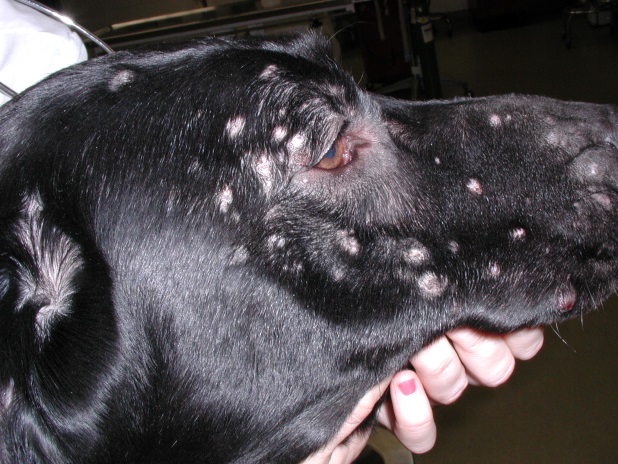
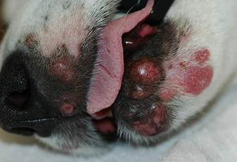
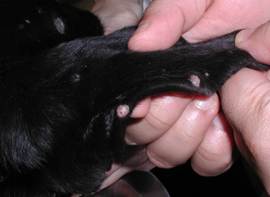
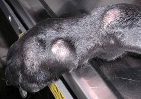
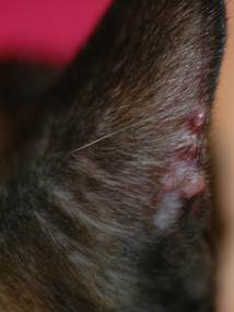
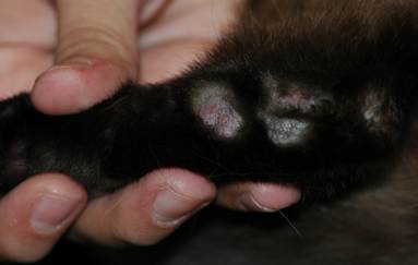
- Know! Lesions are usually multiple and typically affect the head (especially the muzzle, and the periocular region), the pinnae and the paws. Lesions are characterized by firm, painless, non-pruritic, dermal plaques and nodules. Rarely, the lesions can ulcerate and become secondarily infected.
- Remember! Cytology of aspirates obtained from intact nodules and plaques is an easy and fast test and could be diagnostic if a relevant microorganism or neoplastic cells are found. However, an infectious cause cannot be ruled out if no organisms are found on cytology.
- Know! Perform aseptic bacterial (aerobic and anaerobic) and fungal tissue cultures to rule out infectious granulomatous dermatitis. Cultures are invariably negative in this condition if the samples are collected aseptically.
- Know! The definitive diagnosis is based on negative tissue culture results and histopathological findings showing granulomatous or pyogranulomatous inflammation without any infectious agents.
- Know that oral glucocorticoid at immunosuppressive doses is the mainstay therapy. If the disease cannot be controlled long-term with low doses of glucocorticoids, add cyclosporine or mycophenolate mofetil to the treatment regimen as a steroid-sparing agent. Azathioprine may be needed for dogs and chlorambucil for cats that are unresponsive or become refractory to cyclosporine and mycophenolate mofetil therapy.
- Remember that long-term therapy is required in most cases.
-
General Considerations
- Idiopathic sterile pyogranuloma/granuloma syndrome is a sterile inflammatory process involving primarily a pyogranulomatous reaction of the dermis and less commonly the subcutaneous adipose panniculus.
- It is an uncommon disease of dogs and rare disease of cats.
- As the name “idiopathic” indicates, the cause or trigger factor inducing the pyogranulomatous inflammation is currently unknown.
- Good response to corticosteroid or other immunomodulatory therapy supports a sterile process.
Important Facts
- The sterile pyogranulomatous reaction is typically present in the dermis, but it can extend to the subcutaneous adipose panniculus.
- Idiopathic sterile pyogranuloma/granuloma syndrome uncommonly affects dogs and rarely cats.
- As the name “idiopathic” indicates, the cause or trigger factor is not currently known.
-
Clinical Signs
- The doberman pincher, Weimaraner, collie, boxer, dachshund, golden retriever, Chinese shar-pei, pit bull terrier, and English bulldog appear to be predisposed but the disease can develop in any breed.
- No sex or age predilection has been reported; however, it usually affects adult dogs.
- Dogs
- Lesions are often multiple and typically affect the head (especially the muzzle and the periocular region), pinnae and paws. The trunk is less often affected.
- Lesions are characterized by non-pruritic, firm, painless, erythematous or of normal skin color, dermal papules, plaques and nodules.
- The lesions may become alopecic, ulcerated, and secondarily infected, especially the ones on the paws.
-
- Cats
- Firm, erythematous to violaceous to orange-yellow papules, nodules and/or plaques develop on the head, muzzle and pinnae and, less commonly on the paws, legs and trunk.
- Cats
-
-
- Lesions are rarely pruritic.
- The affected dogs and cats are usually healthy otherwise.
-
Important Facts
- Non-pruritic, painless, firm, dermal papules, plaques and nodules are usually multiple and most commonly affect the head, pinnae and paws of dogs and cats.
- The lesions may become alopecic, ulcerated, and secondarily infected, especially the ones on the paws.
- Affected animals are usually healthy otherwise.
-
Differential Diagnoses
- The differential diagnoses include infectious granulomatous and pyogranulomatous disorders (e.g. bacteria, fungus, oomycete causes), sterile nodular panniculitis, cutaneous reactive histiocytosis, foreign body reaction and neoplasia.
-
Diagnosis
- The diagnosis is based on excluding the diseases in the differential diagnoses list as appropriate.
- The patient’s detailed history and clinical signs are important in prioritizing the differentials.
- Cytology of aspirates collected from nodules and plaques is an easy and fast test to evaluate the type of inflammatory infiltrate and the presence or absence of microorganisms.
- Avoid collecting samples from eroded or ulcerated lesions and from draining tracts as these lesions are often secondarily infected.
- Keep in mind that microorganisms are not always seen on cytology, so a negative cytology does not rule out an infectious cause.
- Bacterial (i.e. aerobic and anaerobic) and fungal cultures and skin biopsies are important in determining if the inflammatory process is sterile, when an infectious cause cannot be identified via cytology.
- Bacterial and fungal tissue cultures (not swab cultures) are recommended. The samples need to be taken aseptically to avoid contamination. It is important to sample the core of the lesion, so an incision biopsy may be needed as a punch instrument may not reach the lesion core.
- Consult with the laboratory to determine the best way to submit the samples for culture.
- Bacterial culture from the surfaces of eroded and ulcerated lesions will likely yield non-diagnostic growth because bacterial overgrowth or infection is often associated with these lesions.
- An incisional or excisional biopsy is the ideal sample for histopathology as it guarantees the inclusion of the epidermis, dermis and subcutaneous adipose tissue. Avoid sampling eroded and ulcerated lesions as these lesions will likely be secondarily infected.
- If possible, submit the samples to a pathologist with experience in dermatohistopathology. It is important to include a detailed history, physical examination findings and, ideally, pictures of the case.
- Histopathological findings reveal a nodular to diffuse granulomatous to pyogranulomatous dermatitis and/or panniculitis. Special stains for microorganisms should be requested to investigate if infectious agents are present.
- The diagnosis is based on excluding the diseases in the differential diagnoses list as appropriate.
Important Facts
- The diagnosis is based on ruling out the diseases on the list of differential diagnoses as appropriate.
- The patient’s history and clinical signs help prioritize the diseases in the list of differentials.
- Cytology of samples from intact nodules and plaques is an easy and fast test; however, the absence of microorganisms does not rule out an infectious cause.
- Bacterial and fungal cultures and histopathology are important to determine if the inflammatory process is sterile.
- Tissue samples from the core of the lesions should be ideally collected for bacterial and fungal cultures.
- An incisional or excisional biopsy of an intact nodule and plaque are the ideal samples for histopathology as they include the whole skin and the subcutaneous fat tissue. Special stains to investigate microorganisms should be requested.
- Contact the laboratory to determine the best way to submit samples
-
Treatment
- Oral glucocorticoid is the mainstay therapy. The response is typically rapid if immunosuppressive doses are used.
- Oral prednisone or prednisolone has shown to be effective in many cases. Start at the dose of 2.2 mg/kg q 24h until the lesions have regressed (typically 2 to 4 weeks), and then reduce to the lowest effective dose, which should be given on alternate days.
- The following are drugs that can be used as steroid-sparing agents when the disease cannot be solely controlled long-term with a low dose of glucocorticoid.
- Cyclosporine at 5-10mg/kg q 24h. It can take 4-6 weeks for full clinical response. In some cases, the addition of cyclosporine to the treatment regimen may allow the discontinuation of glucocorticoids.
- Mycophenolate mofetil at 20-40 mg/kg q 24h divided into 2 to 3 dosages. A full clinical response may not be seen before 2-6 weeks. It is an option in cases that cyclosporine was not tolerated or was not effective.
- Azathioprine at 1.0-2.2 mg/kg q 24h may be needed for dogs that are unresponsive or become refractory to cyclosporine and mycophenolate mofetil therapy. After remission is achieved try to reduce the dose to alternate-day therapy. It may take 4-6 weeks before azathioprine starts working.
- A complete blood cell count and serum chemistry profile are recommended every 2 weeks for the first 8-12 weeks to monitor for bone marrow suppression and hepatotoxicity.
- Do not use this drug in cats because they are very prone to the azathioprine-induced bone marrow suppression. Chlorambucil at the dose of 0.1 to 0.2 mg/kg q 24h to 48h should be tried for refractory cases.
- Tetracycline and niacinamide have been reported to work in mild cases as sole therapy. However, the authors practice antibiotic stewardship and try to avoid an antibiotic drug with immunomodulatory properties to treat sterile inflammatory diseases.
- The recommended dosage of tetracycline and niacinamide is 250 mg of each 3 times daily for dogs < 10 kg and 500 mg of each 3 times daily for dogs > 10 kg.
- Doxycycline at 5-10 mg/kg q 12h can be used if tetracycline is not available.
- The trial period should be 8 weeks. Consider another treatment modality if no improvement is noticed after this time.
- Relapses are common when treatment is discontinued.
- Oral glucocorticoid is the mainstay therapy. The response is typically rapid if immunosuppressive doses are used.
Important Facts
- Oral glucocorticoid at immunosuppressive doses is the mainstay therapy.
- If the disease cannot be controlled long-term with low doses of glucocorticoids, add cyclosporine or mycophenolate mofetil to the treatment regimen as a steroid-sparing agent.
- Azathioprine may be needed for dogs and chlorambucil for cats that are unresponsive or become refractory to cyclosporine and mycophenolate mofetil therapy.
- For all immunosuppressive drugs, the ultimate goal is to find the lowest possible dose that controls the disease long-term with minimal side effects.
- Relapses are common when therapy is interrupted.
References
Gross TH, Ihrke PJ, Walter EJ et al. Skin Diseases of the Dog and Cat: Clinical and Histopathologic Diagnosis. 2nd edn. Oxford: Blackwell Publishing, 2005; 320-323.
McKeever PJ, Nuttall T, Harvey RG. A Color Handbook of Skin Diseases of the Dog and Cat. 2nd edn. London: Manson Publishing Ltd., 2009; 82-83.
Miller WH, Griffin GE, Campbell KL. Muller & Kirk Small Animal Dermatology. 7th edn. St Louis: Elsevier Inc., 2013; 704-706.
Panich R, Scott DW, Miller WH. Canine cutaneous sterile pyogranuloma/granuloma syndrome: a retrospective analysis of 29 cases (1976-1988). J Am Anim Hosp Assoc 1991; 27:519-528.
Rosthien E, Scott DW, Riis RC. Tetracycline and niacinamide for the treatment of sterile pyogranuloma/granuloma syndrome in a dog. J Am Anim Hosp Assoc 1997; 33:540-543.
Schissler J. Sterile pyogranulomatous dermatitis and panniculitis. Vet Clin Small Anim 2019: 49:27-36.
Scott DW, Buerger RG, Miller WH. Idiopathic sterile granulomatous and pyogranulomatous dermatitis in cats. Vet Derm 1990; 1:129-137.
Torres S. Sterile nodular dermatitis in dogs. Vet Clin North Am Small Anim Pract 1999; 29:1311-1314.
