3. Canine Claw Disorders
Learning Objectives
- Know that symmetrical lupoid onychitis, idiopathic onychodystrophy and idiopathic onychomadesis are the currently recognized primary claw disorders of dogs.
- Know that symmetrical lupoid onychitis is not a common problem but is currently considered the most common claw disorder of dogs.
- Know that in cases of symmetrical lupoid onychitis, pain (onychalgia) is the main complain and owners will report that dogs typically lick at their paws and do not allow their paws to be touched.
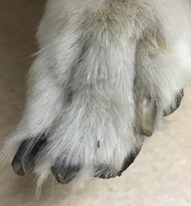
- Know that dogs with symmetrical lupoid onychitis typically present with multiple claws of various paws affected. The clinical signs include one or more of the following: onycholysis (detachment of the claw plate from the claw bed), onychorrhexis (fragmentation of the distal claw plate), onychoschizia (splitting of claw plate starting distally), onychomadesis (sloughing of claws), purulent exudate associated with areas of claw splitting and paronychia (inflammation of the nail fold).
- Know that pain, paronychia, onycholysis, onychoschizia and exudate do not typically precede the dystrophy and claw sloughing in cases of idiopathic onychodystropy and idiopathic onychomadesis; which can be helpful in differentiating these diseases.
- Be aware! The diagnosis of these claw disorders should be based on a characteristic history, clinical signs and ruling out primarily trauma, dermatophytosis (rare) and neoplasia (rare). A claw biopsy is rarely done because the histopathological changes associated with lupoid onychitis are not pathognomonic and, at this time, we do not know the histopathological changes associated with idiopathic onychodystrophy and idiopathic onychomadesis.
- Make sure to treat the secondary bacterial infections appropriately!
- Know that high doses of essential fatty acids (omega 3 + omega 6), a tetracycline and niacinamide, pentoxifylline, gelatin, and biotin are safe treatment options and should be tried first in all cases of primary nail disorders. However, practice antibiotic stewardship and try to avoid using antibiotic with immunomodulatory properties, such as tetracyclines, to treat inflammatory diseases.
- Know that prednisone/solone, azathioprine or cyclosporine can be tried for severe and refractory cases of symmetrical lupoid onychitis.
- Make sure to tell owners that there is no treatment that works for all cases and it may take months for improvement to be noticed. In addition, mention that claw dystrophy may never completely resolve and the main sign of treatment success in symmetrical lupoid onichitis is lack of pain/discomfort.
-
General Considerations
- Canine claw disorders can be primary, where they are the sole problem, or secondary, where they are associated with other disorders.
- Primary canine claw disorders include symmetrical lupoid onychitis, idiopathic onychodystrophy, and idiopathic onychomadesis.
- Symmetrical lupoid onychitis, also known as symmetrical lupoid onychodystrophy, is currently the most recognized claw disorder of dogs.
- The various diseases associated with secondary claw disorders include familial dermatomyositis and other ischemic dermatopathies, drug eruption, nutritional deficiencies, superficial necrolytic dermatitis, epidermolysis bullosa, ergotism, thallotoxicosis, linear epidermal nevi, and disseminated intravascular coagulation.
- Secondary claw disorders will not be discussed in this section. Information on some of these diseases can be found elsewhere in this eBook.
- It is important to define the terminology used to describe claw abnormalities.
- Onychitis: Inflammation somewhere in the claw unit
- Onychalgia: Claw pain
- Onychomalacia: Softening of the claw
- Onychodystrophy: Abnormal claw formation
- Onychomadesis: Sloughing of the claw
- Onychorrhexis: Longitudinal striations associated with breaking/brittleness of the claw plate
- Onychoschizia: Splitting or lamination of the claw usually starting distally
- Onycholysis: Separation of the claw from the claw bed
- Paronychia: Inflammation of the claw fold (i.e. soft tissue surrounding the claw)
Important Facts
- Canine claw disorders can be primary, where they are the sole problem, or secondary, where they are associated with other disorders.
- Symmetrical lupoid onychitis, idiopathic onychodystrophy and idiopathic onychomadesis are primary claw disorders.
- Symmetrical lupoid onychitis, also known as symmetrical lupoid onychodystrophy, is the most recognized claw disorder of dogs.
-
Pathogenesis
- Symmetrical lupoid onychitis
- The pathogenesis is unknown, but studies conducted in Gordon setters and Bearded collies suggest that genetics likely plays a role at least in some breeds.
- Idiopathic onychodystrophy and idiopathic onychomadesis
- As the name “idiopathic” indicates the pathomechanism of these claw disorders is currently unknown. However, mineral abnormalities have been detected in the claw plates of dogs with idiopathic onychomadesis.
- Symmetrical lupoid onychitis
-
Clinical Signs
- Symmetrical lupoid onychitis:
- The reported age of onset in dogs ranges from 2 to 7 years with a mean of 4 years of age.
- No sex predilection has been reported.
- Breeds reported to be more commonly affected include the German shepherd dog, English setter, and Norwegian Gordon setter; however, this claw disorder has been recognized in many other breeds.
- The problem may initially affect only 1 to 3 claws but ,typically, within few weeks to months many claws will become affected.
- Pain (onychalgia) is often the main complaint. Many owners will report that their dogs lick at the paws and do not allow them to be touched.
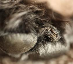
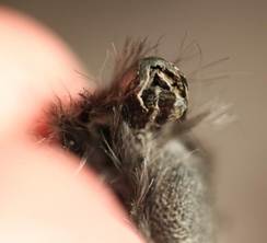
- On physical examination, one or more of the following signs can be present: missing claw plates (onychomadesis), broken and/or split claw plates (onychorrhexis/onychoschizia), separation of the claw plate from the claw bed (onycholysis), presence of sanguineous-purulent exudate from split/broken claw plate sites, and inflammation of the claw fold (paronychia).
- Symmetrical lupoid onychitis:
-
-
- Secondary bacterial infection occurs often; therefore, make sure to perform cytology of exudate.
- After the claw has sloughed, the newly formed claw is dystrophic and characterized by being short, dry, brittle, misshapen and soft.
- Differential diagnoses include the following: onychomycosis (typically caused by dermatophytes), trauma, idiopathic onychomadesis or onychodystrophy (usually inflammation is not present in the absence of a secondary infection), autoimmune/immune-mediated diseases (e.g. pemphigus foliaceus, bullous pemphigoid, systemic lupus erythematosus, ischemic dermatopathies, cold agglutinin disease, drug reaction, vasculitis – typically other clinical signs will be present with these diseases), and neoplasia of the claw.
- Idiopathic onychodystrophy and idiopathic onychomadesis:
- An important feature to differentiate idiopathic onychodystrophy and idiopathic onychomadesis from symmetrical lupoid onychitis is that these nail disorders are not typically preceded by the signs of onychitis described above (i.e. licking at paws, significant discomfort when touching the paws, purulent exudate associated with sites of split/broken claw plates, onycholysis). Paronychia may be noted but is not generally present.
- Idiopathic onychodystrophy:
- Abnormal claw formation (onychodystrophy) is the initial sign.
- It has been recognized more frequently in cocker spaniels, dachshunds, Rhodesian ridgebacks, Siberian huskies and Welsh terriers.
- Older dogs appear to be predisposed
- Idiopathic onychomadesis:
- Sloughing of claws is not preceded by other signs.
-
-
-
-
- It has been recognized more frequently in German shepherd dogs, whippets, and English springer spaniels.
- The newly re-grown claw plate may be short, misshapen, dry and soft as in onychitis.
-
-
Important Facts
- Pain is the main sign of symmetrical lupoid onychitis and owners will report that their pets typically lick at the paws and do not allow the paws to be touched.
- One or more of the following signs can be noticed with symmetrical lupoid onychitis: missing claw; broken and/or split claw plates; separation of the claw plate from the claw bed; presence of sanguineus-purulent exudate from slit/broken claw sites; and inflammation of the claw fold.
- Secondary bacterial infection occurs often with symmetrical lupoid onychitis; therefore, make sure to perform cytology of exudate.
- An important feature to recognize in idiopathic onychodystrophy and idiopathic onychomadesis that differentiates these diseases from symmetrical lupoid onychitis is that claw dystrophy and loss of the claw plate, respectively, are not typically preceded by signs of onychitis (i.e. licking at paws, significant discomfort when touching the paws, purulent exudate associated with sites of split/broken claw plate).
-
Diagnosis
- The diagnosis of claw diseases is based on a suggestive history and characteristic clinical signs.
- A claw biopsy (P3 and its associated claw) can be performed to support a clinical diagnosis of symmetrical lupoid onychitis. However, because it is an invasive procedure and the histopathological findings are not pathognomonic, biopsy of affected nails is not often done. In addition, most cases of idiopathic onychodystrophy and onychomadesis have not been biopsied; therefore, we do not currently know if the histopathological findings associated with these disorders will be meaningful.
- Dermatophyte culture of fragments from deformed claw plates should be performed to rule out onychomycosis caused by dermatophytes if the index of suspicion for this disease is high.
- Perform culture and susceptibility if bacteria have been identified on cytology and the patient will need systemic antibiotics.
Important Facts
- The diagnosis of nail disorders is based on a suggestive history and characteristic clinical signs.
- Biopsy of P3 and its associated claw is an invasive procedure, and the histopathological findings of lupoid onychitis are not pathognomonic; therefore, biopsy of affected nails is not often done. Moreover, the histopathological characteristics of idiopathic onychodystrophy and idiopathic onychomadesis are currently unknown.
- Dermatophyte culture of fragments from misshapen, brittle claw plates should be performed to rule out onychomycosis caused by dermatophytes, if the index of suspicion for this disease is high.
- Perform culture and susceptibility if secondary bacterial infection is present and the patient needs systemic antibiotics.
-
Treatment
- Secondary bacterial infection is a common complication of symmetrical lupoid onychitis. Treat any secondary bacterial infection with the appropriate antibiotic and for the adequate period of time.
- Claw trimming and filing should be part of the treatment regimen.
- The combination of omega-3/omega-6 fatty acids is an important part of the treatment regimen for these primary claw disorders.
- The dose is empirical but at least double the manufacturer’s recommended dose or give at least 100 mg/kg/day of the combined EPA and DHA concentrations.
- It should be used as maintenance therapy and improvement may only be noticed after 2 months of therapy.
- A tetracycline and niacinamide are often used concurrently with fatty acids for dogs with symmetrical lupoid onychitis because of their immunomodulatory properties.
- The recommended dose is 250mg of each three times daily for dogs < 10kg and 500mg of each for dogs >10 kg. After remission is noted one can try reducing the treatment frequency to twice daily.
- Doxycycline at 5mg/kg twice daily can be used in place of tetracycline if tetracycline is not available.
- It is important to consider that doxycycline is an antibiotic often used to treat resistant Staphylococcus skin infections and its use for other reasons should be avoided.
- The recommended dose is 250mg of each three times daily for dogs < 10kg and 500mg of each for dogs >10 kg. After remission is noted one can try reducing the treatment frequency to twice daily.
- Pentoxifylline at the dose of 15mg/kg to 20mg/kg twice daily is typically used in combination with other therapies, especially for cases of lupoid onychitis.
- Oral prednisolone/prednisone at the dose of 1 mg/kg daily can be used in more severe cases of lupoid onychitis. Make sure to start reducing the dose as soon as possible with the goal of administering it at the lowest possible dose on an alternate-day regimen. It can be eventually discontinued in many cases.
- Azathioprine or cyclosporine can be considered for refractory cases of symmetrical lupoid onychitis.
- In refractory cases, total P3 amputation of all affected claws may be required.
- Gelatin at 10g (1 capsule) every 12 hours or one packet of Knox gelatin/7 kg per day can be tried as adjunctive therapy.
- Biotin at 5mg/kg/day, orally, can be also used as adjunctive therapy.
- Make sure to tell pet owners that response to therapy indicates resolution of pain and discomfort in cases of lupoid onychitis.
- Claw plates may never regrow completely normal.
- It may take a long time for normal or slightly dystrophic claw plates to regrow when treatment is successful.
Important Facts
- Omega-3/omega-6 fatty acids (high doses) and a tetracycline and niacinamide are the most common treatments prescribed for these cases. However, try to avoid an antibiotic with immunomodulatory effects, such as tetracyclines, to treat a non-infectious disease to avoid antibiotic resistance.
- Gelatin and biotin can be tried as adjunctive therapy.
- Glucocorticoids. azathioprine, or cyclosporine can be used for more severe cases of symmetrical lupoid onychitis.
- In primary nail disorders refractory to therapy, total P3 amputation of all affected claws should be considered as a treatment option.
- Tell owners that (i) dogs with symmetrical lupoid onychitis, response to therapy indicates resolution of pain; (ii) claw dystrophy improve but may never completely resolve in most cases of primary nail disorders; (iii) it may take months before any significant improvement can be noted with primary nail disorders.
References
Auxilia ST, Hill PB, Thoday KL. Canine symmetrical lupoid onychodystrophy: a retrospective study with particular reference to management. J Small Animal Pract 2001; 42:82-87.
Bergvall K. treatment of symmetrical onychomadesis and onychodystrophy in five dogs with omega-3 and omega-6 fatty acids. Vet Dermatol 1998; 9:263-268.
Boord MJ, Griffin CE, Rosenkrantz WS. Onychectomy as a therapy for symmetric claw and claw fold disease in the dog. J Am Anim Hosp Assoc 1997; 33: 131-138.
Gershony LC, Belanger JM, Hytonen MK et al. Novel locus associated with symmetrical lupois onychodystrophy in the Bearded collie. Genes 2019; doi:10.3390/genes10090635.
Mckeever, PJ, Nuttall T, Harvey RG. A color handbook of skin diseases of the dog and cat. 2nd ed. London: Manson Publishing Ltd, 2009; 269.
Miller WH, Griffin GE, Campbell KL. Muller & Kirk Small Animal Dermatology. 7th ed. St Louis: Elsevier Inc., 2013; 734-737.
Mueller RS. Diagnosis and management of canine claw diseases. Vet Clin North Am: Small Anim Pract 1999; 29: 1,357–1,371.
Mueller R, Rosychuk RA and Jonas LD. A retrospective study regarding the treatment of lupoid onychodystrophy in 30 dogs and literature review. J Am Anim Hosp Assoc 2003; 39: 139-150.
Mueller RS, Friend S, Shipstone M et al. Diagnosis of canine claw disease – a prospective study of 24 dogs. Vet Dermatol 2000; 11:133-141.
Steimer T, Bauer A, Kienzle E et al. Canine symmetrical lupoid onychomadesis in bearded collies. Vet Dermatol 2019; DOI: 10.1111/vde.12779.
Scott DW and Miller WH. Disorders of the claw and claw bed in dogs. Comp Cont Educ Pract Vet 1992; 14:1,448-1,458.
Scott DW, Rousselle S and Miller WH. Symmetrical lupoid onychodystrophy in dogs: A retrospective analysis of 18 cases (1989-1993). J Am Anim Hosp Assoc 1995; 31: 194-201.
Ziener ML, Bettenay SV and Mueller RS. Symmetrical onychodystrophy in Norwegian Gordon and English setters. Vet Dermatol 2008; 19: 88-94.
Ziener ML, Nødtvedt, A. A treatment study of canine symmetrical onychomadesis (symmetrical lupoid
onychodystrophy) comparing fish oil and cyclosporine supplementation in addition to a diet rich in omega-3 fatty acids. Acta Vet. Scand 2014; 56: 66.
Warren S. Claw disease in dogs: Part 1 – anatomy and diagnostic approach. Companion Anim 2013; 18(4): 165-231.
Warren S. Claw disease in dogs: Part 2 – diagnosis and management of specific claw diseases . Companion Anim 2013; 18(5): 226-231.
Wilbe, M., Ziener, M.L., Aronsson, A. et al. DLA class II alleles are associated with risk for canine symmetrical lupoid onychodystropy (SLO). PLoS ONE 2010; 5: e12332.
