6. Panniculitis
Learning Objectives
- Know! Panniculitis is a multifactorial inflammatory condition of the subcutaneous fat tissue. It is uncommon in dogs and rare in cats.
- Review the possible causes of panniculitis. Keep in mind that idiopathic, sterile nodular panniculitis is likely the most common type of panniculitis you will find in general practice and the inflammation is limited to the subcutaneous fat tissue. Typically in infections forms, the inflammation also affects the dermis.
- Know! Clinical signs can vary depending on the etiology of panniculitis. Sterile nodular panniculitis of unknown cause is more frequently seen in dachshunds, miniature poodles and collies but any breed of dog can be affected. There is no breed predilection reported in cats.
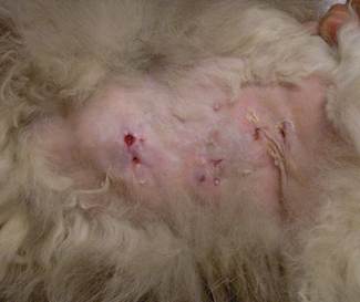
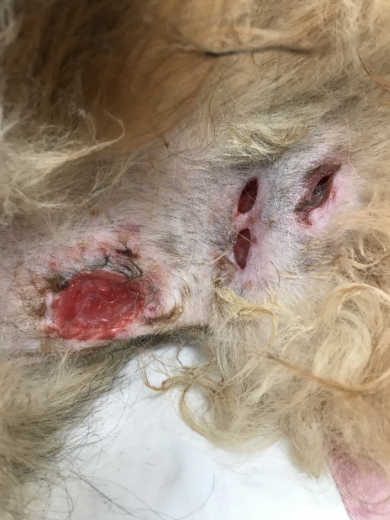
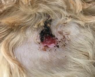
- Know! Panniculitis is characterized by single or multiple deep-seated cutaneous nodules that can be firm or soft and may eventually ulcerate and drain an oily brown to bloody exudate. Ulcerated and draining nodules can become secondarily infected
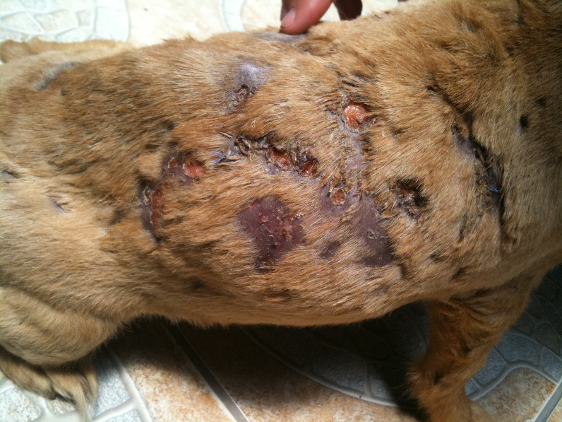
- Know that multiple nodules more frequently localized to the trunk and neck are typically seen with idiopathic sterile nodular panniculitis in dogs, but cats usually have single lesions.
- Remember that animals with multiple nodules can have fever, depression, lethargy, anorexia, vomiting and/or abdominal pain.
- Know that cats that eat diets rich in polyunsaturated fatty acids (PUFAs) such as, red tuna and cod liver oil can have a relative deficiency of vitamin E and develop a disease called pansteatitis. This is an inflammation of the subcutaneous and abdominal fat. These cats have systemic signs.
- Know! Fine needle aspiration of intact nodules for cytology, tissue fungal and bacterial cultures to rule out/in an infectious cause and skin biopsy are all important in the pursuit of the diagnosis. Perform an excision or incision biopsy of intact nodules instead of a punch biopsy for histopathology and tissue culture. Make sure to collect the sample for tissue culture aseptically. Punch biopsies of nodules often do not include subcutaneous fat tissue.
- Remember to perform a CBC, chemistry profile and abdominal ultrasound mainly in cases with systemic signs to rule systemic disease, especially pancreatitis.
- Know that single lesions of idiopathic sterile nodular panniculitis can be surgically removed; however, observe the lesion for at least 6 months to determine if new lesions will develop. Multiple lesions should be managed with immunosuppressive doses of oral glucocorticoids. Azathioprine can be added to the treatment regimen for refractory canine cases and chlorambucil for feline cases. Other glucocorticoid-sparing agents include cyclosporine and mycophenolate mofetil.
- Remember! Cats with pansteatitis have to be treated with vitamin E supplement and the diet has to be balanced.
- Remember that some cases can undergo remission without long-term therapy; however, most cases will require lifelong therapy and, in these cases, the lowest possible dosage of immunosuppressive therapy that keeps the disease under control should be the goal.
-
General Considerations
- Panniculitis is inflammation of the subcutaneous fat tissue also known as panniculus adiposus.
- Various etiologic agents can lead to panniculitis. We will focus on a common form of panniculitis called “sterile nodular panniculitis”.
Important Facts
- Panniculitis is inflammation of the subcutaneous fat tissue.
- Various etiologic agents can trigger panniculitis.
-
Cause and Pathogenesis
- Lipocytes (fat cells) can be damaged by many factors resulting in the release of lipids, which undergo hydrolysis to glycerol and fatty acids. Fatty acids are potent inflammatory agents.
- Post-injection panniculitis:
- It is uncommon in cats and rare in dogs. However, it is probably underdiagnosed.
- It has been associated with various vaccines or injection of medications such as antibiotics (e.g. long acting benzathine penicillin and tylosin).
- It is postulated that the inflammation results from a combination of foreign body and hypersensitivity reactions.
- Traumatic panniculitis:
- Occurs when blunt trauma, chronic pressure, or decreased blood supply induces focal ischemia.
- Infectious panniculitis:
- Occurs when bacterial, fungal, protozoal, viral and parasitic agents become established in the panniculus inducing an inflammatory reaction.
-
- Pancreatic diseases:
- Pancreatitis and pancreatic neoplasms have been associated with panniculitis.
- The pathogenesis is unknown. It is postulated to be triggered by the presence of lipase and amylase in the subcutaneous fat as has been demonstrated in humans with pancreatitis. However, the mechanism by which these enzymes damage the adipose tissue or arrive at sites distant from the pancreas is unknown. It is possible that phospholipase A and trypsin facilitate the penetration of capillary walls and fat cells by lipase and co-lipase.
- Vitamin E deficiency
- Vitamin E is an important biological antioxidant and its deficiency can lead to peroxidation of fat tissue and ultimately panniculitis.
- Cats can have a disease called pansteatitis due to severe vitamin E deficiency.
- The most commonly reported cause is feeding cats diets rich in polyunsaturated fatty acids (PUFA) such as canned red tuna and excessive cod liver oil supplementation.
- Diets high in PUFA produce large quantities of free radicals increasing the oxidative stress and thus the body demand for Vitamin E. If the amount of Vitamin E does not match the amount of PUFA, peroxidation of fat cell membranes will occur and eventually pansteatitis.
- Immune-mediated:
- Occurs with immune-mediated vascular diseases such as systemic lupus erythematosus and reactions to drugs.
- Potassium bromide has been associated with panniculitis in dogs.
- Erythema nodosum-like panniculitis is a septal panniculitis associated with vascular damage due to systemic hypersensitivity reactions.
- Occurs with immune-mediated vascular diseases such as systemic lupus erythematosus and reactions to drugs.
- Idiopathic sterile nodular panniculitis:
- Encompasses the sterile inflammatory diseases of the panniculus that have unknown etiologies.
- Examples: sterile nodular panniculitis and sterile pedal panniculitis of German shepherd dogs.
- Idiopathic sterile nodular panniculitis is probably the most common form of panniculitis.
- Encompasses the sterile inflammatory diseases of the panniculus that have unknown etiologies.
- Pancreatic diseases:
Important Facts
- Panniculitis is a multifactorial condition and possible etiologies include post-injection, traumatic, infectious, nutritional, pancreatic diseases, immune-mediated and idiopathic.
-
Clinical Signs – Idiopathic Sterile Panniculitis
- Dogs – Idiopathic Sterile Nodular Panniculitis (ISNP) and Sterile Pedal Panniculitis of the German shepherd dog:
- There is no age or sex predilection, but some case series reports of ISNP mentioned the age of onset for most dogs to fall between 3 to 5 years.
- Dachshunds, miniature poodles and collies appear to be predisposed to develop ISNP but it has been described in various breeds.
- Lesions of ISNP are characterized by well-demarcated to ill-defined deep-seated cutaneous nodules that can be single or multiple.
- Animals with ISNP or panniculitis associated with systemic mycotic infections are more likely to have multiple lesions.
- Nodules can vary from a few millimeters to several centimeters in diameter and can be of normal skin color, erythematous or red blue.
- Some nodules are firm, others are soft and may ulcerate and drain an oily brown or blood-tinged exudate. Significant fibrosis may be associated with chronic lesions as they try to heal.
- There may be evidence of scarring in the area of ulcerated and draining nodules. These ulcerated lesions are often complicated with a secondary staphylococcal infection.
- Nodules may or may not be painful.
- Lesions associated with ISNP are most commonly located over the ventrolateral neck and trunk.
- Fever, depression, lethargy and/or anorexia are more likely to be present in dogs with multiple lesions.
- Dogs – Idiopathic Sterile Nodular Panniculitis (ISNP) and Sterile Pedal Panniculitis of the German shepherd dog:
-
-
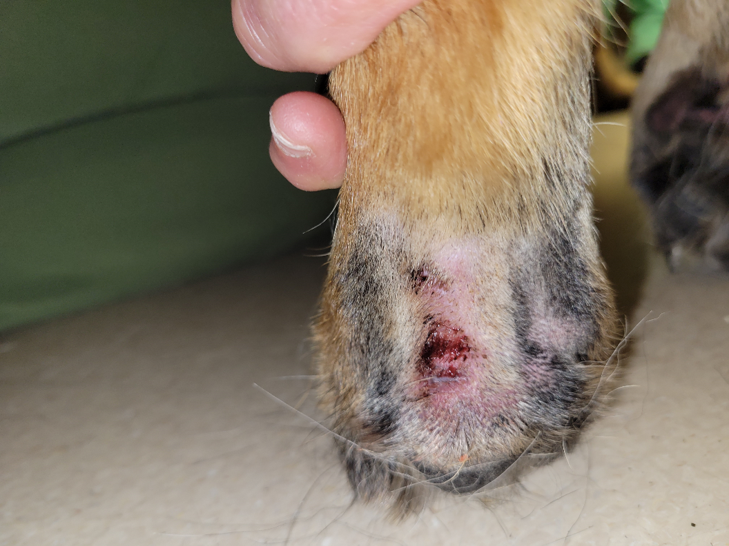
- Sterile pedal panniculitis of the German shepherd dog is characterized by deep and ill-defined erythematous nodules that eventually form draining tracts.
- They are typically located adjacent to the footpads and dorsal to the midline of the carpal or tarsal pads.
- Sterile pedal panniculitis of the German shepherd dog is characterized by deep and ill-defined erythematous nodules that eventually form draining tracts.
-
-
- Cats:
- No age, sex or breed predilection has been described.
- Most cases of idiopathic sterile nodular panniculitis in cats are associated with a single lesion, which is typically localized to the ventral abdomen or ventrolateral thorax.
- Pansteatitis is characterized by the presence of multiple firm nodules diffusely distributed in the subcutaneous and abdominal mesenteric fat tissues. The nodules are painful to the touch and rarely fistulate.
- Cats with multiple lesions will often have systemic signs such as pyrexia, depression, lethargy, anorexia, vomiting and/or abdominal pain. Systemic signs are always present in pansteatitis.
- Cats:
Important Facts
- Clinical signs will vary according to the etiology of panniculitis.
- Lesions of panniculitis are characterized by deep-seated nodules that can be single or multiple.
- Dogs with idiopathic sterile nodular panniculitis or systemic mycotic infections are more likely to develop multiple lesions.
- Systemic signs may be present in dogs and cats with multiple lesions, and they are always present in cats with pansteatitis.
- Some nodules are firm, others are soft and nodules may eventually ulcerate and drain an oily brown- or blood-tinged exudate.
- Ulcerated nodules are likely to become secondarily infected.
- Nodules may or may not be painful.
- Vomiting and abdominal pain can be present in cases associated with pancreatitis.
- Sterile pedal panniculitis of the German shepherd dog is characterized by ill-defined and erythematous nodules that often develop draining tracts. They are typically located in the metatarsal and metacarpal regions and dorsal to the tarsal and carpal pads.
-
Diagnosis
- A thorough history and clinical signs may help identify the triggering cause.
- The diagnosis of sterile nodular panniculitis can only be confirmed after any other cause has been ruled out.
- Fine needle aspiration (FNA) of nodules is easy to perform and must be performed in all cases. It may show infectious agents that could be causing the panniculitis. However, keep in mind that a negative cytology does not rule out an infectious cause.
- In idiopathic sterile nodular panniculitis, the FNA findings include neutrophils, foamy macrophages, lipid and fat cells but no microorganisms.
- The presence of large numbers of eosinophils may indicate injection-induced lesions.
- Direct smear of exudate from draining lesions is not an ideal sample because it may be associated with a secondary bacterial infection.
- Tissue cultures of non-ulcerated nodules for aerobic and anaerobic bacteria, mycobacteria and fungal organisms should be performed if suspecting of panniculitis. No growth is expected in cases of idiopathic sterile nodular panniculitis.
- It is important to make sure that the subcutaneous fat is included in the sample, thus a punch instrument is not adequate if large/deep lesions are sampled. Make sure to sterilize the selected site before collecting the sample. The authors remove the epidermis with a sterile blade to guarantee sample sterility.
- Avoid sampling ulcerated lesions as they will likely yield Staphylococcus sp. or other bacteria causing a secondary infection or contaminating the lesion.
- Histopathology is an important part of the diagnostic pursuit. An excision or incisional biopsy is the recommended technique because a punch biopsy does not include the subcutaneous fat tissue in most cases.
- Histopathological findings are characterized by inflammation of the subcutaneous fat that may be pyogranulomatous, granulomatous, suppurative, eosinophilic, necrotizing or fibrotic.
- Request special stains for mycobacteria, fungal and other microorganisms to rule out infectious agents.
- Polarized light examination is indicated to rule out foreign bodies.
- Histopathological findings are characterized by inflammation of the subcutaneous fat that may be pyogranulomatous, granulomatous, suppurative, eosinophilic, necrotizing or fibrotic.
- In animals with systemic signs and multiples lesions, a CBC, biochemical profile including lipase (i.e. pancreatic lipase immunoreactivity) and amylase, and abdominal ultrasound should be performed to help rule out systemic diseases, especially pancreatitis. Leukocytosis with neutrophilia and mild non-regenerative anemia are often seen in animals with idiopathic sterile nodular panniculitis associated with multiple lesions and systemic signs.
Important Facts
- A thorough history and clinical signs will help to identify the triggering cause.
- Fine need aspiration (FNA) of intact nodules is simple to perform and must be performed in all cases.
- Direct smear of exudate from draining lesions is not an ideal sample because it may be associated with a secondary bacterial infection.
- Tissues cultures of non-ulcerated nodules for aerobic and anaerobic bacteria, mycobacteria, and fungal organisms should be performed to rule out infectious causes.
- Histopathology is an important part of the diagnostic pursuit. An excision or incisional biopsy is recommended because a punch biopsy does not include the subcutaneous fat tissue in most cases.
- Polarized light examination is indicated to rule out foreign bodies.
- A CBC, chemistry profile and abdominal ultrasound should be performed when the animal presents with systemic signs to try to identify a systemic disease, especially pancreatitis.
-
Treatment
- If the panniculitis is due to infectious agents, appropriate treatment for the specific agent should be instituted.
- The treatment options provided below are in reference to idiopathic sterile nodular panniculitis.
- Solitary lesions may be removed surgically and ideally by a board certified or experienced surgeon if the lesion is large or located in an area where it will be challenging to remove.
- One should take into consideration that new lesions will likely develop post-surgical removal. The best approach in these circumstances is to observe the disease behavior for at least 6 months before considering removing a single lesion.
- Animals with multiple sterile lesions respond well to systemic corticosteroids. Prednisolone at the dose of 2.0 mg/kg q 24h for dogs and 4.0 mg/kg q 24h for cats are typically efficacious and should be given until the lesions have regressed. Other oral glucocorticoids can be used if prednisolone does not work.
- Some animals will enter long-term or permanent remission after the treatment is discontinued, albeit this is rare.
- If lesions recur, long-term prednisolone should be instituted. Find the lowest possible dose given every-other-day that maintains the disease controlled.
- The following are drugs that can be used as steroid-sparing agents when the disease cannot be solely controlled long-term with a low dose of oral glucocorticoid.
- Cyclosporine at 5-10mg/kg q 24h. It can take 4-6 weeks for full clinical response. In some cases, the addition of cyclosporine to the treatment regimen may allow the discontinuation of glucocorticoids.
- Mycophenolate mofetil at 20-40 mg/kg q 24h divided into 2 to 3 dosages. A full clinical response may not be seen before 2-6 weeks. It is an option in cases that cyclosporine was not tolerated or was not effective.
- Azathioprine at 1.0-2.2 mg/kg q 24h may be needed for dogs that are unresponsive or become refractory to cyclosporine and mycophenolate mofetil therapy. After remission is achieved try to reduce the dose to alternate-day therapy. It may take 4-6 weeks before azathioprine starts working.
- A complete blood count and serum chemistry profile are recommended every 2 weeks for the first 8-12 weeks to monitor for the development of bone marrow suppression and hepatotoxicity.
- Do not use this drug in cats because they are prone to the azathioprine-induced bone marrow suppression. Chlorambucil at the dose of 0.1 to 0.2 mg/kg q 24h to 48h should be tried for refractory cases.
- Tetracycline and niacinamide have been reported to work in mild cases as sole therapy. However, the authors practice antibiotic stewardship and try to avoid an antibiotic drug with immunomodulatory properties to treat sterile inflammatory diseases.
- The recommended dosage of tetracycline and niacinamide is 250 mg of each 3 times daily for dogs < 10 kg and 500 mg of each 3 times daily for dogs > 10 kg.
- Doxycycline at 5-10 mg/kg q 12h can be used if tetracycline is not available.
- The trial period should be 8 weeks. Consider another treatment modality if no improvement is noticed after this time
- Cats with pansteatitis due to eating diets rich in polyunsaturated fatty acids (PUFA), respond well to oral vitamin E at the dose of 13.5 IU/kg q 24h. The excess dietary content of PUFA should be corrected.
- If the panniculitis is due to infectious agents, appropriate treatment for the specific agent should be instituted.
Important Facts
- Solitary lesions may be removed surgically by a board certified or experienced surgeon. Observe the lesions for at least 6 months before surgically removing them as new lesions may develop.
- Oral glucocorticoid at immunosuppressive doses is the mainstay therapy.
- If the disease cannot be controlled long-term with low doses of glucocorticoids, add cyclosporine or mycophenolate mofetil to the treatment regimen as a steroid-sparing agent.
- Azathioprine may be needed for dogs and chlorambucil for cats that are unresponsive or become refractory to cyclosporine and mycophenolate mofetil therapy.
- For all immunosuppressive drugs, the goal is to find the lowest possible dose that controls the disease long-term with minimal side effects.
- Relapses are common when therapy is interrupted.
- Vitamin E supplementation at the dose of 13.5 IU/kg/day is used to treat feline pansteatitis in conjunction with dietary correction.
References
Contreary CL, Outerbridge CA, Affolter VK et al. Canine sterile nodular panniculitis: a retrospective study of 39 cases. Vet Dermatol 2015; DOI: 10.1111/vde.12247
German AJ, Foster AP, Holden D et al. Sterile nodular panniculitis and pancreatitis in three weimaraners. J Small Anim Pract 2003; 44:449-455.
Gross TH, Ihrke PJ, Walter EJ et al. Skin Diseases of the Dog and Cat: Clinical and Histopathologic Diagnosis. 2nd edn. Oxford: Blackwell Publishing, 2005; 538—558.
Ha-Jung K, Min-Hee K, Jung-Hyun K et al. Sterile panniculitis in dogs: new diagnostic findings and alternative treatments. Vet Dermatol 2010; DOI: 10.1111/j.1365-3164.2011.00957.x
Hendrick MJ, Dunagan CA. Focal necrotizing granulomatous panniculitis associated with subcutaneous injection of rabies vaccine in cats and dogs: 10 cases (1988-1989). J Am Vet Med Assoc 1991; 198:304-305.
Hughes D, Goldschmidt MH, Washabau RJ, et al. Serum alpha 1-antitrypsin concentration in dogs with panniculitis. J Am Vet Med Assoc 1996; 209:1582-1584.
Koutinas AK, Miller WH, Kritsepi M et al. Pansteatitis (Steatitis, “Yellow Fat Disease”) in the Cat: A Review Article and Report of Four Spontaneous Cases. Vet Dermatol 1993; 3:101 – 106.
McKeever PJ, Nuttall T, Harvey RG. A Color Handbook of Skin Diseases of the Dog and Cat. 2nd edn. London: Manson Publishing Ltd., 2009; 88-89.
Miller WH, Griffin GE, Campbell KL. Muller & Kirk Small Animal Dermatology. 7th edn. St Louis: Elsevier Inc., 2013; 701-704.
Niza M, Vilela CL, Ferreira LMA. Feline pansteatitis revisited: hazards of unbalanced home-made diets. J Fel Med Surg 2003; 5: 271-277.
O’Kell AL, Inteeworn N, Diaz SF et al. Canine sterile nodular panniculitis: a retrospective study of 14 cases. J Vet Intern Med 2010; 24:278-284.
Paterson S. Sterile idiopathic pedal panniculitis in the German shepherd dog – clinical presentation and response to treatment of four cases. J Small Anim Pract 1995; 36:498-501.
Scott DW, Anderson WI. Panniculitis in dogs and cats: a retrospective analysis of 78 cases. J Am Anim Hosp Assoc 1988; 24:551-559.
Torres SM. Sterile nodular dermatitis in dogs. Vet Clin North Am Small Anim Pract 1999; 29: 1311-1323.
Yamagishi C, Momoi Y, Kobayashi T et al. A retrospective study and gene analysis of canine sterile panniculitis. J Vet Med Sci 2007; 69: 915-924.
