4. Canine Uveodermatological Syndrome
Learning Objectives
- Know! Canine uveodermatological syndrome is rare and believed to be associated with an autoimmune attack against melanocytes and its associated antigens. Therefore, it makes sense that the heavily pigmented tissues such as the uveal tract, skin, and mucous membranes are primarily involved.
- Know! Akitas have been reported to have an increased risk to develop uveodermatological syndrome. However, other breeds can be affected.
- Know! Most animals are in young adulthood when they are first affected but they can be as old as 12 years of age by the time the disease is diagnosed or as young as 9 months old.
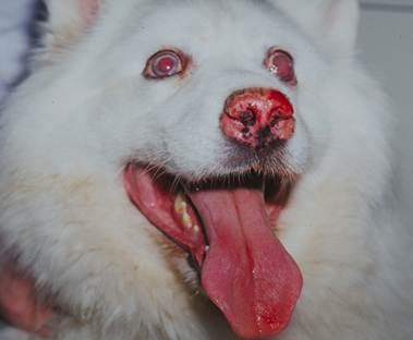
- Know! Ocular signs are usually present first and include bilateral lymphogranulomatous panuveitis. Later, retinal detachment, posterior synechiae with secondary glaucoma, and cataracts may develop. Acute blindness may develop if the inflammatory process is not controlled.
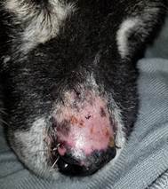
- Know! The cutaneous signs are characterized by depigmentation of the skin and/or hair, erythema, alopecia and scaling involving the eyelids, muzzle, nasal planum, lips, scrotum, anus, vulva, hard palate and footpads. Erosion of affected areas is a less common sign.
- Be aware! The diagnosis is based on a characteristic history, clinical signs and histopathological findings.
- Be aware! Treatment with immunosuppressive/immunomodulatory drugs must be aggressive and instituted early to prevent blindness.
- Know! Oral glucocorticoids and/or azathioprine or cyclosporine are typically used, and treatment must be maintained long-term at the lowest possible dose that controls the disease with minimal side effects.
- Know! Dermatologists and ophthalmologists must work together to achieve the best results in managing this disease.
-
General Considerations
- Canine uveodermatological syndrome, also known as Vogt-Koyanagi-Harada like syndrome, is a rare condition that is thought to represent an autoimmune attack against melanocytes or melanocyte-associated antigens.
- In humans with Vogt-Koyanagi Harada syndrome, antimelanocyte antibodies and a cell-mediated hypersensitivity reaction (TH-1 and TH-17) against tyrosinase peptide antigens have been demonstrated and suggested to play a role in disease development. Similar mechanisms may also occur in dogs.
- Heavily pigmented tissues such as the uveal tract, skin, and mucous membranes are primarily involved.
- Unlike the condition in people, auditory and meningeal abnormalities have not been convincedly reported in the dog, with few anecdotal reports published.
Important Facts
- Canine uveodermatological syndrome is a rare condition that is thought to represent an autoimmune attack against melanocytes or melanocyte-associated antigens.
- Heavily pigmented tissues such as the uveal tract, skin, and mucous membranes are primarily involved.
-
Clinical Signs
- Breeds reported with the disease include the Akita (most commonly affected), Samoyed, Siberian husky, Alaskan malamute, Australia shepherd, basset hound, dachshund, Old English sheepdog, Brazilian fila, chow chow, Shetland sheepdog, white German shepherd, Irish setter, Ainu, Shiba Inu, Bernese mountain dog and Labrador retriever.
- Akitas, Samoyeds and Siberian huskies are suspected to be predisposed to uveodermatological syndrome.
- In one study, Akitas with a specific dog lymphocyte antigen (DLA) – – – DQA1*00201) showed a significantly higher relative risk to develop the disease than Akitas with other DLA class II alleles.
- The reported median age at initial ophthalmologic visit was 4.1 years (range: 9 months to 12 years).
- Male dogs were overrepresented in a retrospective stud y of 50 cases.
- Ocular signs usually precede skin lesions and consist of bilateral lymphogranulomatous panuveitis. Initial signs of panuveitis may include photophobia, blepharospasm, lacrimation, corneal edema, and conjunctival congestion. Later, retinal detachment, posterior synechiae with secondary glaucoma, and cataracts may develop. Bilateral vision loss was reported in 57% of dogs in a retrospective study. Some dogs may recover their vision.
- The reported median and mean time-lapse between the development of skin and ocular lesions were 12 and 20 weeks (range: 4 days to 3 years).
- Bilateral ocular involvement should be expected unless uveal pigmentation is asymmetrical.
- Skin and hair abnormalities typically follow the onset of uveitis, but they may occasionally occur concurrently and rarely precede the uveitis. They consist of the following:
- Depigmentation of the hair (leukotrichia) and skin (leukoderma) that often involve the eyelids, muzzle, nasal planum and lips and, less frequently the scrotum, anus, vulva, hard palate and paw pads.
- Onychomadesis (loss of claw plate) and hyperkeratosis of paw pads occur less often.
- Breeds reported with the disease include the Akita (most commonly affected), Samoyed, Siberian husky, Alaskan malamute, Australia shepherd, basset hound, dachshund, Old English sheepdog, Brazilian fila, chow chow, Shetland sheepdog, white German shepherd, Irish setter, Ainu, Shiba Inu, Bernese mountain dog and Labrador retriever.
-
-
- Erythema, alopecia and scaling can be associated with the skin depigmentation.
- In rare cases, erosions and crusting typically develop in the periocular area, eyelid, around the nasal planum, and around the mouth.
-
-
- In contrast to the disease in people, meningeal and auditory systems involvement have not been convincedly described in dogs.
Important Facts
- Akita dogs have been reported to have an increased risk of developing uveodermatological syndrome and account for about 80% of the cases reported in the literature.
- Most affected animals are young adults.
- Ocular signs usually precede skin lesions and consist initially of bilateral lymphogranulomatous panuveitis.
- Retinal detachment, posterior synechiae, glaucoma, cataracts and acute blindness may occur if the inflammatory process is not arrested.
- Cutaneous signs include depigmentation of the skin and hair, alopecia, erythema, scaling, and less often erosion and crusting involving the eyelids, periocular region, muzzle, nasal planum, lips, scrotum, anus, vulva, hard palate, and/or footpads.
-
Diagnosis
- The diagnosis is based on a compatible history, characteristic clinical signs and supportive skin biopsy findings.
- Histopathological findings, early in the disease, reveal a pronounced lichenoid inflammatory infiltrate of predominantly macrophages, lymphocytes and plasma cells, at the dermal-epidermal junction and decreased epidermal melanin and melanocytes. Pigmentary incontinence is marked.
Important Facts
- The diagnosis of uveodermatological syndrome is based on a compatible history and clinical signs and supportive histopathological findings.
- Skin biopsy results reveal a pronounced lichenoid inflammatory reaction of predominantly large histiocytes at the dermal-epidermal junction and decreased epidermal melanin and melanocytes.
-
Treatment
- To prevent blindness, a consultation with or a referral to an ophthalmologist is important to initiate aggressive therapy as soon as possible.
- Topical or subconjunctival corticosteroids and topical cycloplegic drugs are beneficial in patients with anterior uveitis.
- Oral prednisolone at the dose of 1 to 2 mg/kg daily is recommended until remission. Long-term alternate day therapy is often needed to maintain remission.
- Azathioprine at the dose of 2.2 mg/kg q 24h PO with tapering after clinical resolution to 0.5 mg/kg q 24h PO may allow for a reduction of the corticosteroid dose. It is combined with oral prednisolone at least during the lag-phase of 4-6 weeks.
- In some patients, it may be possible to discontinue the oral corticosteroids and rely on azathioprine alone.
- Cyclosporine at 5mg/kg/day and topical tacrolimus (e.g. 0.1% Protopic®) may be a treatment option but reports of efficacy have been anecdotal at this time. It is combined with oral prednisolone at least during the lag-phase of 2-4 weeks.
- In some patients, it may be possible to discontinue the oral corticosteroids and rely on cyclosporine alone.
- In successfully treated cases, various degrees of skin re-pigmentation will occur.
- Affected dogs will need to be maintained on treatment long-term.
- It is very important that dermatologists and ophthalmologists work together on a long-term basis to increase the change of successfully managing this disease.
Important Facts
- Aggressive immunosuppressive treatment should be instituted as early as possible to avoid blindness.
- Topical or subconjunctival corticosteroids and topical cycloplegics are beneficial in patients with anterior uveitis.
- Oral prednisolone and/or azathioprine or cyclosporine will be required to treat the severe inflammatory process associated with this condition.
- Long-term immunosuppressive therapy using the lowest possible dosage that controls the disease with minimal side effects is needed to maintain remission.
- It is very important that dermatologists and ophthalmologists work together on a long-term basis to increase the chances of successfully managing this condition.
References
Angles JM, Famula TR, Pedersen NC. Uveodermatologic (VKH-like) syndrome in American Akita dogs is associated with an increased frequency of DQA1*00201. Tissue Antigens 2005; 66:656-665.
Carter WJ, Crispin SM, Gould DJ et al. An immunohistochemical study of uveodermatologic syndrome in two Japanese Akita dogs. Vet Ophthalmol 2005; 8:17-24.
Gross TH, Ihrke PJ, Walter EJ et al. Skin Diseases of the Dog and Cat: Clinical and Histopathologic Diagnosis. 2nd edn. Oxford: Blackwell Publishing, 2005; 266-268.
Laus JL, Sousa MG, Cabral VP et al. Uveodermatologic syndrome in a Brazilian Fila dog. Vet Ophthalmol 2004;7:193–196.
McKeever PJ, Nuttall T, Harvey RG. A Color Handbook of Skin Diseases of the Dog and Cat. 2nd edn. London: Manson Publishing Ltd., 2009; 214-215.
Miller WH, Griffin GE, Campbell KL. Muller & Kirk Small Animal Dermatology. 7th edn. St Louis: Elsevier Inc., 2013; 465-466.
Morgan RV. Vogt-Koyanagi-Harada syndrome in humans and dogs. Comp Cont Educ Pract Vet 1989; 11:1211-1218.
Tham HL, Linder KE, Olivry T. Autoimmune diseases affecting skin melanocytes in dogs, cats and horses: vitiligo and the uveodermatological syndrome: a comprehensive review. BMC Vet Res 2019; 15:251.
Zarfoss MK, Tusler CA, Kass PH et al. Clinical findings and outcomes for dogs with uveodermatologic syndrome. J Am Vet Med Assoc 2018; 252: 1263-1271.
