1. Sebaceous Adenitis – Dogs and Cats
Learning Objectives
- Know! Sebaceous adenitis is an inflammatory disease that targets the skin sebaceous glands.
- Know! It is most often seen in standard poodles, akitas, samoyeds, English springer spaniels, vizslas and Havaneses dogs but any breed can be affected. It has shown to be inherited in Akitas and standard poodles and transmitted as an autosomal recessive trait.
- Know! The pathomechanism is unknown but it is important to educate the pet owner that it is a controllable but non-curable disease. Pet owners should also know that genetics likely play a role in disease development and pets with this disease should not be bred.
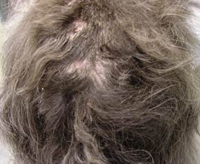
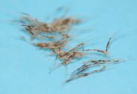
- Remember! The hallmark clinical sign is follicular casting, which is the presence of scales adhered/glued to groups of hair shaft. Hairs epilate easily and the dogs initially develop hypotrichosis and eventually alopecia. The scales also adhere to the skin surface of affected areas. Lesions can develop at any skin site that has hair because the skin sebaceous glands are connected to hair follicles.
- Remember! Diagnosis is based on the patient’s signalment and characteristic history and clinical signs. However, skin biopsies are required to confirm the presumptive clinical diagnosis.
- Know! Differential diagnoses include other diseases associated with follicular casting, scaling and hypotrichosis/alopecia and include demodicosis, dermatophytosis, follicular dysplasia, bacterial folliculitis, nutritional deficiencies, leishmaniasis, and primary seborrhea. Make sure to perform the appropriate diagnostic tests to rule in/out these diseases as appropriate.
- Remember! Topical therapy is a very important part of the treatment regimen and should be included in treatment protocols of all cases as sole or combined therapy.
- Know! Oral cyclosporine is currently considered the most effective treatment; however, it works best when combined with topical therapy. Other treatment options that can be used in the place of cyclosporine include oral vitamin A, retinoids, essential fatty acids, glucocorticoids and oclacitinib.
- Be aware! The prognosis is generally good but chronic cases can be difficult to control.
-
General Considerations
- Sebaceous adenitis is a skin disorder caused by an inflammatory response directed against sebaceous glands ultimately resulting in their destruction.
- It is common in dogs but it is rarely reported in cats.
- The pathogenesis of sebaceous adenitis is unknown. Theories include the following: (i) an inherited developmental defect of the sebaceous glands; (ii) an immune mediated or autoimmune reaction directed against one or more components of the sebaceous glands; (iii) an abnormal keratinization of the sebaceous gland ducts with subsequent obstruction resulting in retention and eventual leakage of sebaceous glands material resulting in an inflammatory response towards the glands; and (iv) an abnormality in lipid metabolism affecting keratinization and sebum production.
Important Facts
- Sebaceous adenitis is caused by an inflammatory response directed against the sebaceous glands of the skin.
- The pathogenesis of sebaceous adenitis is currently unknown.
-
Signalment
- Clinical signs usually appear in young adult to middle aged dogs (1 to 5 years), with a possible male predilection.
- Breeds reported to have sebaceous adenitis include the standard poodle, Akita, Samoyed, vizsla, German shepherd, English springer spaniel, Lhasa apso, chow chow, Rottweiler, and Havanese dogs. However, other breeds can be affected.
- An autosome recessive mode of inheritance was reported in standard poodles and Akitas. Multiple genes may be involved.
-
History and Clinical Signs
Dogs:
-
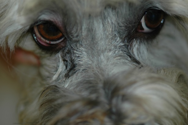
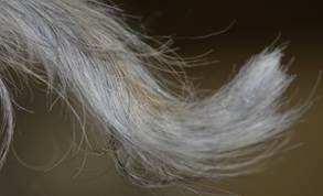
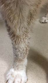
- The most commonly affected areas include the dorsal trunk, neck, head, dorsal aspect of the muzzle, legs, ears – specially the pinnae, and tail. The paws tend to be spared but can be also affected. It can become generalized.
-
- Earlier signs may be limited to mild scaling and erythema.
- In some dogs, the color of the hair coat may change to become darker or lighter. The hair shaft pattern may also change from curly to wavy or straight.
-
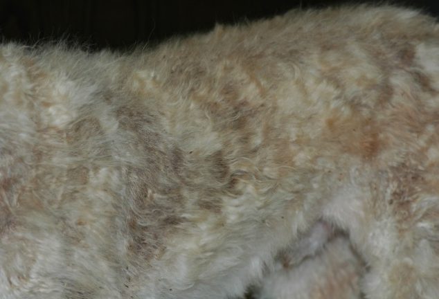
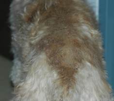
- As the disease progresses variable degrees of hypotrichosis, alopecia, erythema, and scaling will develop. The hair coat becomes dull, brittle and eventually falls off.
-
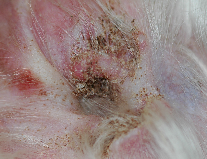
- Ear canals may be affected with dry adhered keratinaceous debris and may be the initial or only presenting sign in some dogs. Some dogs will develop otitis externa.
-
- Affected areas will have various degrees of scaling. Scales adhere to the skin surface and especially to groups of hair shafts. This is called “follicular casting” and is the disease hallmark.
-
- Hairs epilate very easily.
-
- Secondary bacterial folliculitis (papules and pustules) is often present. Bacterial furunculosis may also occur.
- Pruritus may be present and will vary in severity, being most commonly observed in association with bacterial folliculitis.
- Short-coated breeds, such as vizslas, will have slightly different clinical presentation.
- The main differentiating feature is the lesion pattern, which is characterized by annular areas of scales and alopecia that tend to coalesce to form larger areas.
- The scales are fine, white and do not adhere to the skin.
- As for other breeds, follicular cast (i.e. scales adhered to hair shafts) is the disease hallmark.
- Bacterial folliculitis is rare.
Cats:
-
- Lesions typically affect the head, pinnae and neck but can extend to the dorsal trunk and become generalized.
- Clinical signs are usually associated with multifocal annular areas of scaling, crusts, broken hairs, and hypotrichosis to alopecia. Similar to dogs, follicular casting is the disease hallmark.
Important Facts
- Clinical signs of sebaceous adenitis usually appear in young adult to middle aged dogs, with a possible male predilection.
- Breeds reported with sebaceous adenitis include the standard poodle, Akita, Samoyed, vizsla, German shepherd, English springer spaniel, Lhasa apso, chow chow, Rottweiler, and Havanese dogs. However, any breed can be affected.
- An autosome recessive mode of inheritance was reported in standard poodles and Akitas
- The clinical presentation varies slightly between dogs with long and short hair coats.
- Follicular casting (i.e. scales adhered to hair shafts) is the disease hallmark.
- Sebaceous adenitis is rare in cats and the reported clinical signs are similar to that of dogs.
-
Differential diagnoses:
- Differential diagnoses include diseases that develop follicular casting, scaling, and alopecia such as bacterial folliculitis, demodicosis, dermatophytosis, primary idiopathic seborrhea, follicular dysplasia, leishmaniasis and nutritional deficiencies.
- Skin scraping, trichogram, and fungal culture are indicated, if justified, to rule out the diseases in the list of differential diagnoses. Cytology is recommended to investigate secondary infections.
-
Diagnosis
- A tentative diagnosis is based on the patient’s signalment, characteristic history and clinical signs.
- Skin biopsy is required to confirm a presumptive clinical diagnosis.
- The main histopathological feature is a mixed inflammatory infiltrate targeting the sebaceous glands.
- In chronic cases, few or no sebaceous glands will be present and the inflammatory process will be minimal. Peri-follicular fibrosis may be present.
- A biopsy sample of different lesions at various stages of disease development should be obtained.
Important Facts
- A tentative diagnosis is based on the patient’s signalment, characteristic history and clinical signs.
- Skin biopsy is required to confirm a presumptive clinical diagnosis.
- Biopsy samples of different lesions at various stages of development should be obtained.
- Differential diagnoses include bacterial folliculitis, demodicosis, dermatophytosis, primary idiopathic seborrhea, leishmaniasis and follicular dysplasia.
-
Treatment
- Pet owners have to understand that sebaceous adenitis is a non-curable disease, and the goal of therapy is to control the symptoms and prevent secondary skin infections.
- In general, the response to therapy varies from one patient to the next.
- Topical therapy is a very important part of the treatment regimen of all cases.
- Milder forms of sebaceous adenitis may be solely controlled with topical keratolytic agents, emollients and lipids directed at restoring the skin sebum and removing the scales.
- Antiseborrheic shampoos (sulfur, salicylic acid, tar) and emollient rinses are used as needed.
- Antiseborrheic mousses, sprays or spot-on products containing lipids (e.g. phytosphingosine, ceramides) and essential fatty acids can be helpful.
- Oil baths or topical sprays or rinses containing 50% to 75% propylene glycol mixed in water or even vegetable oil such as olive oil can be applied every 1 to 3 days as needed to help loose up the adherent scales.
- Oil treatment is typically followed by an antiseborrheic shampoo to help remove excess oil after at least 30 minutes of oil contact time.
- The frequency of topical treatments (e.g. bath, spray, mousse, spot-on etc.) will vary (e.g. daily to weekly) according to the severity of symptoms and the client’s ability to follow your recommendations.
- Oral cyclosporine (5 mg/kg once daily) is currently the preferred treatment for sebaceous adenitis, particularly severe cases, as it usually leads to an increase in sebaceous glands and clinical improvement in most cases.
- A study showed that oral cyclosporine combined with topical treatment or topical therapy alone have better results than cyclosporine as sole therapy. This study demonstrates the importance of topical therapy in the treatment of sebaceous adenitis.
- The most common adverse effects of cyclosporine include vomiting and diarrhea. Other less common side effects include gingival hyperplasia, psoriasiform lichenoid-like dermatosis (i.e. an unusual reaction to staphylococcal infection), hypertrichosis, and papilloma virus infection.
- The disease can flare during treatment even when the treatment regimen has not changed.
- In this situation, investigate the following: (i) compliance is not a concern, (ii) the commercial cyclosporine has not been changed from a brand name to a generic drug or to a different generic product, and (iii) the cyclosporine has been given with food as food can reduce its bioavailability.
- For mild cases or cases where owners cannot afford cyclosporine – vitamin A (retinol) at 400 to 1,000 IU/kg PO per day can be tried. Make sure to monitor for the development of keratoconjunctivitis sicca (i.e. Schirmer tear test).
- Isotretinoin has been effective to treat sebaceous adenitis in dogs. It belongs to the retinoid group, which comprises drugs related to vitamin A. This drug is tightly regulated in the USA and difficult to obtain.
- The recommended dose is 1 mg/kg once to twice daily with improvement occurring in about 6 weeks. Thereafter, try to reduce the treatment frequency to every-other-day or the dose to 0.5 mg/kg once daily
- Monitor the patient closely for potential side effects. They include vomiting or diarrhea, hepatotoxicity, increased serum triglyceride concentrations, keratoconjunctivitis sicca and teratogenicity.
- Perform a Schirmer tear test and chemistry profile periodically during treatment.
- A short-acting oral glucocorticoid such as prednisolone can be tried if the biopsy results show severe inflammation, and the owners do not want to try cyclosporine. However, it is often not effective as sole therapy.
- Start with 1 mg/kg/day and reduce to the lowest possible dose regimen that maintains the disease controlled.
- A case report showed the efficacy of oclacitinib (Apoquel®) for the treatment of sebaceous adenitis in a Rottweiler dog. Cyclosporine combined with oral prednisolone did not work after 4 weeks of therapy. The treatment protocol was changed to oclacitinib (0.7 mg/kg/day) and oral prednisolone (0.5 mg/kg/day) and after 6 weeks of therapy all lesions resolved. The dog was maintained on oclacitinib at 0.6 mg/kg/day and prednisolone at 0.2 mg/kg q 72h without recurrence during a 12 months follow-up period.
- There are a few reports of successful therapy with a combination of a tetracycline and niacinamide. However, the authors practice antibiotic stewardship and avoid the use of antibiotics with immunomodulatory properties to treat inflammatory diseases.
- High oral doses of essential fatty acids (omega-3 and omega-6), found in many commercial fatty acid supplements and evening primrose oil can be added to the treatment regimen as an adjective of topical and systemic therapy.
- If bacterial folliculitis is present, shampoos containing an antibacterial agent (e.g. benzoyl peroxide, chlorhexidine, ethyl lactate) should be prescribed.
- If systemic antibiotic therapy is required because of a generalized and severe secondary pyoderma or topical therapy was not effective as sole therapy, make sure to choose a tier one (i.e. first choice) antibiotic (refer to Bacterial Skin Diseases-Canine Pyoderma in Volume 1).
- Recheck the patient to determine if the infection is resolved before discontinuing therapy.
- Advice owners to not breed the affected pet because there is strong evidence in support of a genetic component playing a role in disease development.
- Cats with sebaceous adenitis may respond to similar therapies; however, there is limited literature information.
-
Prognosis:
- The prognosis is generally good, but owners have to understand that sebaceous adenitis is a non-curable disease.
- Dogs with severe dermal fibrosis and destruction of sebaceous glands on histopathology have the poorest prognosis for therapeutic response.
Important Facts
- Topical therapy is a very important part of the treatment regimen of sebaceous adenitis and can be used as sole therapy or in combination with other treatments.
- Oral cyclosporine is currently the preferred treatment for sebaceous adenitis, and it should be discussed with owners as a treatment option, especially for severe cases.
- Examples of treatment options that can be tried instead of cyclosporine include vitamin A, retinoids, oclacitinib, and oral glucocorticoids.
- High doses of essential fatty acids can be tried as adjunctive therapy.
- Identify and properly treat secondary infections.
- Advise owners to not breed the affected pet because there is strong evidence in support of genetics playing a role in disease development.
- The dogs with severe dermal fibrosis and destruction of sebaceous glands have the poorest prognosis for therapeutic response.
References
Frazer MM, Schick AE, Lewis TP et al. Sebaceous adenitis in Havanese dogs: A retrospective study of the clinical presentation and incidence. Vet Dermatol 2022; 22:267-274.
Lam ATH, Outerbridge CA, Gericota B et al. Oral vitamin A as an adjunctive treatment for canine sebaceous adenitis. Vet Dermatol 2011; 22:305-311.
Linek M, Boss C, Haemmerling R et al. Effects of cyclosporine A on clinical and histologic abnormalities in dogs with sebaceous adenitis. J Am Vet Med Assoc 2005; 226:59-64.
Lortz J, Favrot C, Mecklenburg L et al. A multicentre placebo-controlled clinical trial on the efficacy of oral ciclosporin A in the treatment of canine sebaceous adenitis in comparison with conventional topical treatment. Vet Dermatol 2010; 21:593-601.
Nuttall T, Reece D and Roberts E. Life-long diseases need life-long treatment: long-term safety of ciclosporin in canine atopic dermatitis. Vet Rec 2014; 174: 3-12.
Perez-Aranda M, Yotti C, Perez J et al. Successful treatment of sebaceous adenitis with oclacitinib and low-dose prednisolone in a dog. Vet Dermatol 2023: DOI: 10.1111/vde.13216.
Pye C. Canine sebaceous adenitis. Can Vet J 2021; 62:293-296.
Sousa CA. Sebaceous adenitis. Vet Clin Small Anim 2006; 36:213-249.
Tevell EH, Bergvall K, Egenvall A. Sebaceous adenitis in Swedish dogs, a retrospective study of 104 cases. Acta Vet Scand 2008; 50:11-18.
White SD, Rosychuk RAW, Scott KV et al. Sebaceous adenitis in dogs and results of treatment with isotretinoin and etretinate: 30 cases (1990-1994). J Am Vet Med Assoc 1995; 207:197-200.
