2. Familial Canine Dermatomyositis
Learning Objectives
- Know! Dermatomyositis is a familial disease reported in the collie, Shetland sheepdog, Beauceron shepherd, Belgian Tervuren and Portuguese water dog. The mode of transmission in collies and Shetland sheepdogs appear to be autosomal dominant with variable expressivity.
- Know! Familial dermatomyositis is classified under ischemic dermatopathies. Other diseases in this category include: (i) juvenile onset dermatomyositis-like disease in atypical breeds, (ii) post rabies vaccine panniculitis, (iii) generalized vaccine-associated ischemic dermatopathy and (iv) generalized idiopathic ischemic dermatopathy.
- Know! All ischemic dermatopathies are treated similarly.
- Know! Familial dermatomyositis is most often seen in collies and Shelties.
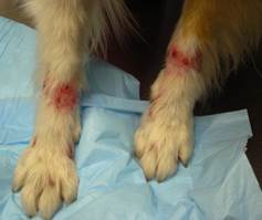
- Know! Signs usually occur before 6 months of age, but it has been reported in adult animals. Alopecia, erythema, scales, erosions/ulcers, and crusting are typically present on the dorsal aspect of the muzzle, around the eyes, ear margins, tarsal and carpal areas, digits and tip of the tail. Rarely, vesicles are present.
- Be aware! Nail abnormalities may also develop in some cases.
- Remember! Muscle involvement typically occurs after the development of skin lesions and the severity of muscle lesions correlates positively with the severity of the skin lesions. The muscles of mastication and distal extremities are usually involved.
- Know! Severely affected dogs have difficulty in eating, drinking, swallowing and may have growth retardation, megaesophagus, lameness, widespread muscle atrophy and infertility.
- Remember! Pruritus is not a feature of this disease, but it may develop with secondary bacterial infections.
- Know how to diagnose dermatomyositis. Would you consider including an EMG, muscle biopsy, serum IgG and immune complex concentrations in your diagnostic plan?
- Know that the disease waxes and wanes and mild cases may spontaneously resolve.
- Know that mild cases may only need vitamin E and marine lipid supplements and they can be part of the treatment regimen of any case.
- Be aware! Severe cases will typically require oral glucocorticoids, cyclosporine, or oclacitinib. Pentoxifylline is often used as sole therapy in mild cases or combined with these drugs in severe cases.
- Be aware! Topical 0.1% tacrolimus ointment can be added to the treatment regimen, mostly moderate to severe cases.
- Remember! Despite the fact that a tetracycline and niacinamide combination have been effective to treat some cases of familial dermatomyositis, practice antibiotic stewardship and avoid using an antibiotic with immunomodulatory properties to treat a non-infectious disease.
- Remember! Lesions worsen with sunlight exposure and trauma; therefore, measures to avoid these complicating factors should be part of every treatment regimen.
- Remember! Advice owners to not breed affected dogs.
-
General Considerations
- Familial canine dermatomyositis is a hereditary inflammatory disease of the skin and muscle.
- Based on the patient’s clinical signs and histopathological findings, familial dermatomyositis is considered an ischemic dermatopathy.
- Other diseases classified as ischemic dermatopathies include (i) juvenile onset dermatomyositis-like disease in atypical breeds, (ii) post rabies vaccine panniculitis, (iii) generalized vaccine-associated ischemic dermatopathy and (iv) generalized idiopathic ischemic dermatopathy.
- All forms of ischemic dermatopathies are managed similarly.
Important Facts
- Familial dermatomyositis is a hereditary inflammatory disease of the skin and muscle.
- It is considered an ischemic dermatopathy.
- Other forms of ischemic dermatopathies exist and are managed similarly to familial dermatomyositis.
-
Cause and Pathogenesis
- The etiopathogenesis of familial dermatomyositis is currently unknown.
- It is familial in collies, Shetland sheepdogs, Beauceron shepherds, Belgian Tervurens, and Portuguese water dogs. Breeding studies in Collies support an autosomal dominant mode of inheritance with variable expressivity and a study in Shetland sheepdogs suggests a similar mode of transmission.
- Juvenile onset dermatomyositis-like disease has been reported in many other breeds such as the Kuvasz, German shepherd dog, Welsh corgi, Lakeland terrier, Rottweiler, and chow chow. However, it is currently not known if the disease also has a familial basis in these breeds.
- A study investigating the role of genetics in collies and shelties with dermatomyositis showed primary candidate polymorphisms in three conserved regions on chromosomes 10 and 31 supporting the presence of genetics in disease development in these two breeds.
Important Facts
- The pathogenesis is unknown.
- A familial history has been reported not only in collies and Shetland sheepdogs but also in Beauceron shepherds, Belgian Tervurens and Portuguese water dogs.
- The mode of transmission in collies and Shetland sheepdogs appears to be autosomal dominant with variable expressivity.
- Various other breeds can develop clinical signs and histopathological findings compatible with dermatomyositis. In these other breeds, it is referred to as dermatomyositis-like disease.
-
Signalment
- No sex predilection has been reported.
- Lesions usually develop before 6 months of age but can occasionally develop in adults.
- Breeds overrepresented in the category of familial dermatomyositis are the collie and Shetland sheepdog.
-
Clinical Signs
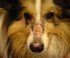
- Initial lesions are characterized by erythema, scaling and mild crusting. Lesions eventually evolve to develop hyper- or hypopigmentation and scarring alopecia. Ulcers/erosions are also commonly present, especially in severely affected dogs. Vesicles may be occasionally found.
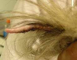
- Some dogs will develop nail abnormalities such as onychomadesis, onychoschizia and onychorrhexis.
- Lesions are typically present on the face (especially the dorsal aspect of the muzzle, around the eyes and ear margins), carpal and tarsal areas, digits and tip of the tail. Oral and footpad involvement is seen more often in Beaucerons.
- Initial lesions are characterized by erythema, scaling and mild crusting. Lesions eventually evolve to develop hyper- or hypopigmentation and scarring alopecia. Ulcers/erosions are also commonly present, especially in severely affected dogs. Vesicles may be occasionally found.
-
- The rate of development and progression of lesions is quite variable as they often wax and wane and may undergo spontaneous resolution.
- Myositis typically occurs after the development of skin lesions and correlates with the severity of skin lesions. It is characterized by atrophy of the muscles of mastication and distal extremities in most cases.
- Cases with mild dermatomyositis may have focal and asymptomatic myositis or muscles may not be affected.
- Severely affected dogs will have difficulties in eating, drinking and swallowing. Some dogs will also have megaesophagus.
- Pruritus and pain are generally not features of the disease; however, pruritus may develop with secondary skin infections.
Important Facts
- Familial dermatomyositis is most prevalent in collies and Shetland sheepdogs.
- Signs usually develop before 6 months of age.
- The most common lesions are erythema, scales, crusting, ulcers and scarring alopecia localized to the dorsal aspect of the muzzle, around the eyes, ear margins, carpal and tarsal areas, digits and tip of the tail.
- Nail abnormalities may also develop in some cases.
- Lesions may spontaneously resolve.
- Myositis typically occurs after the development of skin lesions and correlates with lesion severity. It is usually asymptomatic and characterized by post-inflammatory atrophy of the muscles of mastication and distal extremities.
- Pruritus and pain are generally not features of the disease.
-
Differential Diagnoses
- Differential diagnoses include demodicosis, dermatophytosis, bacterial folliculitis, discoid lupus erythematosus and epidermolysis bullosa simplex if vesicles are present.
-
Diagnosis
- A presumptive clinical diagnosis can be made based on signalment and characteristic history and clinical signs. It is also important to rule out the diseases on the differential diagnoses list as appropriate.
- Biopsy of the affected skin is required to confirm the diagnosis. Muscle biopsy is recommended to determine if muscle involvement is present. However, it is rarely done.
- Properly chosen skin biopsy samples reveal a cell-poor interface dermatitis at the dermal-epidermal junction with vacuolated and/or apoptotic basal cells. Atrophic hair follicles and perifollicular fibrosis are commonly noted and may be the only findings in chronic lesions. Vasculitis is occasionally present.
- Biopsy samples of affected muscles show a mixed-cell infiltration with muscle fiber degeneration, scarring, and atrophy.
- Needle electromyogram (EMG) of affected muscles is not routinely done. Supportive findings include fibrillation potentials, positive sharp waves, and bizarre high-frequency discharges.
- Severely affected dogs may have non-regenerative anemia. If the patient has myositis the serum creatinine levels may be slightly elevated.
- Serum concentration of IgG and circulating immune complexes are usually elevated in the face of normal IgA and IgM levels. However, serum immunoglobulin levels are nonspecific, so this test is not routinely done.
Important Facts
- The presence of erythema, scaling, crusts, erosions/ulcers and scarring alopecia on the face, tail and distal extremities in a collie or Shetland sheepdog (or other breeds) before 6 months of age should increase the index of suspicion for familial dermatomyositis.
- Demodicosis and dermatophytosis are the main differential diagnoses and should be ruled out.
- Skin biopsies are necessary to confirm a presumptive diagnosis.
- Muscle biopsies, EMG and serum immunoglobulin levels can be also performed. However, because the results are inconsistent and nonspecific and these tests will add to the client’s cost, clinicians do not typically request them.
-
Treatment
- As lesions of dermatomyositis can wax and wane it is difficult to determine the effectiveness of any particular treatment.
- No treatment may be needed for relatively mild cases as they may spontaneously resolve. However, areas more severely affected may heal with scarring alopecia.
- Oral vitamin E (200 to 800 IU/day) or marine lipid supplements (100 mg per kg of body weight of combined EPA and DHA) may provide some improvement for mild cases.
- Prednisolone (1-2 mg/kg/day) has been used in more severe cases.
- Reduce the dosage to the lowest effective dose, which should be given every-other-day. Be aware that glucocorticoid therapy may worsen the already existent muscle atrophy.
- Oclacitinib (Apoquel®) has been reported to effectively control four dogs with juvenile-onset ischemic dermatopathy, which resembles dermatomyositis.
- The initial dose ranged from 0.4 to 0.7 mg/kg twice daily. It was given along with a tapering regimen of prednisolone. The prednisolone was discontinued in all dogs and the oclacitinib dose was reduced to a once-daily maintenance regimen.
- A dose as high as 1.0 mg/kg daily may be needed to control dermatomyositis or other ischemic dermatopathies long-term.
- Inform the pet owner that oclacitinib is not labeled to treat ischemic dermatopathies.
- Perform CBC and chemistry profile before starting therapy. Repeat CBC regularly during the first year to monitor for potential side effects (i.e. bone marrow suppression).
- Pentoxifylline (Trental®) is often used for the management of ischemic dermatopathies, either alone or in combination with medications.
- It improves tissue oxygenation and has anti-inflammatory properties.
- A dosage of 25 to 30 mg/kg, twice daily has been proposed. It must be given with food to prevent vomiting.
- There is a lag period of 2 to 3 months before clinical benefits are noted.
- Anecdotal reports suggest that tetracycline and niacinamide may be beneficial for some cases.
- Dose: 250 mg of each 3 times daily for dogs < 10kg and 500 mg of each for dogs > 10 kg.
- Perform a two-month trial before determining efficacy.
- It can be used in combination with prednisolone and pentoxifylline for severe cases.
- The authors practice antibiotic stewardship and avoid the use of antibiotics with immunomodulatory properties to treat inflammatory diseases.
- Topical 0.1% tacrolimus ointment applied twice daily to affected areas has shown to ameliorate lesions and should be considered in combination with one or more of the treatments mentioned above.
- Oral cyclosporine is not used often to treat familial dermatomyositis or other ischemic dermatopathies. However, because it is a calcineurin inhibitor like tacrolimus, it should be considered as a treatment option for this disease.
- In a retrospective study of 117 dogs with ischemic dermatopathies other than familial dermatomyositis, oral cyclosporine was prescribed in 17% of the dogs.
- The average dose was 5.5mg/kg/per day but, unfortunately, the response to therapy was not reported.
- In a retrospective study of 117 dogs with ischemic dermatopathies other than familial dermatomyositis, oral cyclosporine was prescribed in 17% of the dogs.
- Dogs with familial dermatomyositis should not be used for breeding regardless of the severity of clinical signs.
- Lesions worsen with sunlight exposure and trauma; therefore, measures to avoid these complicating factors should be part of every treatment regimen.
- Scarred alopecia is a sequel in most cases.
Important Facts
- Mild cases can resolve spontaneously.
- Therapy is only symptomatic, and mild cases may only need vitamin E and marine lipid supplements.
- Pentoxifylline is often used for ischemic dermatopathies either alone or combined with immunomodulatory drugs.
- Severe cases will typically require immunomodulatory/immunosuppressant drugs such as oral glucocorticoids, oclacitinib, cyclosporine, topical tacrolimus.
- Lesions worsen with sunlight exposure and trauma; therefore, measures to avoid these complicating factors should be part of every treatment regimen.
- The condition often follows its own course when the animal reaches maturity, with some dogs getting progressively better and others getting progressively worse.
- Advice owners to not breed affected animals.
References
Backel KA, Bradley CW, Cain CL et al. Canine ischemic dermatopathy: a retrospective study of 117 cases (2005-2016). Vet Dermatol 2019; DOI: 10.1111/vde.12772.
Bresciani F, Zagnoli L, Fracassi F. Dermatomyositis-like disease in a Rottweiler. Vet Derm 2014; 25:229-232.
Campbell KL, Lowe AD, Lichtensteiger CA. Dermatomyositis in three Portuguese water dog littermates. Vet Dermatol 2008; 19:69.
Evans JM, Nooral RE, Tsai KL, et al. Beyond the MHC: A canine model of dermatomyositis shows a complex pattern of genetic risk involving novel loci. PLoS Gen 2017; DOI:10.1371/journal.pgen.1006604.
Hargis AM, Mundell AC. Familial canine dermatomyositis. Comped Contin Educ 1992; 14:855.
Miller WH, Griffin GE, Campbell KL. Muller & Kirk Small Animal Dermatology. 7th edn. St Louis: Elsevier Inc., 2013; 585-587.
Levy BJ, Linder KE and Olivry T. The role of oclacitinib in the management of ischemic dermatopathy in four dogs. Vet Dermatol 2019; DOI: 10.1111/vde.12743.
Nesbitt G.E. & Ackerman L.J. Miscellaneous Canine Skin Diseases. In: Canine and Feline Dermatol.ogy:Diagnosis and Treatment. Veterinary Learning Systems, Trenton, New Jersey, 1998, p 314-353.
Wahl JM, Clark LA, Skalli O et al. Analysis of gene transcript profiling and immunobiology in Shetland sheepdogs with dermatomyositis. Vet Dermatol 2008; 19:52-58.
