19. Rabies Vaccine Associated Focal Vasculitis and Panniculitis
Learning Objectives
- Be suspicious of rabies-associated vasculitis and panniculitis when presented with a small-breed dog with a focal area of alopecia with or without hyperpigmentation, erythema and scaling localized on the site of vaccination.
- Be aware that some dogs will develop generalized lesions.
- Remember! Lesions only develop 2 to 6 months after vaccination and sometimes even longer.
- Know that pentoxifylline solely or in combination with tacrolimus ointment can be a good treatment option for localized lesions when owners require an intervention.
- Know! Generalized cases will require immunosuppressive therapy such as glucocorticoids, oclacitinib, azathioprine and cyclosporine.
- Be aware! Lesions can recur if the dog is revaccinated.
-
General Considerations
- Vasculitis and panniculitis associated with rabies vaccine is typically limited to the site of vaccination but, in rare occasions, it can become generalized.
- Small breeds such as miniature or toy poodles, Yorkshire terrier, Bichon frize, Shih Tzu, Maltese, Shetland sheepdog, and Pomeranian appear to be predisposed.
Important Facts
- Vasculitis and panniculitis associated with rabies vaccine is typically limited to the site of vaccination but, in rare occasions, it can become generalized
- Small breeds are predisposed.
-
Cause and Pathogenesis
- It is hypothesized that vaccine antigen induces a vasculitis and pannicultis, which leads to ischemia and atrophy of follicular structures resulting in alopecia. The vasculitis is believed to be due to a type-III hypersensitivity reaction in which antigen-antibody complexes deposit in the endothelium of small vessels, activate complement and injury the cell walls. In addition, predominantly lymphocytic and multifocal inflammation of the subcutaneous adipose tissue also develops in these cases.
- Rabies antigen and IgG have been demonstrated in the cell walls of blood vessels associated with skin lesions.
Important Facts
- The vasculitis is thought to be caused by a type-III hypersensitivity reaction to rabies vaccine antigens.
- Moreover, predominantly lymphocytic inflammation of the subcutaneous adipose tissue is characteristic of cutaneous rabies vaccine reaction.
-
Clinical Signs
- Skin lesions are generally focal and develop on the injection site (e.g. the lateral thigh, shoulder). Lesions are characterized by roughly circular areas of alopecia with or without mild to moderate erythema, scaling and hyperpigmentation. The affected site may feel thicker and firmer to the touch.
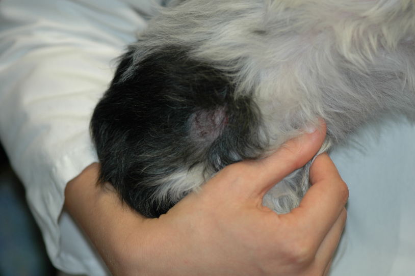
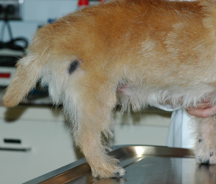
-
- Infrequently, a generalized form can be seen involving not only the injection site but also the face, pinnae, footpads, tip of the tail, and skin overlying boney prominences. In these cases, ulcerations may be present with the alopecic areas.
- Focal alopecic lesions are asymptomatic in most cases.
Important Facts
- Skin lesions are typically localized to the injection site but can be generalized in some cases.
- In general, lesions are characterized by alopecia, with or without mild to moderate erythema, scaling and hyperpigmentation.
-
Diagnosis
- A careful history will indicate vaccination about 2-6 months previously.
- The characteristic clinical signs will support the diagnosis.
- Differential diagnoses for focal alopecic lesion include demodicosis, dermatophytosis, alopecia areata and cicatricial alopecia.
- Skin biopsy is not needed if the history and clinical signs are characteristic and demodicosis and dermatophytosis have been ruled out.
- Characteristic findings include ischemic changes including atrophy of hair follicles, collagen pallor and smudging. Moreover, nodular accumulations of primarily lymphocytes are present in the adipose tissue (i.e. panniculus adipose) and often surround blood vessels. Basophilic amorphous foreign material may be present.
Important Facts
- The history of vaccination about 2-6 months before and the characteristic focal area of alopecia on the vaccination site should increase the clinician’s index of suspicion of rabies vaccine associated vasculitis and pannicultis.
- Demodicosis and dermatophytosis are examples of other causes of focal alopecia and should be ruled out.
- Skin biopsy should be performed in the case of unusual clinical presentation or to confirm the presumptive clinical diagnosis.
-
Treatment
- Benign neglect for asymptomatic focal lesions.
- Pentoxifylline has been effective in some cases and should be tried when the owner is concerned with aesthetics.
- This drug has the ability to increase red blood cell deformability, which will increase tissue perfusion and oxygenation. Pentoxifylline also has an anti-inflammatory effect, which can be beneficial.
- The recommended dose ranges from 15-25 mg/kg q 8-12h. A treatment trial of 2 to 5 months is recommended before assessing the response.
- Surgical excision or tacrolimus ointment with or without pentoxifylline are also options for localized lesions.
- Generalized cases will require immunosuppressive therapy such as glucocorticoids, oclacitinib, azathioprine and cyclosporine.
Important Facts
- Benign neglect for cases of asymptomatic focal lesions.
- Pentoxifylline and/or tacrolimus ointment can be used for focal lesions where owners want treatment.
- Surgical excision of the alopecic site is also an option for focal lesions.
- Generalized cases will require immunosuppressive therapy.
-
Revaccination
- The effects of revaccination are generally unknown because many of these patients are not revaccinated.
- There are anecdotal reports of recurrence and development of new lesions with revaccination.
- If rabies vaccination is not required by law and the patient is not exposed to risky situations to acquire rabies, it is reasonable to not recommend revaccination.
- Revaccination should be avoided in generalized cases.
References
Miller WH, Griffin GE, Campbell KL. Muller & Kirk Small Animal Dermatology. 7th edn. St Louis: Elsevier Inc., 2013; 485-488.
Wilcok BP, Yager JA. Focal cutaneous vasculitis and alopecia at sites of rabies vaccination in dogs. J Am Vet Med Assoc 1986; 188:1174-1177.
