14. Acral Lick Dermatitis – Canine
Learning Objectives
- Know! The pathogenesis of acral lick dermatitis is unknown and it is one of the most frustrating dermatologic conditions to manage. If the primary trigger/cause is not identified and removed or satisfactorily controlled, most cases recur at the same site or different sites.
- Know! Many factors have been suggested to trigger the constant licking/chewing cycle that leads to the development of the acral lick lesion, including allergies (atopic dermatitis, food allergy, flea allergic dermatitis, contact dermatitis), bacterial folliculitis, foreign bodies, neuropathy, local trauma, and joint or bone diseases. Moreover, it is known that behavior is a primary factor or a contributing factor in acral lick dermatitis. Always look for an underlying condition when managing this disease!
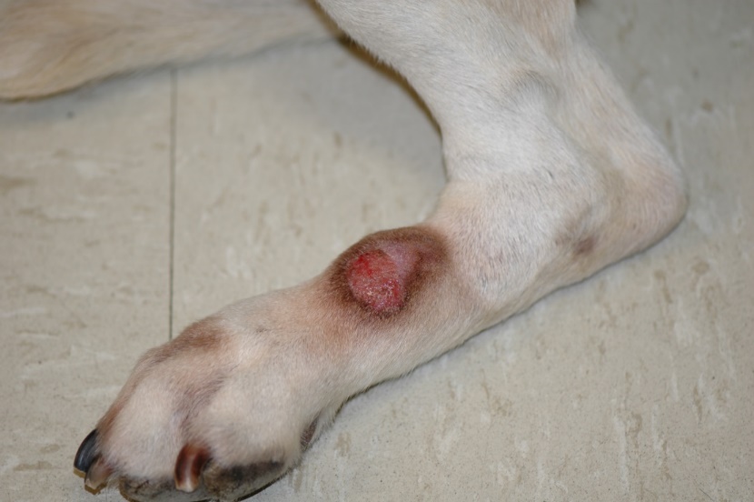
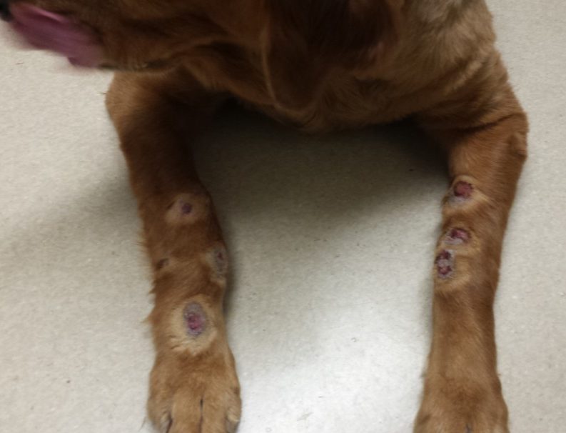
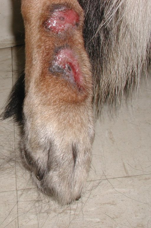
- Know! Lesions typically develop at middle age and large dog breeds are overrepresented. Acral lick lesions are usually single but multiple lesions can be present. They generally develop on the dorsal aspect of the extremities and are characterized by alopecic, erythematous, eroded or ulcerated, firm, nodules, and plaques. However, initial lesions are subtle and often not seen by the owner because they are covered with hair.
- Know! The diagnosis is based on the pet’s detailed history, characteristic clinical signs and elimination of other primary causes. The minimum database should include skin scrapings, cytology of exudate expressed from deep tissue, and bacterial culture from tissue or deep exudate sample. Biopsy for histopathology is performed if the clinical presentation is unusual, or you suspect neoplasia. If joint or bone involvement is suspected, radiographs should be taken.
- Know! Management should include preventing the dog from licking and/or chewing by placing a device (e.g. Elizabethan collar, basket muzzle, etc.); treating the lesion and treating the obsessing licking/chewing behavior, which even when not the primary problem is a contribution factor.
- Remember! No single treatment works for all cases and a plan must be delineated for each dog and owner. It is recommended to include a behaviorist in the design of the patient’s treatment plan.
-
General Considerations
- Canine acral lick dermatitis, also known as lick granuloma or acral pruritic nodule, is a chronic, inflammatory, self-inflicted lesion that develops on one or more distal legs. The lesion results from excessive and persistent licking and/or chewing.
- It is considered a common disease accounting for 2.9% of 559 dermatological cases seen in general practice in one study.
- Acral lick dermatitis is generally a challenging disease to manage because, in many cases, it is difficult to identify the triggering factor(s) and to treat the condition successfully with no recurrences. This situation often leads to frustration on the part of the pet owner and veterinary in charge of the case.
- Before diagnosing a case as purely psychogenic in origin, various underlying primary causes should be considered. However, many cases may have a psychological component.
Important Facts
- Acral lick dermatitis is a chronic, inflammatory, self-inflicted lesion that develops on one or more distal legs of dogs.
- Various underlying causes should be considered before diagnosing a case as purely psychogenic in origin.
-
Etiologic Factors
- Many factors have been suggested to trigger the compulsive licking/chewing that will eventually lead to the development of acral lick dermatitis. These include allergies (atopic dermatitis, food allergy, flea allergic dermatitis, contact allergy), parasitic disorders (scabies, demodicosis), bacterial folliculitis, foreign body, neuropathy, local trauma, venipuncture, catheter site reaction, and joint or bone diseases.
- It is important to know that most cases are associated with a secondary deep bacterial infection, which contributes to the perpetuation of the licking and chewing behavior.
- In the past, many dermatologists thought that cases presenting with acral lick lesions were associated with primary psychogenic causes such as stereotypic or obsessive-compulsive disorder, separation anxiety, noise phobia, boredom, stress, and attention seeking disorder. Today, however, it is believed that other causes (listed above) can also trigger this disease and should be pursued before considering a case purely psychogenic in origin.
- Nevertheless, it is important to know that many cases have a psychogenic component. In fact, it has been estimated that up to 70% of dogs with acral lick dermatitis may have anxiety-related conditions.
- It is also important to keep in mind that independent of the precipitating factor, once the lesion has developed it will contribute to the perpetuation of the compulsive licking/chewing behavior. This likely occurs because excessive licking/chewing can cause the production and release of endorphins, making the animal feel better. Moreover, endorphins produce an analgesic effect that decreases the dog’s pain perception. This process essentially addicts the dog to the licking and/or chewing activity, making it compulsive.
- In addition, dogs with little social interaction (e.g. live completely outdoors) or stimulation, (e.g. confined in crates or kept chained) may be predisposed to develop acral lick dermatitis.
- Many factors have been suggested to trigger the compulsive licking/chewing that will eventually lead to the development of acral lick dermatitis. These include allergies (atopic dermatitis, food allergy, flea allergic dermatitis, contact allergy), parasitic disorders (scabies, demodicosis), bacterial folliculitis, foreign body, neuropathy, local trauma, venipuncture, catheter site reaction, and joint or bone diseases.
Important Facts
- Many factors have been suggested to predispose or cause acral lick dermatitis, including allergies (atopic dermatitis, food allergy, flea allergic dermatitis, contact allergy), psychogenic causes, bacterial folliculitis, foreign body, neuropathy, local trauma, and joint or bone diseases.
- It is important to know that most cases are associated with a secondary deep bacterial infection, which contributes to the perpetuation of the licking and chewing behavior.
- Once developed, the acral lick lesion will perpetuate the compulsive licking/chewing behavior even when the triggering cause has been addressed, emphasizing the importance of also treating the lesion.
-
Clinical Signs
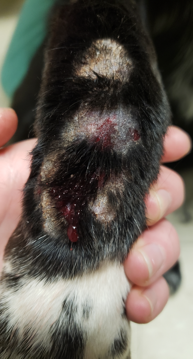
- Acral lick lesions evolve over time. The initial lesion is characterized by an area of erythema, scaling, with or without a small erosion and crusting. This lesion may not be noticed by the owner until it becomes chronic.
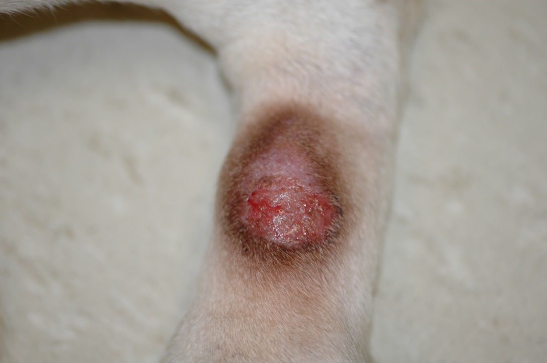
- A chronic lesion is characterized by a firm, alopecic, eroded to ulcerated nodule or plaque. The marked dermal fibrosis associated with chronic lesions is responsible for the firm characteristic of the lesion.
- Lesions are typically oval or circular but may have a linear configuration if associated with a previous wound.
- The most frequently affected sites are the dorsal carpal and metacarpal regions. However, the lesion can also develop on the tarsus and metatarsus areas and lateral surfaces. The term “acral” means extremity.
-
- The lesion is self-inflicted and caused by the dog’s constant licking and chewing at the distal leg.

-
- Lesions are usually single but can be multiple.
-
- Large breeds are overrepresented such as the doberman pincher, Labrador retriever, golden retriever, boxer, Dalmatian, Irish setter, Weimaraner, German shepherd, and Great Dane.
-
- Acral lick dermatitis can occur at any age but develops most commonly in dogs older than 5 years of age.
- It is important to know that the lesions are often associated with superficial and, more importantly, deep bacterial infections, which should be addressed as part of the treatment regimen.
-
- The constant licking/chewing and secondary bacterial folliculitis may eventually result in rupture of affected hair follicles (i.e. furunculosis) leading to foreign body reactions triggered by free keratin in the dermis. This will aggravate the inflammatory process and contribute to the perpetuation of the licking/chewing cycle.
-
-
- Acral lick lesions tend to recur, either at the same or different sites, if the trigger factor or cause cannot be identified and controlled.
- A large lesion adjacent to a joint that has been present for years may be associated with arthritis or ankylosis of the underlying joint, which can contribute to perpetuating the obsessive licking/chewing behavior.
- Not uncommonly, lesions of acral lick dermatitis that have been present for months to years will result in the development of an underlying reactive periostitis.
Important Facts
- Acral lick lesions are created and maintained by constant licking and/or chewing at the distal extremity.
- The constant licking/chewing will eventually lead to the development of an alopecic, erythematous, firm, eroded or ulcerated nodule or plaque, which characterizes a chronic acral lick lesion.
- An acral lick dermatitis lesion is usually single but it can be multiple.
- Large breeds are overrepresented, and lesions typically develop in middle age dogs.
- Acral lick lesions tend to be recur, either at the same or different sites.
-
Differential Diagnoses
- Differential diagnoses for the skin lesion include calcinosis circumscripta, neoplasia (e.g. mast cell tumor), dermatophyte kerion, other deep fungal infection, oomycete infection, and acral mutilation syndrome.
-
Diagnosis
- The diagnosis of acral lick dermatitis is based on the patient’s detailed history, characteristic clinical signs and elimination of other possible causes.
- A thorough history is crucial to help identify the triggering factor/cause.
- The minimum database should include skin scrapings, cytology of exudate expressed from deep tissue and bacterial culture from tissue (ideally) or deep exudate samples.
- Deep bacterial infection is present in most cases (90% of cases in one study).
- A study showed that bacteria cultured from the surface exudate samples differ from the ones cultured from the deep tissue, indicating the need for deep bacterial cultures.
- Tissue culture is preferred. However, if owners cannot afford tissue culture or deny it because it is an invasive procedure, clean the eroded/ulcerated surface and squeeze the skin to allow deep exudate to surface and sample it.
- Select a non eroded or non ulcerated area for tissue culture. A 2-3 mm punch biopsy instrument can be used. Surgically prep the selected area and remove the epidermis with a sterile blade to avoid any possible contamination of the sample with surface bacteria. Submit the tissue in either a Port-A-Cul tube or a sterile tube with a small amount of non bacteriostatic, sterile saline.
- Biopsy of the lesion for histopathology is important if the clinical presentation is unusual and if the index of suspicion for neoplasia is high.
- Fungal culture should be performed if a deep fungal infection is suspected.
- If joint or bone involvement is suspected, radiographs should be taken. Secondary periosteal reaction can develop when lesions are present for months to years. It is important to note that arthritis can be the triggering cause in some dogs.
- If there is a history of generalized pruritus (either seasonal or non seasonal) allergy workup should be performed.
- The diagnosis of acral lick dermatitis is based on the patient’s detailed history, characteristic clinical signs and elimination of other possible causes.
Important Facts
- Diagnosis of acral lick dermatitis is based on the pet’s detailed history, clinical signs, and elimination of other primary causes.
- The minimum database should include skin scrapings, cytology of exudate expressed from deep tissue, and bacterial culture from tissue or deep exudate sample.
- Biopsy of the lesion for histopathology should be performed in unusual clinical presentation or if neoplasia is suspected.
- Fungal culture should be performed if a fungal infection is suspected.
- If there is a history of generalized pruritus (either seasonal or non seasonal) allergy workup should be performed.
- If joint or bone involvement is suspected, radiographs should be taken. Secondary periosteal reaction is often present when lesions are present for months to years. Moreover, arthritis can be the triggering cause in some cases.
-
Treatment
- In most cases, a guarded prognosis for lesion resolution and no recurrence is given to dogs with acral lick dermatitis because the trigger or cause is often difficult to identify.
- If the underlying condition can be determined and removed or satisfactorily controlled, the prognosis improves dramatically.
- Even if an underlying problem has been identified and successfully treated, concurrent treatment of the lesion is essential.
- Treat the secondary infection:
- It is important to treat the frequently associated secondary deep bacterial infection with appropriate long-term (6 to 8 weeks or longer) antibiotic therapy. Remember to practice antibiotic stewardship and select a first tier antibiotic based on culture and susceptibility results (e.g. cephalexin, cefadroxil, amoxicillin clavulanic acid).
- Ideally, antibiotic duration should be based on a negative tissue culture that should be taken when the lesion stops decreasing in size. If the owner does not want a biopsy done, use the criterion of discontinuing therapy when the lesion stops reducing in size. In addition, when this goal is achieved, squeeze the lesion to try to extrude deep exudate. If no exudate is noticed, you can stop therapy. If exudate is extruded, perform cytology and repeat the culture and susceptibility test.
- Treat the lesion:
- Stop the lick/chew cycle by recommending a device to prevent licking/chewing.
- Physical devices must be tailored to the dog. It may take multiple attempts until the right physical device for a patient is selected.
- Some examples include Elizabethan collar, BiteNot collar, socks or booties, DogLegg, bandages, and basket muzzle.
- Some topical products can also help prevent the dog from licking/chewing.
- Examples include bitter apple and a 1:2 mixture of HEET and bitter apple applied to the lesion and surrounding skin 2-3 times daily. HEET contains capsaicin that may deplete the nerve ends of substance P resulting in the decrease of neuropathic pain and neuropathic pruritus. Another option is LickGard ointment. This product contains natural ingredients and has a strong smell and undesirable taste for the dog.
- If using a device that covers an eroded or ulcerated lesion (e.g. bandage, sock, booty, DogLegg), make sure the owner removes it daily and knows how to recognize signs of infection/overgrowth, as a closed environment tends to promote bacterial infection/overgrowth.
- Physical devices must be tailored to the dog. It may take multiple attempts until the right physical device for a patient is selected.
- Synotic® is the best anti-inflammatory agent for this condition because it contains DMSO and fluorinated steroid (fluocinolone acetonide). DMSO (dimethyl sulfoxide)will help the fluorinated steroid penetrate the extensively fibrotic lesion. Recommend applying Synotic® at least 3 times daily.
- Stop the lick/chew cycle by recommending a device to prevent licking/chewing.
- Treat the patient’s mind as it is known that behavior is a primary factor or a contributing factor in the development and/or perpetuation of acral lick dermatitis. The authors recommend that you consult with a behaviorist to develop the best treatment plan for your patient that includes behavioral modification and/or pharmacological intervention.
- A change in the dog’s lifestyle may be beneficial.
- Take the dog to as many walks as possible on a daily basis. Running is even better for athletic, larger dogs.
- Take the dog to work if possible – human companionship is beneficial.
- Avoid confining the dog into a cage, kennel or run – boredom and frustration can trigger or perpetuate the obsessive licking/chewing.
- Acquire a companion for the dog – be aware that depending on the dog’s personality this approach may not work or can make things worth.
- Behavioral/Psychogenic therapy. Below are several drugs that have been used to treat acral lick dermatitis with variable results. However, consult with a behaviorist to delineate the best treatment plan for your patient.
- Tricyclic antidepressants (TCAs):
- Amitriptyline (Elavil) 1-2 mg/kg orally twice daily.
- Imipramine (Tofranil) 0.5 – 1.0 mg/kg orally twice daily.
- Clomipramine (Anafranil) 2-4 mg/kg orally twice daily.
- Selective serotonin re-uptake inhibitor (SSRI):
- Fluoxetine (Prozac) 0.1 mg/kg/day to 1 mg/kg orally once daily (expensive).
- Anxiolytic drugs can be tried if the TCAs or SSRIs does not work satisfactorily. Side effects are common (e.g. drowsiness):
- Phenobarbital 2.2 – 6.6 mg/kg orally twice daily.
- Diazepam (Valium) 0.2 mg/kg orally twice daily.
- Hydroxyzine (Atarax) 2.2 mg/kg orally twice times daily.
- Tricyclic antidepressants (TCAs):
- Endorphin blockers (expensive):
- Naltrexone (Trexan®) 2.2 mg/kg orally once daily. In a study, 7 of 10 dogs responded well, but 4 dogs relapsed when the drug was discontinued. Long-term control may be accomplished with reduced doses or longer intervals between doses. No significant side effects have been seen. The drug is expensive.
- Endorphin substitution:
- Hydrocodone (Hycodan®) at 0.25 mg/kg three times daily improved all three treated dogs within 3 weeks.
- A change in the dog’s lifestyle may be beneficial.
- Avoid surgical excision because dehiscence is common, and the lesion will recur or new lesions will develop if the trigger/cause is not identified and removed or controlled.
- Other treatment options associated with variable results:
- CO2 laser therapy has the advantage of sterilizing the lesion, as the tissue is vaporized, and of reducing postoperative pain. It can also remove deep pyogranulomatous tracts and seal nerve ending, potentially reducing some of the sensations triggering the licking/chewing.
- It should only be considered as a treatment option if the trigger/cause has been identified and removed or satisfactorily controlled; otherwise, the lesion will likely recur, or new lesions will develop.
- Radiation therapy has been used to treat acral lick dermatitis with some satisfactory results. It is believed that X-ray irradiation destroys nerve endings stopping the licking/chewing cycle. Efficacy has been shown to correlate with the size and duration of the lesion. Large chronic lesions are much less likely to respond favorably.
- It should only be considered as a treatment option if the trigger/cause has been identified and removed or satisfactorily controlled. Otherwise, the lesion will likely recur, or new lesions will develop.
- Acupuncture has been anecdotally reported to work for some cases, but more studies need to be performed before this treatment modality can be recommended.
- Low-level laser therapy (LLLT):
- LLLT (laser combined with light-emitted diodes [LEDs]) is a noninvasive therapy that has been used to stimulate wound healing and to relieve inflammation, pruritus and pain. The mechanism of action is unknown, but it is thought to induce cytochrome c oxidase within the mitochondria to produce ATP, which alters factors associated with cell survival, proliferation, tissue repair and regeneration.
- A randomized, double blinded, sham-controlled trial investigated the effect of LLLT in treating acral lick dermatitis. Seven dogs received ten LLLT treatments (three nonconsecutive treatments for two weeks and two nonconsecutive days for two weeks) and six dogs received the same protocol of sham-therapy (device was off). All dogs received systemic antibiotic therapy based on culture and susceptibility and trazodone. No significant difference in licking intensity, lesion size and lesion thickness between the dogs that received LLLT therapy and the dogs that received sham therapy. However, there was a significant increase in hair regrowth in the treatment group compared to controls.
- More randomized, double-blinded, controlled trials need to be conducted before considering LLLT as an effective treatment modality for dogs with acral lick dermatitis.
- CO2 laser therapy has the advantage of sterilizing the lesion, as the tissue is vaporized, and of reducing postoperative pain. It can also remove deep pyogranulomatous tracts and seal nerve ending, potentially reducing some of the sensations triggering the licking/chewing.
Important Facts
- If the underlying condition can be determined and removed or satisfactorily controlled, the prognosis for resolving the lesion without recurrence improves dramatically.
- Even if the primary cause has been identified and successfully managed, concurrent treatment of the acral lick lesion is essential.
- There are many treatment modalities to manage the acral lick lesion and break the itch/chew cycle. However, the results are variable.
- It is known that behavior is a primary factor or a contributing factor in acral lick dermatitis; therefore, behavioral modification and/or psychopharmacologic therapy will be needed.
- Consult with a behaviorist to delineate the best treatment plan for the patient that includes behavioral modification and/or pharmacological intervention.
- The treatment regimen should be tailored to the dog and the owner.
References
Azra R. Using CO2 laser on acral lick granulomas. Vet Pract News 2016; 32-33.
Budgin J and Flaherty M. Alternative therapies in veterinary dermatology. Vet Clin North Am Small Anim Pract 2013; 43:189-204.
Dodman NH, Shuster L, White SD, et al. Use of narcotic antagonists to modify stereotypic self-licking, self-chewing, and scratching behavior in dogs. J Am Vet Med Assoc 1998; 193:815-819.
Goldberger E and Rapoport JL. Canine acral lick dermatitis: response to anti-obsessional drug clomipramine. J Am Anim Hosp Assoc 1991; 22: 179-182.
Hill PB, Lo A, Eden CA et al. Survey of the prevalence, diagnosis and treatment of dermatological conditions in small animals in general practice. Vet Rec 2006; 158: 533-539.
Hewson CJ, Luescher UA, Parent JM, et al. Efficacy of clomipramine in the treatment of canine compulsive disorder. J Am Vet Med Assoc 1998; 213:1760-1766.
Looney AL, Rothstein E. Use of acupuncture to treat psychodermatosis in the dog. Canine Pract 1998; 23:18.
Lundeberg T, Bondesson L and Tomas M. Effect of acupuncture on experimentally induced itch. Br J Dermatol 1987; 117: 771-777.
MacDonald J. Acral lick dermatitis. In: Bonagura JD, editor. Kirk’s current veterinary therapy XV. Small animal practice. Philadephia WB Saunders; 2014. E-Book Chapter 33.
McKeever PJ, Nuttall T, Harvey RG. A Color Handbook of Skin Diseases of the Dog and Cat. 2nd edn. London: Manson Publishing Ltd., 2009; 62–64.
Miller WH, Griffin GE, Campbell KL. Muller & Kirk Small Animal Dermatology. 7th edn. St Louis: Elsevier Inc., 2013; 650-653.
Overall KL, Dunham AE. Clinical features and outcome in dogs and cats with obsessive -compulsive disorder: 126 cases (1989-2000). J Am Vet Med Assoc 2002; 221: 1445-1452.
Owen LN. Canine lick granuloma treated with radiotherapy. J Small Anim Pract 1989; 30: 454-456.
Rivers B, Walters P, McKeever P. Treatment of canine acral lick dermatitis with radiation therapy – 17 cases (1979-1991). J Am Anim Hosp Assoc 1993; 29:541-544.
Schnedeker AH, Cole LK, Diaz SF et al. Is low-level laser therapy useful as an adjunctive treatment for canine acral lick dermatitis? A randomized, double-blinded, sham-controlled study. Vet Dermatol 2020; DOI: 10.1111/vde.12921.
Shumaker AK, Angus JC, Coyner KS, et al. Microbiological and histopathological features of canine acral lick dermatitis. Vet Dermatol 2008; 19:288-298.
Shumaker AK. Diagnosis and treatment of canine acral lick dermatitis. Vet Clin Small Anim 2019; 49: 105-123.
Sueda KLC. Canine compulsive disorder. Clinician;s Brief 2019; 17: 57-62.
Virga V. Self-directed behaviors in dogs and cats. Vet Med 2005; 100:212-222.
Virga V. Behavioral dermatology. Vet Clin North Am Small Anim Pract 2003; 33: 231-251.
White SD. Naltrexone for treatment of acral lick dermatitis in dogs. J Am Vet Med Assoc 1990: 196:1073-1079.
Wynchank D and Berk M. Fluoxetine treatment of acral lick dermatitis in dogs: a placebo-controlled randomized double blind trial. Depress Anxiety 1998; 8: 21-23.
