10. Cutaneous Adverse Drug Reaction
Learning Objectives
- Remember! Cutaneous adverse drug reaction (CADR) can be non-immunologically or immunologically mediated. The non-immunologically mediated form can be predictable, which is usually related to the drug dose and pharmacological activity (e.g. overdose, drug-drug interactions), and non-predictable, which is typically dose-unrelated and not associated with the drug activity (e.g. idiosyncratic, intolerance; pseudo-allergy). The pathomechanism of immunologically mediated reactions is not completely understood but may involve types I to IV hypersensitivities.
- Know! CADR develop when drugs are administered by any route (i.e. orally, parentally, topically or by inhalation).
- Remember! Any drug can induce a cutaneous adverse reaction. The ones more frequently reported in dogs and cats are trimethoprim-potentiated sulfas, penicillins, cephalosporins, non-steroidal anti-inflammatories, and topical agents.
- Know! CADR can mimic any dermatosis, which makes the diagnosis more challenging!
- Know! Generally, clinical signs develop within 1 to 3 weeks after initiation of therapy, but it may take months in some cases (e.g. rabies vaccination).
- Know! History is very important in diagnosing CADR. Clinical signs are not very helpful because they are quite variable and can mimic any dermatosis. Skin biopsy can be performed to rule out other skin disorders. Generally, clinical signs resolve or improve within 7 to 14 days after discontinuation of the drug. However, in some cases it may take weeks to months before any improvement is observed. Scoring systems have been developed to help with the diagnosis.
- Remember! Provocative exposure is not indicated because it can be life-threatening.
- Know! Discontinue and avoid further use of the suspected drug or any drugs with similar chemical structure. Corticosteroids, cyclosporine or other immunosuppressive drug should be used carefully to prevent sepsis in cases with extensive skin surface area ulceration. Intravenous human immunoglobulin should be considered for severe cases or cases refractory to glucocorticoids or other immunomodulatory/immunosuppressive therapies. Administer supportive care as needed for each patient.
-
General Considerations
- Cutaneous adverse drug reaction (CADR) is a predictable (dose-dependent) or unpredictable (idiosyncratic) reaction to any drug administered orally, parentally, topically or by inhalation.
- It is uncommon in dogs and cats.
- Despite being the most common form of adverse drug reaction, CADR was reported in only 2% of all dogs and 1.6% of all cats seen at a university hospital in the USA during a period of 7 years. However, these frequencies are likely an underestimate because veterinarians do not often relate skin diseases with a drug reaction, and it is difficult to prove a causation.
- CADR can mimic any dermatosis.
- More extreme manifestations of CADR may include erythema multiforme or toxic epidermal necrolysis.
Important Facts
- Cutaneous adverse drug reaction (CADR) is an unintended, predictable or unpredictable reaction to any drug administered orally, parentally, topically or by inhalation.
- It is reportedly an uncommon condition in dogs and cats, but it is likely underestimated.
- CADR can mimic any dermatosis.
-
Cause and Pathogenesis
- Any drug can induce a cutaneous adverse reaction. In dogs and cats, some of the reported triggers include antibiotics (e.g. potentiated sulfas, penicillins, cephalosporins), vaccines (mainly rabies and distemper), non-steroidal anti-inflammatory drugs (e.g. carprofen), phenothiazines (e.g. acepromazine), anti-helminthic drugs (e.g. levamisole and diethylcarbamazine), anti-epileptic drugs (e.g. phenobarbital, potassium bromide, zonisamide), monoclonal antibodies (e.g. felinized anti-nerve growth factor (frunevetmab)) and topical medications.
- The pathomechanisms can be classified into immunologically and non-immunologically mediated.
- Non-immunologically mediated:
- It can be predictable when related to the drug pharmacological activity (e.g. overdose, drug-drug interactions).
- Examples: (i) non-inflammatory alopecia, skin atrophy and calcinosis cutis associated with the inappropriate use of glucocorticoids; (ii) non-inflammatory alopecia associated with the use of cytotoxic substances etc.
- It can be unpredictable and typically non-dose dependent (e.g. idiosyncratic, pseudo-allergic, intolerance). This type is often related to the patient’s genetic makeup that results in metabolic or enzymatic deficiencies. It can also be the result of the patient’s reduced renal or hepatic function.
- It can be predictable when related to the drug pharmacological activity (e.g. overdose, drug-drug interactions).
- Immunologically mediated:
- These are non-dose dependent and are unpredictable.
- All forms of hypersensitivity reactions have been implicated but the pathomechanism is not completely understood:
- Type I or IgE-mediated hypersensitivity – can manifest as urticaria, angioedema, pruritus.
- Type II or antibody (IgM and IgG)-mediated hypersensitivity – can manifest as vasculitis with signs of edema, erythema, erosions and ulcerations.
- Type III or immune-complex mediated hypersensitivity – as type II, it can manifest with signs of vasculitis.
- Type IV or cell-mediated hypersensitivity – the best examples in this category are Steven Johnson Syndrome (SJS) and Toxic Epidermal Necrolysis (TEN).
- It requires previous exposure (sensitization).
- Signs may develop several days after first exposure to the drug.
- The reaction observed is not related to the dose or pharmacological action of the drug.
- Minute quantities of drug can trigger an allergic reaction.
- Non-immunologically mediated:
- Drugs can induce a cutaneous reaction when administered by any route:
- Oral, injectable, topical or by inhalation.
- Intravenous administrations are more likely to induce anaphylactic reactions.
- CADR can occur after a single or multiple drug administrations, but forms associated with a hypersensitivity reaction require previous exposure.
- In humans, it appears that intermittent and repeated administrations of a drug are more sensitizing than uninterrupted treatments.
Important Facts
- CADR can be classified into non-immunologically and immunologically mechanisms.
- Non-immunologically mediated is typically predictable, dose-dependent and related to the drug activity (e.g. overdose, drug-drug interactions). It can also be unpredictable and usually dose-independent and related to the patient’s genetic makeup that results in metabolic or enzymatic deficiencies.
- The pathomechanism of the immunologic type is not completely understood but can involve all four types of hypersensitivity reaction and require previous drug exposure. In addition, the reaction is not dose-dependent or related to the pharmacological action of the drug.
- CADR can occur secondary to drug administration by any route.
-
Clinical Signs
- The clinical signs are variable and mimic many other dermatoses.
- Lesions may involve the skin and/or mucous membranes.
- There is no age or sex predilection.
- Some breeds are more prone to develop specific types of CADR:
- Local injection reactions (especially rabies): miniature poodle, bichon frisé, Yorkshire terrier, silky terrier, Pekingese, and Maltese terrier. Note that these are all small-size breeds.
-
-
- Reaction to sulfonamides: doberman pincher, miniature schnauzer, and Samoyed.
- Reactions to herbal, citrus or coal tar shampoo known as superficial suppurative necrolytic dermatitis: miniature schnauzer.
- CADR typically occurs in 1 to 3 weeks after commencement of therapy. However, in some cases (e.g. rabies vaccination) the reaction may only occur months after the drug is administered.
- Lesions typically resolve after 1 to 2 weeks of drug discontinuation but rarely the eruption persists for weeks to months after the triggering drug is discontinued (e.g. rabies vaccine reaction).
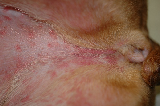
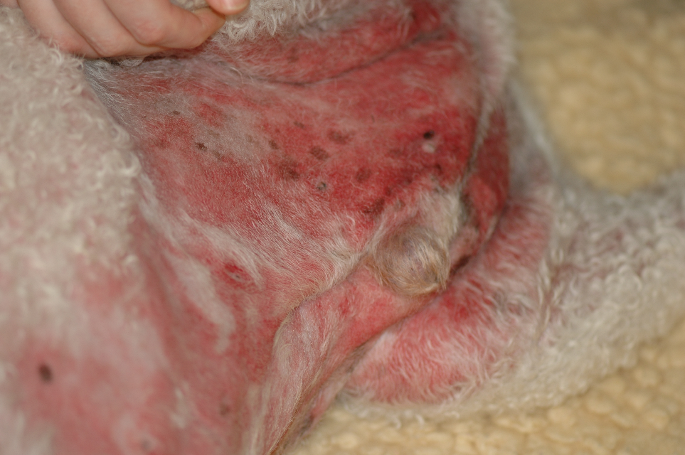
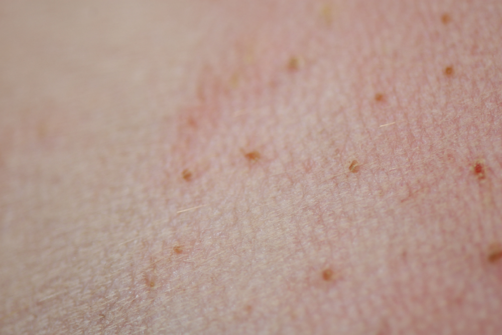
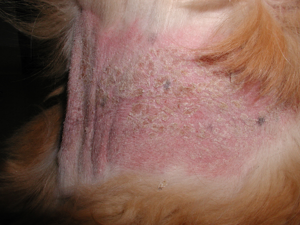
- Below are examples of the various clinical presentations of CADR:
- Urticaria and angioedema.
-
-
-
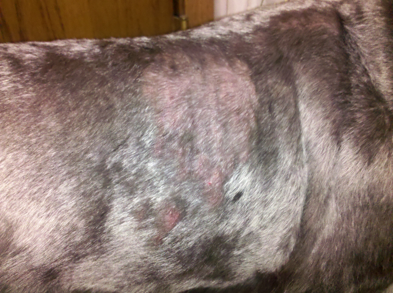
- Maculopapular eruptions – macules and papules with or without pruritus.
-
-
-
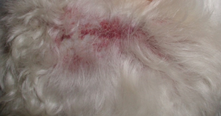
- Erythematous single to coalescing macules and/or papules with edema – the best example is an acute sterile eosinophilic dermatitis most commonly affecting the face, pinnae and trunk of dogs. Systemic signs such as vomiting, diarrhea, and lethargy may be present and precede the skin lesions in some cases. This condition is referred to as Canine Acute Eosinophilic Dermatitis with Edema or Wells’–like syndrome. Drugs have been considered likely triggers in various cases.
-
-
-
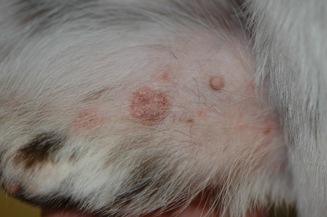
- Erythematous papules, plaques and nodules with or without the presence of pustules, edema and ulceration – the best example is a condition called Canine Sterile Neutrophilic Dermatosis. Systemic signs such as fever, diarrhea, arthritis, lethargy and lymphadenopathy have been reported and often precede skin lesions. Drugs, especially non-steroidal anti-inflammatories, have been considered the culprit in some cases. This disease can be fatal.
-
-
-
- Fixed drug eruption – focal to multifocal areas of sharply demarcated erythematous lesions that recur at the same location upon re-challenge.
- Erythroderma and exfoliation – in other words, diffuse erythema associated with scaling. This reaction pattern has been associated with topical or systemic medications.
-
-
-
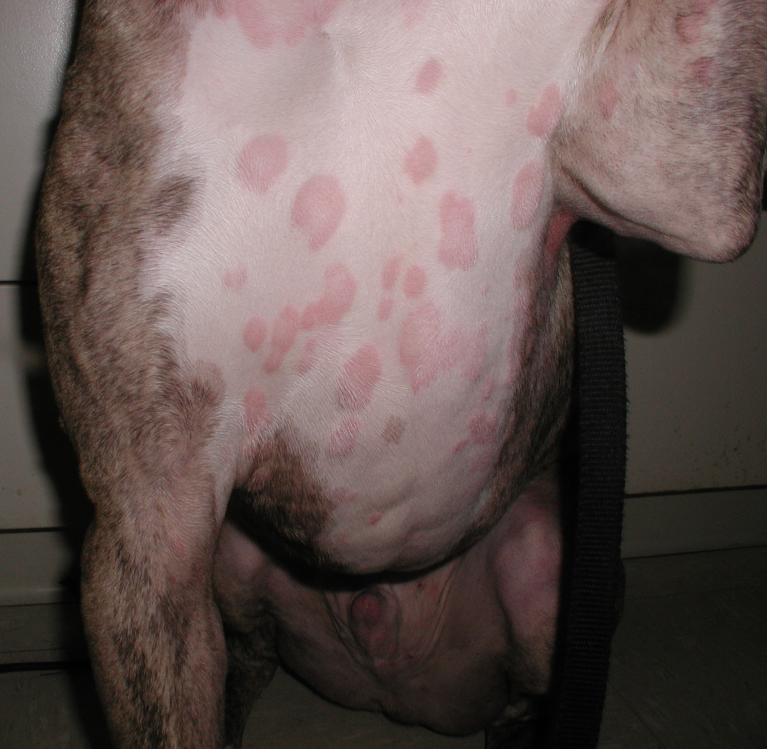
- Purpuric macules and plaques – hemorrhage is typically present in the dermis. The condition has been associated with thrombocytopenia or vascular damage (vasculitis).
-
-
-
- Vesicobullous and erosive/ulcerative lesions – mimic immune-mediated or autoimmune diseases such as pemphigus vulgaris, autoimmune sub-epidermal bullous diseases, Steven Johnson syndrome, toxic epidermal necrolysis and erythema multiforme.
-
-
-
- Pustules and yellowish to tan crusts mimicking pemphigus foliaceous.
-

-
-
- Erythematous plaques associated with fine scales – an example is a condition associated with cyclosporine therapy called psoriasiform lichenoid-like dermatitis.
-
-
-
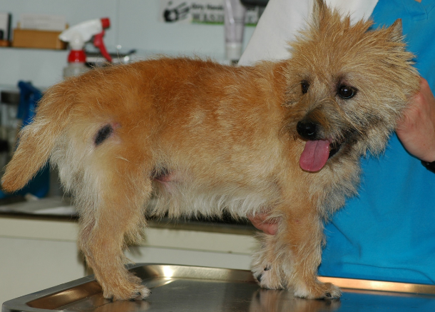
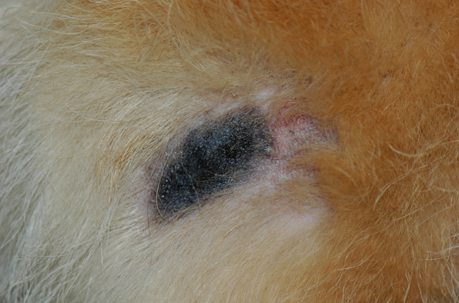
- Focal to multifocal, demarcated areas of alopecia and ulceration associated with necrosis – an example is the vasculitis caused by high doses of itraconazole (≥ 10mg/kg/day). Similar lesions can also be seen at sites of vaccine or other drugs injections.
-

-
-
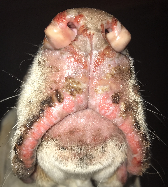
- Generalized erythematous papules that may coalesce to form plaques, and diffuse marked erythema in miniature schnauzers is likely a condition named “superficial suppurative necrolytic dermatitis”. Affected schnauzers typically present with fever, depression and pain. The condition has been reported to develop about 24-72h after the topical application of herbal or citrus or coal tar shampoo or anti-parasitic collar (e.g. imidacloprid + flumethrin). It can be fatal.
-
-
-
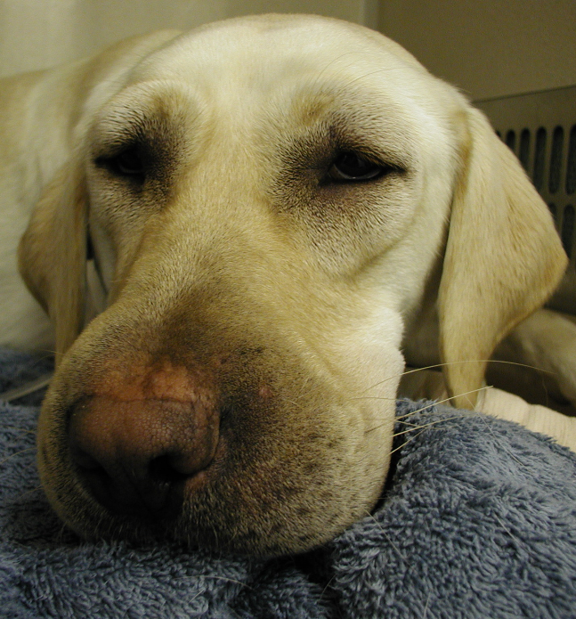
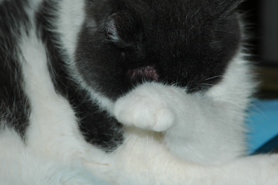
- Facial and neck pruritus – a classic example is methimazole treatment in cats.
-

Important Facts
- There is no age or sex predilection, but some breeds are prone to develop specific cutaneous drug reactions.
- Lesions typically develop within 1 to 3 weeks after initiation of therapy, but some reactions may only occur months after the drug is administered (e.g. rabies vaccine reaction).
- Lesions typically resolve after 1 to 2 weeks of drug discontinuation.
- The clinical signs and distribution of lesions are variable and mimic many dermatoses.
- Lesions may involve the skin and/or mucus membranes.
- The cutaneous lesions can manifest as one or more of the following lesions: urticaria and angioedema, diffuse erythema, exfoliation, purpura, papules, plaques, erosions, ulcerations, and pustules among others.
-
Diagnosis
- A detailed drug history is crucial in diagnosing CADR.
- Ask the pet owner about any oral (including supplements), topical (including ocular), injectable (including vaccines), or inhaled medication.
- Remember that the signs may only develop several days to months after the drug administration and may occasionally last for weeks after drug discontinuation.
- Physical examination findings are not very helpful because CADR can mimic any dermatosis.
- Histopathological findings are quite variable and not helpful in supporting a presumptive diagnosis. However, histopathology is important to rule out other skin diseases.
- Scoring systems to help correlate the administered drug(s) with the patient’s clinical signs have been developed. Modifications of these scoring systems have been made for use in dogs and cats. Please refer to Hinn et al, 1998 (Drug Exposure Score – DES), Koch T et al 2016 (Naranjo) and Mauldin 2006 (modified score system) for description of these scoring systems.
- A detailed drug history is crucial in diagnosing CADR.
Important Facts
- History is the most important clue in diagnosing CADR.
- Clinical signs are not very helpful because CADR can mimic any dermatosis.
- Histopathological findings are quite variable and are not helpful in supporting a diagnosis but they can be helpful in ruling out other dermatosis.
- Resolution or improvement of clinical signs usually occurs 1 to 2 weeks after drug discontinuation. However, the clinical signs may persist months after drug withdraw.
- Provocative exposure is not recommended because it can be life-threatening.
- Scoring systems are available to help with the diagnosis and have been modified for use in dogs and cats.
-
Treatment
- Discontinue and avoid further use of the suspected drug.
- Drugs of similar chemical structure to the suspected ones should be avoided because cross-reactivity can occur.
- Clinical signs are treated symptomatically.
- Oral and short-acting corticosteroids are indicated on a short-term basis to decrease inflammation and pruritus.
- Cyclosporine, pentoxifylline or other immunosuppressive drugs can be considered for cases associated with immunologically mediated reactions, if glucocorticoids are not effective.
- Use carefully corticosteroids or other immunosuppressive therapy in cases with severe, extensive ulcerative lesions. These drugs have the potential to predispose to bacterial sepsis.
- Intravenous human immunoglobulin is a treatment option for severe cases or cases refractory to other immunomodulatory or immunosuppressive therapy.
Important Facts
- Discontinue and avoid further use of the suspected drug or any drugs with similar chemical structure.
- Corticosteroids or other immunosuppressive or immunomodulatory therapy can be considered on a short-term basis to decrease inflammation and/or pruritus.
- Intravenous human immunoglobulin is an option for severe or refractory cases.
- Supportive treatment is given as needed.
-
Prognosis
- It depends on the severity of the reaction and if the culprit can be identified and removed.
References
Ackermann AL, Frank LA, McEntee MF et al. Erythema multiforme associated with zonisamide in a dog. Vet Dermatol 2015; 26: 391-e89.
Boynosky NA and Stokking LB. Potassium bromide-associated panniculitis. J Small Anim Pract 2014; 55: 640-642.
Hammes K, Vannini I, Nitzl D et al. Canine sterile neutrophilic dermatosis (resembling Sweet’s syndrome) with severe extracutaneous manifestations. Schweiz Arch Tierheilkd 2019; 161(4):231-238.
Hinn AC, Olivry T, Luther PB, et al. Erythema multiforme, Stevens-Johnson syndrome, and toxic epidermal necrolysis in the dog: classification, drug exposure and histopathological correlations. J Vet Allergy Clin Immunol 1998; 6:13-20.
Koch T, Mueller RS, Dobenecker B et al. Cutaneous adverse drug reactions in dogs treated with antiepileptic drugs. Front Vet Sc 2016; doi: 10.3389/fvets.2016.00027.
Mauldin EA. Canine acute eosinophilic dermatitis with edema (Wells-like syndrome). Vet Clin Small Anim 2018; 49: 47-51.
Mauldin EA, Palmeiro BS, Goldschmidt MH. Comparison of clinical history and dermatologic findings in 29 cases with severe eosinophilic dermatitis: a retrospective analysis. Vet Derm 2006; 17:338-347.
Miller WH, Griffin GE, Campbell KL. Muller & Kirk Small Animal Dermatology. 7th edn. St Louis: Elsevier Inc., 2013; 466-472.
Mellor PJ, Roulois AJ, Day MJ et al. Neutrophilic dermatitis and immune-mediated haematological disorders in a dog: suspected adverse reaction to carprofen. J Small Anim Pract 2005; 46: 237-242.
Murayama N, Midorikawa K and Nagata N. A case of superficial suppurative necrolytic dermatitis of miniature schnauzers with identification of a causative agent using patch testing. Vet Dermatol 2008; 19: 395-399.
Nuttall TJ and Malham T. Successful intravenous human immunoglobulin treatment of drug-induced Stevens-Johnson syndrome in a dog. J Small Anim Pract 2004; 45: 357-361,
Storrer A, Mackie JT, Gunew MN et al. Cutaneous lesions and clinical outcomes in five cats after frunevetmab injections. J Fel Med Surg 2023; DOI: 10.1177/1098612X231198416
Trepanier LA. Delayed hypersensitivity reactions to sulfonamides: syndromes, pathogenesis and management. Vet Dermatol 1999; 10: 241-248.
Trotman TK, Phillips H, Fordyce H, et al. Treatment of severe adverse cutaneous drug reactions with human intravenous immunoglobulin in two dogs. J Am Anim Hosp Assoc 2006; 42:312-320.
Vitale CB, Ihrke PJ, and Gross TL. Putative diethylcarbamazine-induced urticaria with eosinophilic dermatitis in a dog. Vet Dermatol 1994; 5: 197-203.
Voie KL, Campbell KL, and Lavergne SN. Drug hypersensitivity reactions targeting the skin in dogs and cats. J Vet Intern Med 2012; 26: 863-874.
Zhou Z, Noland E, Rosser E et al. Sterile neutrophilic dermatitis n a cat associated with a topical plant-derived oil flea preventative. Vet Dermatol 2021; DOI: 10.1111/vde.13019.
