2.10 Vesicular Cutaneous Lupus Erythematosus
-
General Considerations
- Vesicular cutaneous lupus erythematosus is a rare severe, vesicular/ulcerative form of cutaneous lupus erythematosus.
- Onset of disease is usually in the summer months, suggesting a role of UV exposure in the pathogenesis.
Important Facts
- Vesicular cutaneous lupus erythematosus is a rare and severe skin disease.
- It is the only canine cutaneous lupus erythematosus disorder classified as subacute.
- Its onset is usually during the spring and summer months.
-
Clinical Signs
- Signalment: the information was based on a comprehensive review on canine cutaneous lupus erythematosus.
- Breeds at high risk include Shetland sheepdogs, rough collies, and border collies. The crosses of these breeds can also be affected.
- The female to male ratio is 0.9.
- The age at the time of disease onset range from 2 to 11 years; median 5.5 years.
- Lesions:
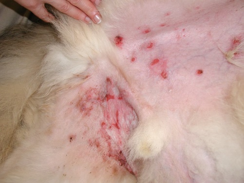
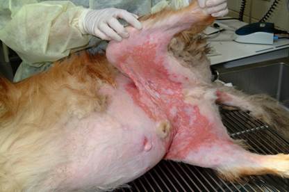
- Flaccid vesicles are initially formed. However, they rupture easily and are replaced by focal to coalescing, annular, polycyclic or serpiginous erosions and ulcerations.
- Signalment: the information was based on a comprehensive review on canine cutaneous lupus erythematosus.

-
- Distribution:
- Lesions usually affect the ventral abdomen, inguinal areas, medial thighs, and axillary regions.
- Distribution:
-
-
- Mucocutaneous junctions, concave aspects of the pinnae, and oral mucosa may be affected in some cases but these lesions are typically minor in extent and severity.
- Lesions are often painful, but are generally non-pruritic.
- Concurrent myositis, confirmed with electromyography, has been reported in one dog.
- Secondary bacterial colonization, bacteremia, and sepsis were described in one dog.
- Systemic signs are usually not associated with this condition.
-
Important Facts
- Shetland sheepdogs, rough collies, border collies and their crossed are predisposed.
- There is no apparent sex predilection.
- Median age at disease onset is 5.5 years.
- Skin lesions are characterized by focal to coalescing, annular, polycyclic or serpiginous erosions and ulcerations usually affecting the ventral abdomen, inguinal areas, medial thighs, and axillae.
- Vesicular cutaneous lupus erythematosus is usually not associated with systemic signs.
-
Diagnosis
- Differential diagnosis include:
- Erythema multiforme minor or major.
- Compatible history and characteristic clinical signs are important parts of the diagnostic equation.
- Cytology:
- No specific findings. No specific findings. It is important to perform cytology to look for secondary bacterial infection/overgrowth.
- Histopathology:
- Subepidermal clefting and vesiculation and lymphocyte-rich interface dermatitis with hydropic degeneration and apoptosis of basal keratinocytes, with or without lymphocytic satellitosis. Lymphocytic interface mural folliculitis may be present.
- Select active borders of erosive and ulcerative lesions to biopsy.
- Direct immunofluorescence or immunohistochemistry:
- Focal or diffuse areas of IgG deposition in the basement membrane was reported in 50% of the dogs.
- History, clinical signs, and histopathological findings are more valuable diagnostic tools.
- Preserve samples in Michel’s solution for immunofluorescence and formalin for immunohistochemistry.
- Indirect immunofluorescence:
- No circulating anti-basement membrane antibodies were identified in dogs tested in a study.
- In a study, ELISA and Immunoblotting (using Hep2 human cell extracts) demonstrated evidence of auto-antibodies to ENA (i.e. extractable nuclear antigens) antigens in most cases (82%). In addition, autoantibodies to Ro/SSA or La/SSA were detected most commonly (55% of dogs).
- Antinuclear antibody (ANA) test:
- Negative.
- Differential diagnosis include:
Important Facts
- Diagnosis should be obtained by a suggestive signalment (i.e. middle-age to older Shetland sheepdogs, rough and border collies) and history (i.e. erosive/ulcerative skin disease with possible onset during Spring and Summer), characteristic clinical signs (i.e. erosions/ulcers on ventral abdomen, inguinal areas, and medial thighs) and histopathological findings (i.e. subepidermal clefting, vesiculation, and lymphocyte-rich interface dermatitis with keratinocyte apoptosis).
- ANA test is negative.
-
Treatment
- One or a combination of the following drugs: (See below “Therapy for Autoimmune Diseases” for dose and specific on treatment regimen).
- Glucocorticoids.
- Azathioprine.
- Cyclosporine.
- Oclacitinib (Apoquel®) – Anecdotal report has shown its good efficacy in treating vesicular cutaneous lupus erythematosus.
- Tacrolimus.
- Mycophenolate mofetil – It was reported to cause remission in one rough collie dog after discontinuation of oral glucocorticoid.
-
Prognosis
- Complete remission is not achieved in all cases, but overall prognosis is guarded to good with appropriate therapy.
References
Bizikova P, Linder KE, Anderson JG. Erosive and ulcerative stomatitis in dogs and cats: which immune-mediated diseases to consider? J Am Vet Med Assoc 2023; doi.org/10.2460/javma.22.12.0573.
Medleau L, Hnilica KA. Chapter 8. Autoimmune and immune-mediated skin disorders. In: Small Animal Dermatology: A color Atlas and Therapeutic Guide 2006. 2nd ed. W.B. Saunders, Missouri, 189-227.
Miller, Griffin and Campbell. Chapter 9. Autoimmune and immune-mediated dermatoses. In: Muller & Kirk’s Small Animal Dermatology 2013. 7th ed., W.B. Saunders, Missouri; 432-462.
Olivry T. Auto-immune skin disease in animals: time to reclassify and review after 40 years. BMC Vet Res 2018; https://doi.org/10.1186/s12917-018-1477-1.
Olivry T, Chan LS. Autoimmune blistering dermatoses in domestic animals. Clin Dermatol 2001; 19(6):750-760.
Olivry T., Jackson HA. Diagnosing new autoimmune blistering skin diseases of dogs and cats. Clin Tech Small Anim Pract 2001; 16: 225-229.
Olivry T, Linder KE, Banovic F. Cutaneous lupus erythematosus in dogs: a comprehensive review. BMC Vet Res 2018; https://doi.org/10.1186/s12917-018-1446-8
