1.7 Sex Hormone Dermatoses – Canine
-
General Considerations
- The canine sex hormone dermatoses are typically associated with hyperestrogenism, hypertestosteronism, or rarely hyperprogesteronism. Some dogs may have normal serum estrogen concentrations and cutaneous hyperestrogenism due to increased numbers of estrogen receptors in the skin.
- Laboratory measurement of sex hormone concentrations may not be helpful in the diagnosis of these disorders because most commercial assays measure only one type of estrogen (e.g. estradiol or estrone), androgen, or progestagen but various hormonal substances are produced by the body. In addition, hormone values fluctuate during the day and reference intervals are determined based on a single sample collection.
- Consider sex hormone dermatoses as a differential diagnosis in any intact animal with an endocrine alopecia when hyperadrenocorticism and hypothyroidism have been ruled out.
Important Facts
- The canine sex hormone dermatoses are associated with hyperestrogenism, hypertestosteronism, or rarely hyperprogesteronism.
- Laboratory measurement of sex hormone concentrations may not be helpful in the diagnosis of these disorders because most commercial assays measure only one type of estrogen (e.g. estradiol or estrone), androgen, or progestagen but various hormonal substances are produced by the body. In addition, hormone values fluctuate during the day and reference intervals are determined based on a single sample collection.
- We usually discourage experimentation with sex hormone supplementation. Once the major endocrinopathies have been ruled out, owners are often content with the knowledge that their pet’s hair loss is cosmetic only.
- Consider a sex hormone dermatosis as possible diagnosis in any intact animal with an endocrine alopecia when hyperadrenocorticism and hypothyroidism have been ruled out.
Hyperestrogenism – Intact females
-
General Considerations
- Sex hormone dermatoses in intact female dogs are typically associated with hyperestrogenism:
- They occur especially in middle age to older dogs.
- They may be associated with cystic ovaries, ovarian neoplasia, or administration of diethylstilbestrol, or other estrogens for the treatment of urinary incontinence or mismating.
- 10% to 20% of the ovarian tumors are malignant and the estrogen-producing ones are typically originated from granulosa-theca cell. These tumors can also secrete progesterone and induce mammary hyperplasia.
- Sex hormone dermatoses in intact female dogs are typically associated with hyperestrogenism:
-
Clinical Signs
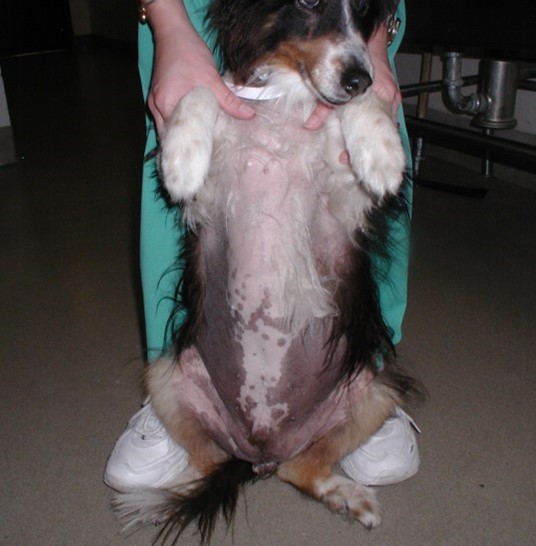
- Bilaterally symmetric alopecia that typically starts in the perineum, groin, and flanks. As with other endocrine alopecias, the head and distal extremities are not affected. Hyperpigmentation is often present.
- Pruritus is absent but it may be present if seborrhea oleosa and/or superficial pyoderma occur.
- Estrus cycle abnormalities (i.e. irregular cycles, prolonged estrus, and nymphomania) often occur, and endometritis or pyometra may be seen.
- The nipples and vulva are enlarged and comedones are usually numerous on the ventrum and vulvar skin.
-
Diagnosis
- The primary differential diagnoses include hypothyroidism and hyperadrenocorticism.
- Definitive diagnosis is based on a characteristic history, typical clinical signs, laboratory test results that rule out other conditions, and response to therapy.
- Elevated blood estrogen concentrations may or may not be present to support the diagnosis.
- Abdominal ultrasound may help identify ovarian tumors or cysts.
-
Treatment
- Ovariohysterectomy.
- A good response is usually evident within 3 months but occasionally may not be seen for 6 months.
- If an ovarian neoplasm is suspected, chest radiographs should be taken before surgery to investigate the presence of metastasis.
Important Facts
- Consider sex hormone imbalance as a differential in any intact animal with an endocrine alopecia where hyperadrenocorticism and hypothyroidism have been ruled out.
- Hyperestrogenism in intact females usually affect middle age (ovarian cysts) to older (functional ovarian tumors) dogs.
- Treatment with estrogens can lead to hyperestrogenism in dogs of any age.
- Bilaterally symmetric alopecia, abnormal estrus cycles, and enlarged vulva and nipples are clinical signs suggestive of hyperestrogenism.
- Diagnosis should be based on a characteristic history, typical clinical signs and laboratory test results that rule out other conditions (e.g. hypothyroidism and hyperadrenocorticism) and response to therapy.
- Elevated blood estrogen concentration is not always present to support the diagnosis.
- Response to ovariohysterectomy is usually evident within 3 months.
Male Feminization Syndrome – Intact males
-
General Considerations
- Male feminization syndrome is the result of hyperestrogenism usually associated with a functional Sertoli cell tumor.
- It has been reported to occur in 24% – 57% of dogs with Sertoli cell tumors.
- Seminomas and interstitial cell tumors can also secrete estrogen and cause feminization syndrome. However, interstitial cell tumors usually produce testosterone and will cause hyperandrogenism and seminomas are usually non-functional tumors.
- Multiple testicular tumors can be present in the same animal.
-
Clinical Signs
- Undescended testicles are 10 times more likely to develop functional Sertoli cell tumors or seminomas.
- 70% of dogs with testicles located in the abdominal cavity will develop Sertoli cell tumor and feminization syndrome.
- Feminization is more likely to occur with larger tumors and tends to be increasingly severe as tumor size increases.
- Approximately 20% of the tumors can be malignant but only about 10% metastasize to the lungs and abdominal lymph nodes.
- Boxers, Shetland sheepdogs, Weimaraners, Cairn terriers, Pekingese, and Collies are predisposed.
- The disease usually affects middle-aged to older dogs.
- Specific Signs:
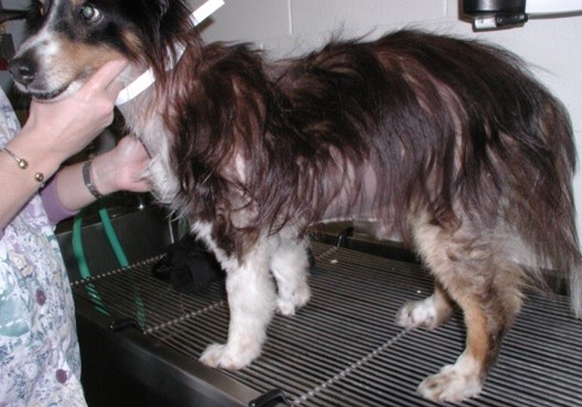
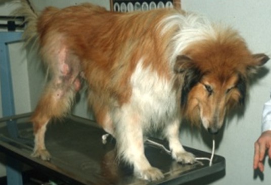
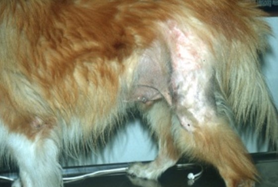
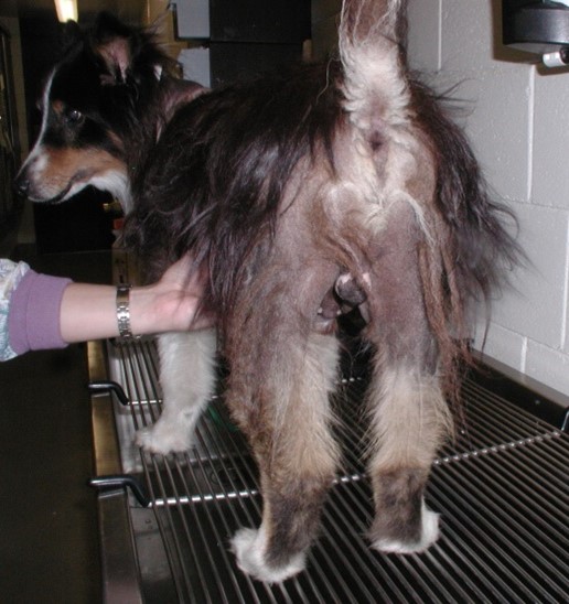
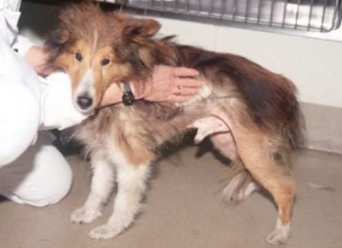
- Bilateral symmetric non-inflammatory alopecia begins in the perineum region and extends to the legs (more often caudal aspect), ventrum, chest, neck, and flanks.
-
-
- Hyperpigmentation may occur and pruritus is only observed if secondary superficial pyoderma develops.
-
-
-
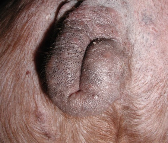
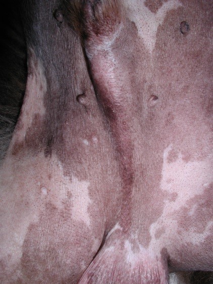
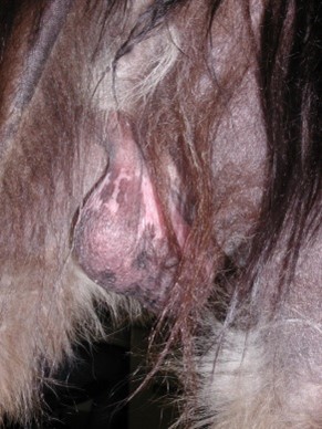
- Linear preputial dermatosis (pigmented line extending from preputial orifice to the scrotum) is a common but not consistent finding associated with estrogen producing tumors.
-
-
-
- Pendulous prepuce, gynecomastia, enlarged nipples, cryptorchidism or mass in scrotal testis are typical signs of male feminization syndrome. Uninvolved testicles are usually atrophied.
-
-
-
- The prostate is often enlarged and infected due to estrogen-induced squamous metaplasia and there may be clinical signs referable to prostatomegaly, prostatitis or both.
- Behavior changes, such as attractiveness to other male dogs, may be present but may not be noticed by the pet owner.
- Estrogen-induced bone marrow suppression (i.e. thrombocytopenia, neutropenia and anemia) is uncommon to rare but is a life-threatening complication.
-
-
Diagnosis
- Differential diagnosis:
- Other endocrine diseases that affect the skin and occur in middle age to older dogs such as hyperadrenocorticism and hypothyroidism.
- Definitive diagnosis should be based on a characteristic history, typical clinical signs, measurement of basal estradiol 17-ß concentration (not always elevated), and response to castration. Recent studies showed that testicular and peripheral blood oestradiol 17-ß concentration is increased in most dogs with Sertoli cell tumors and feminization syndrome compared to normal dogs. Testosterone concentration was reduced in affected dogs. The authors concluded that testosterone/oestradiol ratio where shown to better correlate with the presence of feminization syndrome than each hormone separately since not every affected dog had elevated oestradiol 17- ß concentration.
- Radiographs/Ultrasound:
- Abdomen – Abdominal mass (undescended testis) and sub-lumbar lymphadenopathy (suggests metastases).
- Thorax – Metastases occurs in about 10% of 20% of the dogs with malignant tumors.
- Testicles – Imaging will show a homogenous mass.
- Differential diagnosis:
-
Treatment
- Bilateral castration if metastasis has not occurred. A good clinical response is usually seen within 3 months but it may take up to 6 months in some cases. Remission followed by relapse indicates functional metastatic tissue.
- Although the metastatic rate of these tumors is low, all dogs should be examined carefully before surgery.
- Cisplatin as a single agent can be tried in dogs with metastatic tumors.
- Dogs with blood dyscrasias have a poor prognosis and death may be related to aplastic anemia, thromboembolism, or septicemia.
Important Facts
- Most cases of hyperestrogenism associated with male feminization syndrome and symmetric non-pruritic alopecia are caused by Sertoli cell tumor. However, seminomas and interstitial cell tumors can rarely cause identical clinical signs.
- Sertoli cell tumor and seminomas with feminization syndrome occur more often in dogs with cryptorchidism.
- About 70% of the cases with abdominal testis will develop Sertoli cell tumor and feminization syndrome.
- The size of the tumor correlates with the severity of the feminization syndrome.
- About 20% of the cases are malignant and about 10% of these tumors metastasize to the thorax and/or abdominal lymph nodes.
- Bone marrow suppression due to hyperestrogenism is rare.
- Bilateral symmetric and non-pruritic alopecia begins in the perineum and extends to the ventrum, chest, neck, and flanks. Like other endocrinopathies that affect the skin the head and distal extremities are spared.
- Dogs with hyperestrogenism may show various combinations of feminization signs such as pendulous prepuce, gynecomastia, enlarged nipples, and attraction by other males.
- A testicular mass of various sizes may be present in the scrotum, or an abdominal or inguinal mass may be palpated if the dog has undescended testicles.
- After bilateral castration, clinical response is usually seen within 3 months but it may take 6 months before signs are resolved. Signs return if metastasis occur.
Hyperandrogenism – Intact males
-
General Considerations
- Hyperandrogenism is a rare condition that affects intact male dogs.
- Hypertestosteronemia is typically associated with a functional testicular neoplasia, especially interstitial cell tumor but it can also be idiopathic.
- The excess testosterone increases the activity of the glands stimulated by androgens including the circumanal and tail glands, which have the same tissue (i.e. hepatoid), and the sebaceous glands. This can result in hyperplasia and/or increase secretion of the glands.
-
Clinical Signs
- Cases associated with functional testicular neoplasia will have one or more of the following signs:
- Hyperplasia of the circumanal and tail glands.
- The circumanal glands typically acquire a donut-like shape.
- The area associated with the hyperplasia of the tail gland (proximal tail) becomes bulgy and eventually alopecic and greasy.
- In severe cases, nodules and/or cysts may develop at the affected sites.
- Hyperpigmented macules of the tail glands, perineum, ventral tail, scrotum, and ventral abdomen may develop before, during, or after the circumanal and tail glands hyperplasia.
- A testicular mass is typically palpable but not always.
- Idiopathic cases will present with one or more of the following signs:
- Severe greasy seborrhea and rarely symmetric truncal alopecia.
- The seborrhea is most marked on the face, ears, feet, axilla, and groin.
- Secondary infections are common and will often induce pruritus and aggravate the condition.
- Dogs will often develop hypersexual behavior and aggression to other dogs and/or humans.
- Cases associated with functional testicular neoplasia will have one or more of the following signs:
-
Diagnosis
- Definitive diagnosis should be based on a characteristic history, typical clinical signs, blood testosterone concentrations and response to castration.
- Testicular ultrasound may detect a non-palpable tumor.
-
Treatment
- Castration is the best treatment. Skin lesions start to regress 2-4 weeks after castration.
- Severe glandular hyperplasia may not improve significantly after castration.
- Behavior changes may take longer to resolve.
- Anti-seborrheic shampoos containing degreaser such as benzoyl peroxide, selenium sulfide or tar should be added for cases with seborrhea oleosa.
- Secondary infections need to be treated adequately.
Important Facts
- Canine hyperandrogenism is typically associated with interstitial cell tumor; however, it can be idiopathic.
- Hyperplasia of the circumanal and tail glands are typical signs associated with a testosterone secreting testicular tumor. Pigmented macules can be also associated with the affected areas.
- Severe seborrhea oleosa and rarely symmetric truncal alopecia are signs of idiopathic hyperandrogenism.
- Castration is the treatment of choice but it may take a while for clinical signs to resolve or improve.
References
Miller WH, Griffin GE, Campbell KL. Muller & Kirk’s Small Animal Dermatology. 7th ed. Philadelphia: W.B. Saunders Co., 2013; 532-540 .
Miller WH. Sex hormone-related dermatoses in dogs. In: Kirk RW ed. Current Veterinary Therapy Small Animal Practice. volume X. Philadelphia: W.B. Saunders Co., 1989; 595-606.
