2.4 Pemphigus Erythematosus
-
General Considerations
- Pemphigus erythematosus is a rare disorder.
- It is considered a facial form of pemphigus foliaceus.
- It has some similarities with facial discoid lupus erythematosus and has been considered a crossover between these two diseases:
- Immunofluorescence similar to both pemphigus (intercellular staining) and lupus (staining along basement membrane).
- Presence of a lichenoid ± interface dermatitis on histopathology, which is characteristic of facial lupus erythematosus.
- Low serum titer of antinuclear antibody (ANA) in some affected dogs.
- Lesions are aggravated by UV light.
- The fact that pemphigus erythematosus is a separate disease from pemphigus foliaceus should be considered critically for the following reasons.
- Pemphigus foliaceus has been reported to only affect the face in ~16% of cases.
- Photosensitivity has also been reported in dogs with pemphigus foliaceus.
- The presence of a lichenoid (band-like) ± interface dermatitis in conjunction with a superficial acantholytic epidermal pustule may solely reflect the nasal/paranasal location of the lesions.
- The presence of immunoglobulins and complement at the basement membrane zone by direct immunofluorescence or immunohistochemistry is not a finding specific of lupus and it has also been reported in cases of pemphigus foliaceus.
- The presence of low serum titers of ANA antibodies is not a specific finding and has been reported in about 30% of dogs with pemphigus foliaceus.
- All these facts suggest that pemphigus erythematosus is a facial-predominant form of pemphigus foliaceus and not a separate disease entity
Important Facts
- Pemphigus erythematosus is a rare disorder.
- Lesions are identical to those of pemphigus foliaceus but are localized solely to the face.
- It also has some similarities with facial discoid lupus erythematous including low serum titer of ANA antibodies, lesions are aggravated by UV light, presence of a lichenoid ± interface dermatitis on histopathology, and immunofluorescence staining similar to both pemphigus (intercellular) and lupus (basement membrane).
- There is compelling argument to consider pemphigus erythematosus a facial-predominant form of pemphigus foliaceus and not a separate disease entity.
-
Clinical Signs
- Lesions – Similar to pemphigus foliaceus.
- Primary lesions including pustules and rarely vesicles and bullae are seldom seen because they rupture easily.
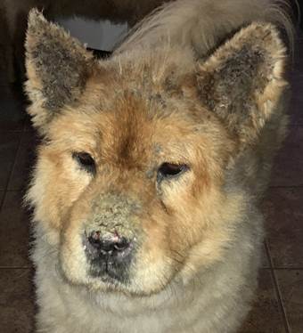
- Secondary lesions are common and include crusts, scales, erosions, alopecia, erythema, and depigmentation.
- Distribution:
- Face (dorsal muzzle; may extend to nasal planum) and pinnae.
- Mucosal sites are not involved.
- Differential diagnoses:
- Pemphigus foliaceus, mucocutaneous pyoderma, pustular dermatophytosis, demodicosis, zinc-responsive dermatosis, eosinophilic pustulosis, and cutaneous drug reaction.
- Lesions – Similar to pemphigus foliaceus.
Important Facts
- Lesions are similar to the ones present in pemphigus foliaceus but the distribution is limited to the face (dorsal aspect of the muzzle [may extend to the nasal planum] and pinnae).
- Mucosal sites are not affected.
-
Diagnosis
- History:
- Lesions may develop suddenly or gradually.
- Lesions become worse with UV light.
- Clinical Signs:
- Pustules, yellowish-brown crusts, erosions, and depigmentation localized to the face only.
- History:
-
- Exclusion of other dermatoses and especially the following:
- Mucocutaneous pyoderma (tends to involve not only the muzzle but also the lips).
- Pemphigus foliaceus (rarely limited to the face).
- Cytological exam of direct smears of content from intact pustules or from exudate present under recent crusts:
- Nondegenerate neutrophils, ± eosinophils, and many acantholytic keratinocytes (hallmark).
- Histopathology:
- Lesions characteristic of pemphigus foliaceus include subcorneal or intraepidermal pustule with neutrophils, ± eosinophils, and acantholytic keratinocytes.
- Lesions typical of lupus erythematosus may be present in some cases.
- Histopathology is currently the most important diagnostic test.
- Direct immunofluorescence or immunohistochemistry:
- Intercellular and/or basement membrane zone (lupus-like) staining of immunoglobulins deposits.
- Low serum titer of ANA antibodies is present in some cases. ANA antibody test is not specific.
- Exclusion of other dermatoses and especially the following:
Important Facts
- History and clinical signs including pustules and crusts on dorsal muzzle and pinnae provide important diagnostic information.
- Cytological exam of direct smear from intact pustules’ content and/or from the exudate present underneath a recent crust reveals nondegenerate neutrophils, ± eosinophils, and many acantholytic keratinocytes (hallmark).
- Histopathology is the most important diagnostic test and reveals subcorneal pustules containing neutrophils, +/- eosinophils, and many acantholytic keratinocytes. In addition, features of lupus erythematosus will be present in some cases.
- Direct and indirect immunofluorescence or immunohistochemistry tests are not frequently used as a diagnostic tool because false negative results are common and these tests are not routinely available.
- Remember! Do not biopsy nasal planum for direct immunofluorescence or immunohistochemistry because this site is often positive in normal dogs (i.e. false positive).
- Low serum titer of ANA antibody test may be present in some dogs. ANA is not a specific test.
-
Treatment
- One or more of the following drugs can be used in the treatment of pemphigus erythematosus – (See “Therapy for Autoimmune Diseases” for dose and specific on treatment protocol).
- Glucocorticoids.
- Azathioprine (do not use it in cats because they will likely develop bone marrow suppression).
- Chlorambucil.
- Mycophenolate mofetil could be considered as a glucocorticoid-sparing agent.
- Tacrolimus, a topical calcineurin inhibitor like cyclosporine, has been reported to work for cases of pemphigus erythematosus. It is applied to focal lesions and can be used as sparing agent of glucocorticoids and azathioprine.
- Oclacitinib (Apoquel®) has been reported to be effective in canine pemphigus foliaceus and may also be efficacious in dogs with pemphigus erythematosus.
- Gold Salts.
- One or more of the following drugs can be used in the treatment of pemphigus erythematosus – (See “Therapy for Autoimmune Diseases” for dose and specific on treatment protocol).
References
Bizikova P, Burrows A. Feline pemphigus foliaceus: original case series and a comprehensive literature review. BMC Vet Res 2019; https://doi.org/10.1186/s12917-018-1739-y.
Carrasco I, Martinez M and Albinyana G. Beneficial effect of oclacitinib in a case of feline pemphigus foliaceus. Vet Dermatol 2021; 32:299-301.
Dalmau A, Ordeix L. Putative pemphigus-like reaction to oral fluralaner in a dog. Vet Dermatol 2023; DOI: 10.1111/vde.13243.
Lopes NL, Campos DR, Machado MA et al. A blinded, randomized, placebo-controlled trial of the safety of oclacitinib in cats. BMC Vet Res 2019; 15:137.
Medleau L, Hnilica KA. Chapter 8. Autoimmune and immune-mediated skin disorders. In: Small Animal Dermatology: A color Atlas and Therapeutic Guide 2006. 2nd ed. W.B. Saunders, Missouri, 189-227.
Miller, Griffin and Campbell. Chapter 9. Autoimmune and immune-mediated dermatoses. In: Muller & Kirk’s Small Animal Dermatology 2013. 7th ed., W.B. Saunders, Missouri; 432-462.
Olivry T. Auto-immune skin disease in animals: time to reclassify and review after 40 years. BMC Vet Res 2018; https://doi.org/10.1186/s12917-018-1477-1.
Olivry T, Chan LS. Autoimmune blistering dermatoses in domestic animals. Clin Dermatol 2001; 19(6):750-760.
Olivry T., Jackson HA. Diagnosing new autoimmune blistering skin diseases of dogs and cats. Clin Tech Small Anim Pract 2001; 16: 225-229.
Olivry T. Linder KE. Dermatoses affecting desmosomes in animals: a mechanistic review of acantholytic blistering skin diseases. Vet Dermatol 2009; 20: 313-326.
Outerbridge CA, Affolter VK, Lyons LA et al. An unresponsive progressive pustular and crusting dermatitis with acantholysis in nine cats. Vet Dermatol 2017; DOI: 10.1111/vde.12501
