2.11 Mucocutaneous Lupus Erythematosus
-
General Considerations
- Mucocutaneous lupus erythematosus is classified as one of the forms of canine chronic lupus erythematosus.
- As the name indicates, the lesions of dogs with mucocutaneous lupus erythematosus develop close to mucosal sites (i.e. juxtamucosal).
Important Facts
- Canine mucocutaneous lupus erythematosus is one of the forms of chronic lupus erythematosus.
- Lesions develop adjacent to mucosal sites (i.e. juxtamucosal).
-
Clinical Signs
- Signalment: the information is based on 37 cases summarized in a comprehensive review paper on canine lupus erythematosus.
- German shepherd dogs and their crosses accounted for most (47%) of the reported cases.
- The reported median age at disease onset was 6 years; range 3 to 13 years. Most cases (71%) in a series of 21 dogs started developing clinical signs between 4 and 8 years of age.
- Female dogs were 1.8 times more likely to develop the disease than male dogs.
- Lesions:
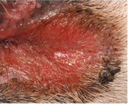
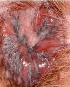
- Characteristic lesions are deep erosions and/or shallow ulcers that do not heal with scarring, which differs from lesions of facial and generalized discoid lupus erythematosus.
- Signalment: the information is based on 37 cases summarized in a comprehensive review paper on canine lupus erythematosus.
Olivry T. BMC Vet Res 2028; 14:132.
Olivry T. BMC Vet Res 2028; 14:132.
-
-
- Crusts can form when the exudate produced by the erosive/ulcerative lesions dries out.
- Hyperpigmentation can be seen at the periphery of erosive/ulcerative lesions or at the site of previous lesions, where the hyperpigmentation can acquire a reticulated pattern.
- Pruritus is typically absent or mild.
- Pain is reported during urination or defecation when lesions are formed on or around the anus and the vulvar area.
- Systemic signs have not been reported.
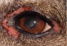
- Distribution:
- Lesions have been reported to develop more frequently on the anal ring or around the anus (67%), on the genitalia or perigenital area (47%), and lips (28%).
-

-
-
- Less often lesions can develop around the eyes (17%), nasal planum (11%), and oral cavity (9%).
- Most reported cases had two or more sites affected, and the lesions were typically distributed in a symmetrical pattern.
-
Important Facts
- German shepherd dogs and their crosses are overrepresented in the reported cases.
- The median age at disease onset was 6 years and females were reported to have the disease almost twice more often than males.
- Lesions are characterized by deep erosions and/or ulcerations localized more commonly on or around the anus, genital or perigenital area, and lips.
- The oral cavity is rarely affected, which helps differentiate this disease from the autoimmune subepidermal blistering diseases.
- Most reported cases had at least two sites affected.
- Systemic signs were absent according to the reported cases.
-
Diagnosis
- Differential diagnoses include the following:
- Mucocutaneous pyoderma, mucous membrane pemphigoid, and erythema multiform minor and major.
- Compatible history and characteristic clinical signs are important parts of the diagnostic equation.
- Cytology:
- Direct smears of the exudate collected from an ulcer, deep erosion, or underneath a crust will yield nonspecific findings.
- This test is important to investigate the presence of secondary infections.
- Histopathology:
- Cell-rich interface dermatitis characterized by damage of basal keratinocytes (i.e. hydropic degeneration/vacuolation, apoptosis with or without satellitosis) and a lymphocytic/plasmacytic superficial dermal infiltrate. The interface dermatitis often extends to the infundibulum wall of hair follicles and it is often patchy or present in limited areas. Less commonly, apoptosis extends to the upper epidermal layers. This finding can lead to the misdiagnosis of erythema multiforme.
- Biopsy the active border of an erosion or ulceration.
- Aim to submit the biopsy samples to a pathologist with interest in dermatohistopatholgy. Make sure to include a detailed history and clinical signs description. If possible, also submit good quality photos.
- Cell-rich interface dermatitis characterized by damage of basal keratinocytes (i.e. hydropic degeneration/vacuolation, apoptosis with or without satellitosis) and a lymphocytic/plasmacytic superficial dermal infiltrate. The interface dermatitis often extends to the infundibulum wall of hair follicles and it is often patchy or present in limited areas. Less commonly, apoptosis extends to the upper epidermal layers. This finding can lead to the misdiagnosis of erythema multiforme.
- Direct immunofluorescence or immunohistochemistry:
- Linear deposition of immunoglobulins along the basement membrane zone.
- Remember! Skin biopsy samples are submitted for these tests (samples should be fixed in Michel’s fixative for immunofluorescence and formalin for immunohistochemistry).
- Sensitivity has been reportedly good; however, negative results can occur.
- CBC, chemistry profile, and urinalysis:
- Used as baseline for monitoring potential side effects associated with therapy.
- Antinuclear antibody (ANA) test:
- Typically negative.
- Differential diagnoses include the following:
Important Facts
- History and clinical signs are important diagnostic information.
- Cytological exam of direct smear from the exudate present underneath a recent crust or from a deep erosion/ulceration yield non-specific findings. However, this test is important to investigate the presence of secondary infection.
- Histopathology is the most important diagnostic test and reveals a cell-rich interface dermatitis.
- Direct immunofluorescence test has shown to have good sensitivity but it is not frequently used as a diagnostic tool because it is not typically available in commercial laboratories.
-
Treatment
- One or more of the following options is recommended: (See “Therapy for Autoimmune Diseases” for dose and specifics on treatment regimens).
- Glucocorticoids.
- Azathioprine (do not use it in cats because they develop bone marrow suppression).
- Chlorambucil.
- Ciclosporin.
- Mycophenolate mofetil.
- Oclacitinib – Complete remission was achieved in two dogs with mucocutaneous lupus erythematosus.
- Doxycycline and niacinamide. Try to avoid this treatment modality to prevent bacterial resistance to antibiotics (practice antibiotic stewardship).
- One or more of the following options is recommended: (See “Therapy for Autoimmune Diseases” for dose and specifics on treatment regimens).
-
Prognosis
- Prognosis is good with therapy, with most cases having partial to complete remission. However, dose reduction or treatment discontinuation many lead to relapses.
References
Bizikova P, Linder KE, Anderson JG. Erosive and ulcerative stomatitis in dogs and cats: which immune-mediated diseases to consider? J Am Vet Med Assoc 2023; doi.org/10.2460/javma.22.12.0573.
Medleau L, Hnilica KA. Chapter 8. Autoimmune and immune-mediated skin disorders. In: Small Animal Dermatology: A color Atlas and Therapeutic Guide 2006. 2nd ed. W.B. Saunders, Missouri, 189-227.
Miller, Griffin and Campbell. Chapter 9. Autoimmune and immune-mediated dermatoses. In: Muller & Kirk’s Small Animal Dermatology 2013. 7th ed., W.B. Saunders, Missouri; 432-462.
Olivry T. Auto-immune skin disease in animals: time to reclassify and review after 40 years. BMC Vet Res 2018; https://doi.org/10.1186/s12917-018-1477-1.
Olivry T, Chan LS. Autoimmune blistering dermatoses in domestic animals. Clin Dermatol 2001; 19(6):750-760.
Olivry T., Jackson HA. Diagnosing new autoimmune blistering skin diseases of dogs and cats. Clin Tech Small Anim Pract 2001; 16: 225-229.
Olivry T, Linder KE, Banovic F. Cutaneous lupus erythematosus in dogs: a comprehensive review. BMC Vet Res 2018; https://doi.org/10.1186/s12917-018-1446-8.
Olivry T, Rossi MA, Banovic F, et al. Mucocutaneous lupus erythematosus in dogs (21 cases). Vet Dermatol 2015; 26: 256–e55
