3.11 Nasal and Digital Hyperkeratosis
-
General Considerations
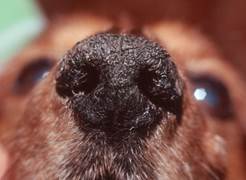
- It is an idiopathic keratinization disorder characterized by excessive accumulation of tightly adhered keratin on the nasal planum or footpads, or both.
- Nasal and digital hyperkeratosis are seen typically in older dogs, probably as a senile change. Some dogs only have nasal or digital disease.
- American cocker spaniels and English springer spaniels appear to be predisposed, although any breed can be affected.
- Keep in mind that older dogs with abnormal weight bearing due to arthritis, for example, may develop footpad hyperkeratosis at the border of the footpads that does not touch the floor.
Important Facts
- Nasal and digital hyperkeratosis are idiopathic keratinization disorders characterized by the accumulation of tightly adhered keratin on the nasal planum or footpads or both.
- Nasal and digital hyperkeratosis occur typically in older dogs, probably as a senile change.
- Any breed can be affected.
-
Clinical Signs
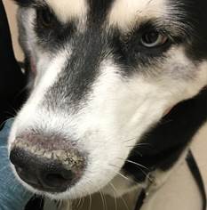
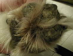
- Lesions may be focal or diffuse and are characterized by tightly adherent thick accumulations of keratin on the nasal planum and/or footpads.
- It involves the footpads of all paws as well as the metacarpal and metatarsal pads.
- Lesions may be focal or diffuse and are characterized by tightly adherent thick accumulations of keratin on the nasal planum and/or footpads.
-
- The skin is usually very dry and cracks and fissures may develop.
- Severe footpad involvement causes lameness.
Important Facts
- Lesions may be focal or diffuse and are characterized by tightly adherent thick accumulations of keratin on the nasal planum or footpads, or both.
- Cracks and fissures may develop with time.
- Severe footpad involvement causes pain and lameness.
-
Diagnosis
- Nasal and/or digital hyperkeratosis in a middle age to old dog without evidence of other clinical signs is diagnostic.
- If other clinical signs either systemic or dermatologic are present, a thorough workup to identify an underlying etiology is necessary.
- All diseases that can cause similar lesions must be considered as differential diagnoses including:
- Hereditary nasal and footpad hyperkeratosis, canine distemper, pemphigus foliaceous, drug reaction, zinc responsive dermatosis, generic dog food dermatosis, and superficial necrolytic dermatosis.
- Biopsy of affected sites should be considered if the disease starts early in life and other symptoms are present. Histopathology will help rule in/out the various diseases in the list of differential diagnoses.
Important Facts
- Nasal and/or digital hyperkeratosis in a middle age to old dog without evidence of other concurrent diseases is diagnostic.
- If other clinical signs either systemic or dermatologic are present, a thorough workup to identify an underlying etiology is necessary.
- If the disease starts at an early age and other symptoms are present, skin biopsy will be important to rule in/out other diseases affecting the nasal planum and footpads.
-
Treatment
- The disease is non-curable and treatment is symptomatic.
- Mild and asymptomatic cases may not need treatment.
- Since there is continuous keratin production, there is a constant need for controlling the accumulation of keratin
- The excess keratin needs to be soften and physically removed in more severe cases associate with discomfort.
- Hydrating the lesions with water or wet dressing may be required to allow the physical removal of excessive keratin.
- In many cases, a topical keratolytic agent in a gel formulation is applied every 12 hours initially and then as needed to keep the condition under control. Examples: Products containing salicylic acid, lactic acid, and urea (e.g. KeraSolv Gel®), topical 0.025 or 0.01% tretinoin gel (e.g. Retin-A®).
- If fissures are present, topical corticosteroids and antibiotics may be needed to control secondary inflammation and secondary infection.
- Continued application of hydrating and moisturizing agents, such as propylene glycol, petroleum jelly, urea or products containing essential oils (e.g. Dermoscent Biobalm®), are usually required at varying frequencies.
Important Facts
- Treatment is symptomatic.
- Mild cases may not require therapy.
- Physical removal of the excess keratin after its softening should be part of the treatment regimen in cases associated with discomfort.
- Daily to as needed application of a topical keratolytic agent in a gel formulation may control the disease long-term.
- If fissures are present, topical products containing corticosteroids and antibiotics may be needed to control secondary inflammation and infection.
- Continued application of hydrating and moisturizing agents, such as propylene glycol, petroleum jelly, urea or essential oils is often required to maintain the disease controlled.
References
Kwochka KW: Primary Keratinization Disorders of Dogs. In: Griffin CE, Kwochka KW, MacDonald JM (eds). Current Veterinary Dermatology. St Louis, Mosby Year Book, 1993; p 176-190.
Kwochka KW: Overview of normal keratinization and cutaneous scaling disorders of dogs. In: Griffin CE, Kwochka KW, MacDonald JM (eds). Current Veterinary Dermatology. St Louis, Mosby Year Book, 1993; p 167-175.
Miller, Griffin and Campbell. Chapter 14. Keratinization defects. In: Muller & Kirk’s Small Animal Dermatology. 7th ed., W.B. Saunders, Missouri, 2013; p 630-646.
Power HT, Ihrke PJ. Synthetic retinoids in veterinary dermatology. Vet Clin North Am: Small Anim Pract, Philadelphia, WB Saunders, 1990; p 1525.
