3.9 Feline Acne
-
General Considerations
- Feline acne is considered a keratinization disorder of facial hair follicles of unknown cause.
- There is no age, sex, or breed predilection.
- Disease severity can fluctuate in a single cat but the disease typically persists throughout the cat’s life.
Important Facts
- Feline acne is considered a keratinization disorder of facial hair follicles.
- The cause is unknown and it occurs in cats of any age, sex, and breed.
- The disease typically persists throughout the cat’s life.
-
Pathogenesis
- The pathogenesis and specific etiology of feline acne are unknown.
- As it occurs in males and females at the same prevalence, circulatory androgenic effects on the sebaceous glands and hair follicles are likely not a primary cause.
- Several factors have been suggested as predisposing or aggravating causes including:
- Poor grooming of the chin.
- Stress.
- Abnormal sebum production.
- Inability of telogen hair to extrude the developing keratosebaceous plug.
- Eating from a plastic bowl.
Important Facts
- The pathogenesis and specific etiology of feline acne are unknown.
- Several factors have been suggested as predisposing or aggravating causes such as, poor grooming habits, stress, abnormal production of sebum, eating from a plastic bowl etc.
-
History and Clinical Signs
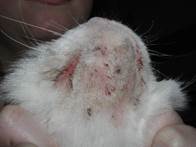
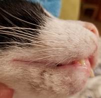
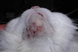
- It usually affects the chin and lower lip but occasionally the upper lips may be involved.
- Comedone is the hallmark of the disease and develops initially.
- Mild cases are asymptomatic and characterized by the presence of comedones and the accumulation of black keratinous debris at the affected sites.

-
- Alopecia, erythema, papules, edema, and crusts can develop in more severe cases.
-
- In some cases, bacterial folliculitis characterized by the presence of pustules may develop.
- Bacteria typically isolated from the lesions include Pasteurella multocita, B-hemolytic Streptococci, and Staphylococcus spp.
- The condition can become quite severe if infected hair follicles rupture (i.e. furunculosis) resulting in extension of the bacterial infection to deep tissues. In these cases, firm nodules or cysts, marked edema, erythema, draining tracts, crusting, and alopecia will develop. Pain and regional lymphadenopathy may be noticed.
-
- Scarring is often a sequel of chronic and deep inflammation.
- Pruritus and pain are uncommon unless there is a secondary deep bacterial folliculitis and furunculosis.
- Occasionally, Malassezia spp. will overgrow at the affected areas. Be suspicious of this condition if the affected sites have a diffuse brownish discoloration.
- Rarely dermatophyte or Demodex spp. infection will be limited to the chin and mimic acne.
Important Facts
- Feline acne usually start at a young age (~ 1 year) and is cyclic during the life of the cat.
- The severity of lesions is variable but, in many cats, the disease is mild and asymptomatic.
- The hallmark sign is the presence of comedones on the chin and lower lip. Lesions can extend to the upper lips and surrounding skin.
- Mild cases are characterized by the presence of comedones and the accumulation of a dark-brown debris giving the cat an appearance of dirty chin.
- Severe cases are associated with secondary bacterial infection and rupture of hair follicles.
- Clinical signs typically associated with severe cases include one or more of the following: papules, nodules, edema, draining tracts, hemorrhagic crusts, severe erythema, and alopecia.
- Severe cases may also be associated with tissue scarring, regional lymphadenopathy, and pain.
- Bacteria typically isolated from secondary infections include Pasteurella multocita, B-hemolytic Streptococci, and Staphylococcus.
- Occasionally Malassezia spp. will overgrow at affected areas.
- Rarely, dermatophyte or Demodex spp. infection will be limited to the chin and mimic acne.
-
Diagnosis
- The history and classical clinical signs are usually diagnostic.
- Cytology is highly indicated to identify secondary Malassezia and bacterial overgrowth/infection.
- If treatment with systemic antibiotic is indicated, bacterial culture and susceptibility will be required to select the appropriate antibiotic.
- Skin scraping, trichogram, and fungal culture should be performed when suspecting of dermatophytosis or demodicosis.
- In cases of marked chin swelling without other lesions, skin biopsy may be needed to rule out eosinophilic granuloma.
Important Facts
- Cytology is highly indicated to identify secondary Malassezia and bacterial overgrowth/infection.
- Skin scraping, trichogram, and fungal culture should be performed when suspecting of dermatophytosis or demodicosis.
- If systemic antibiotic therapy is required, perform bacterial culture and susceptibility to choose the appropriate antibiotic.
- In cases of marked chin edema, biopsy may be needed to rule out eosinophilic granuloma.
-
Treatment
- In mild, asymptomatic cases, treatment usually is not necessary.
- Topical therapy is indicated in all cases requiring treatment to help remove the dark-brown debris and comedones and treat secondary infections.
- Careful clipping of lesional areas will facilitate cleaning and application of topical medications.
- Antimicrobial and keratolytic wipes are often efficacious and convenient options. The affected areas are cleaned once to twice daily until remission or significant improvement is achieved, then the frequency is reduced to 2-3 times weekly.
- If wipes are not effective, antimicrobial and keratolytic/keratoplastic shampoos can be tried.
- Antiseborrheic keratolytic and/or keratoplastic shampoos containing sulfur and salicylic acid can be helpful to remove the dark-brown debris.
- Benzoyl peroxide shampoo can be helpful for its keratolytic, comedolytic or follicular flushing, degreasing, and antibacterial effects. It is important to recommend rinsing the shampoo very well after application to prevent skin irritation.
- Antibacterial shampoos (e.g. chlorhexidine, ethyl lactate) can be used as adjunctive therapy in cases associated with secondary bacterial infection.
- Mupirocin ointment has been reported to be effective in the management of the secondary bacterial infection associated with feline acne.
- Mupirocin has excellent activity against gram-positive cocci, is bactericidal, works well in acid pH, and is not systemically absorbed.
- However, this antibiotic should be selected for severe chronic cases or cases associated with resistant bacterial infection, as it is an important antibiotic used in methicillin-resistant Staphylococcus aureus (MRSA) infections in humans.
- Topical clindamycin or metronidazole ointment can also be tried to treat secondary bacterial infections.
- Ointments or creams containing combined antiinflammatory and antimicrobial agents such as, Otomax® and Mometamax®, may be used when skin inflammation and secondary infections are present.
- Advise owners to use these products per recommendation to avoid undesirable side effects.
- Make sure to recheck the patient in about 2 weeks to evaluate response and determine if the infection has resolved.
- Systemic antibiotic may be required for severe cases with deep bacterial infection that did not respond to topical antibacterial ointments or creams.
- Systemic antibiotic should be selected based on bacterial culture and sensitivity.
- If this is not possible, select first tier antibiotics that are known to be effective against Staphylococcus spp. Examples include first generation cephalosporin (e.g. cephalexin, cefadroxil) and amoxicillin + clavulanic acid.
- In cases with severe inflammation and pruritus, a short course (i.e. 10-14 days) of oral prednisolone (1-2 mg/kg every 24 hour)) may be used.
- Ideally, treat any bacterial infection before starting oral glucocorticoid.
Important Facts
- In mild asymptomatic cases, treatment is usually not necessary.
- Medicated wipes and/or shampoos containing ingredients with one or more of the following properties can be used: keratolytic, keratoplastic, degreasing, comedolytic, follicular flushing, and antibacterial.
- If secondary infection is present, topical antibiotic containing mupirocin, clindamycin, or metronidazole should be tried before considering systemic antibiotic.
- Mupirocin ointment should be saved for severe chronic cases or cases associated with resistant bacterial infection, as it is an important antibiotic used in methicillin-resistant Staphylococcus aureus (MRSA) infections in humans.
- If topical antibiotic therapy was not effective in resolving the secondary bacterial infection, systemic antibiotic should be used and selected based on culture and susceptibility results.
- Make sure to recheck the patient when treating any secondary bacterial infection.
- A short course of oral prednisolone may be needed for cases associate with severe inflammation.
- Ideally, treat any secondary bacterial infection before starting oral glucocorticoids.
References
Jazic E et al. An evaluation of the clinical, cytological, infectious and histopathological features of feline acne. Vet Dermatol 2006; 17(2): 134-140.
Kwochka KW: Primary Keratinization Disorders of Dogs. In: Griffin CE, Kwochka KW, MacDonald JM (eds). Current Veterinary Dermatology. St Louis, Mosby Year Book, 1993; p 176-190.
Kwochka KW: Overview of normal keratinization and cutaneous scaling disorders of dogs. In: Griffin CE, Kwochka KW, MacDonald JM (eds). Current Veterinary Dermatology. St Louis, Mosby Year Book, 1993; p 167-175.
Miller, Griffin and Campbell. Chapter 14. Keratinization defects. In: Muller & Kirk’s Small Animal Dermatology. 7th ed., W.B. Saunders, Missouri, 2013; p 630-646.
White SD et al. Feline acne and results of treatment with mupirocin in an open clinical trial: 25 cases (1994–96).Vet Dermatol 1997; 8(3): 157-164.
