3.10 Canine Ear Margin Dermatosis
-
General Considerations
- Canine ear margin dermatosis is an uncommon idiopathic keratinization defect of the pinna margin.
- Lesions are usually bilateral.
- The cause is unknown.
- Dachshunds appear to be predisposed; however, other breeds can also develop this disease.
Important Facts
- Ear margin dermatosis is an uncommon idiopathic keratinization defect of the pinna margin.
- Lesions are usually bilateral.
- The cause is unknown.
- It can occur in any breed but Dachshunds appear to be predisposed.
-
Clinical Signs
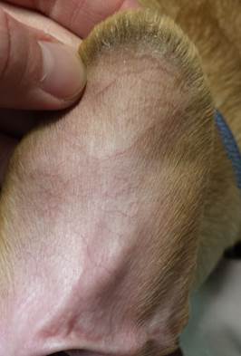
- Kerato-sebaceous material is tightly adhered to the skin surface and especially hair shafts of the concave and convex margins of both pinna forming what is called follicular casts.
-
- Hairs epilate very easily on affected areas and alopecia may develop with time.

-
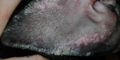
- In severe chronic cases, progression to the development of fissures may occur. Pain is typically present in these cases and fissures often bleed when the animal shakes its head.
- Pruritus is typically absent.
Important Points
- Lesions are characterized by the presence of greasy scales that are tightly adhered to the skin surface and hair shafts of both sides of the margin of the pinna.
- Lesions are typically bilateral.
- In severe cases, the greasy scales form thick layers that affect the whole pinna margin.
- Alopecia and fissures will eventually develop in severe cases.
- Pruritus is usually absent.
-
Differential diagnoses
- If severe pruritus is present, sarcoptic mange should be suspected and multiple skin scrapings and a treatment trial should be performed if scrapings are negative.
- If erythema, crusts, and/or deep erosions to ulcerations are present, vasculitis must be considered.
- Causes of pinna vasculitis include facial or generalized discoid lupus erythematosus, cold agglutinin disease, frostbite, and idiopathic pinna vasculitis.
-
Diagnosis
- The diagnosis of early ear margin dermatosis is based on the patient’s history and presence of the characteristic ear margin lesions.
- Multiple skin scrapings should be performed in cases of moderate to severe pruritus to rule in scabies. If scrapings are negative, perform a parasiticidal trial to rule out scabies.
- A skin biopsy is the most important diagnostic procedure to differentiate the various diseases associated with pinnal vasculitis.
- Additional tests including CBC, chemistry profile, antinuclear antibody (ANA), and Coomb’s should be selected based on the patient’s history and clinical signs.
Important Facts
- The diagnosis of early ear margin dermatosis is based on the patient’s history and clinical signs.
- If pruritus is present, perform multiple skin scrapings to rule in scabies. If scrapings are negative, perform a parasiticidal trial before ruling out scabies.
- If erythema, deep erosions to ulcerations, and/or crusts are present, vasculitis must be considered.
- A skin biopsy should be performed to differentiate the various causes of pinna vasculitis.
- Selection of additional tests should be based on the patient’s history and clinical signs.
-
Treatment
- Ear margin dermatosis is typically not curable but it is a controllable disease.
- Mild forms are usually controlled with topical therapy.
- Topical treatment with antiseborrheic agents (e.g. benzoyl peroxide, tar, sulfur, salicylic acid) will remove the greasy scales (i.e. kerato-sebaceous material)
- The frequency of therapy varies from daily to weekly and should be tailored to each case.
- Topical glucocorticoid creams and/or systemic prednisone (1.1 mg/kg per day) may be indicated to reduce inflammation in severe cases.
- Keep in mind that idiopathic ear margin seborrhea is not associate with clinical inflammation thus, be suspicious of an underlying vasculitis if inflammation is present.
- Pentoxifylline can be tried at the dose of 15-20 mg/kg q 8hrs for 4 to 8 weeks. It has excellent safety margins, improves local oxygenation and has anti-inflammatory properties.
- Surgical removal of affected tissues can be discussed with pet owners in cases refractory to therapy.
- The full list of differential diagnoses should be evaluated before considering such an aggressive therapy.
- Educate the pet owner that lesions may return since we are not treating the cause, which is unknown.
- To reduce the recurrence risk, the tissue should be removed well into the normal portion of the pinnae.
- The surgical procedure will not be effective if the disease is due to an autoimmune disease or vasculitis.
- Affected dogs should not sleep close to forced-air heating ducts, wood stoves, or other dry heat sources as these may exacerbate the condition.
Important Facts
- Ear margin dermatosis cannot be cured and treatment is symptomatic.
- Topical treatment with antiseborrheic agents (e.g. benzoyl peroxide, tar, sulfur, salicylic acid), applied daily to weekly is the mainstay therapy.
- Idiopathic ear margin seborrhea is not associated with clinical inflammation thus, be suspicious of an underlying vasculitis if inflammation is present.
- Pentoxifylline (Trental®) can be tried at the dose of 15-20 mg/kg q 8hrs for 4 to 8 weeks since it is safe, improves blood oxygenation, and has an anti-inflammatory effect.
- Surgical removal of affected tissues can be discussed with pet owners in cases refractory to therapy.
- The full list of differential diagnosis should be considered before instituting such an aggressive therapy.
- The procedure will not be effective if the disease is due to autoimmune disease or vasculitis.
- To reduce the recurrence risk, the tissue should be removed well into the normal portion of the pinnae.
- Affected dogs should not sleep by forced-air heating ducts, wood stoves, or other dry heat sources as these may exacerbate the condition.
References
Kwochka KW: Primary Keratinization Disorders of Dogs. In: Griffin CE, Kwochka KW, MacDonald JM (eds). Current Veterinary Dermatology. St Louis, Mosby Year Book, 1993; p 176-190.
Kwochka KW: Overview of normal keratinization and cutaneous scaling disorders of dogs. In: Griffin CE, Kwochka KW, MacDonald JM (eds). Current Veterinary Dermatology. St Louis, Mosby Year Book, 1993; p 167-175.
Miller, Griffin and Campbell. Chapter 14. Keratinization defects. In: Muller & Kirk’s Small Animal Dermatology. 7th ed., W.B. Saunders, Missouri, 2013; p 630-646.
Power HT, Ihrke PJ. Synthetic retinoids in veterinary dermatology. Vet Clin North Am: Small Anim Pract, Philadelphia, WB Saunders, 1990; p 1525.
