1.3 Hyperadrenocorticism – Feline
Learning Objectives
- Know that hyperadrenocorticism (HAC) is a rare endocrinopathy in cats.
- Learn! Most cases in cats (75% to 85%) are pituitary dependent.
- Learn! About 80% to 90% of cats with HAC have concurrent diabetes mellitus in contrast to only 10% of dogs.
- Know the clinical signs and clinicopathological abnormalities associated with feline HAC.
- Remember! Extremely fragile skin can be a sign of HAC in cats and is reported in about 15% of the cases. The presence of skin fragility in a cat with history and clinical signs characteristic of HAC should increase your index of suspicion for this disease. Skin fragility can also be seen in exogenously administered glucocorticoids and internal diseases in cats.
- Know the endocrine tests used to diagnose feline HAC and to differentiate pituitary- dependent from adrenal-dependent HAC. Pay attention to differences in the tests protocols between dogs and cats. Know how to interpret the results.
- Know that adrenalectomy (unilateral in cases of adrenal-dependent and bilateral in cases of pituitary-dependent HAC) is currently the best treatment option for feline HAC. However, trilostane may show to be a good alternative for long-term management of HAC in cats.
- Know the complications associated with adrenalectomy. Remember! The ability of the owner and clinician to successfully manage the post-surgery adrenal insufficiency is very important in the decision making to decrease the risks of post-surgical complications and death.
- Remember! Medical therapy has had limited success in the management of feline HAC. However, it should be considered as a pre-surgical preparation option and the limited experience with trilostane has shown some good results.
- Know the current options for medical therapy of feline HAC.
-
General Considerations
- Hyperadrenocorticism (HAC) is a rare endocrine disorder in the cat.
- Pituitary-dependent hyperadrenocorticism (PDH) due to a pituitary adenoma or adenocarcinoma occurs in 75% to 80% of the cases and adrenal-dependent hyperadrenocorticism (ADH) occurs in 20% to 25%.
- Iatrogenic HAC is more difficult to induce in the cat than in the dog possibly because cats have lower numbers of high affinity glucocorticoid receptors. Nevertheless, also use great caution when treating cats with glucocorticoids.
- Clinical signs, clinicopathological findings, and radiographic changes are generally less dramatic in cats, which may hinder early diagnosis.
- About 80% to 90% of cats with HAC present with concurrent diabetes mellitus, which contributes to the clinical picture.
Important Facts
- HAC is rare in cats.
- PDH occurs in 75% to 80% and ADH occurs in 20% to 25% of cases.
- About 80% of feline cases are associated with diabetes mellitus compared with 5% to 10% of of dogs with HAC.
-
Clinical Signs
- Signalment:
- Age – The age at disease onset ranges from 6 to 15 years (mean: 11 years).
- Sex – There is no sex predilection although some reports have noted more females affected than males.
- Breed – No breed predilection reported.
- Clinical findings – The percentages below are based on the relatively few cases reported to date.
- Polyuria (PU) and polydipsia (PD) – These signs occur in 80% to 92%% of the cases. They can present before or after the diagnosis of diabetes mellitus. However, in most cats PU and PD develop because of diabetes. The rare cats that do not develop diabetes mellitus, a form of diabetes insipidus (similar to dogs) may explain the PU/PD. However, because cats with HAC rarely have low urine specific gravity, this theory unlikely explains the PU and PD signs. Some non-diabetic cats with HAC and PU/PD also had renal failure and this is a more plausible cause for their PU/PD.
- Distended abdomen – This clinical sign occurs in 64% to 75% of the cases. It occurs because of fat redistribution to the abdominal cavity in addition to muscle wasting and weakness.
- Polyphagia – Increased appetite occurs in about 68% of the cases. This sign can occur before or after the development of diabetes mellitus.
- Non-inflammatory spontaneous alopecia or failure to regrow hair after clipping – This sign develops in 34% to 83% of the cases. Alopecia typically affects one or more of the following sites: trunk, flank, and ventrum.
- Muscle wasting and weakness – These signs occur in 58% to 64% of the cases. They develop due to the catabolic effect of cortisol on proteins.
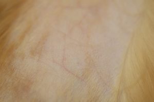
- Thin skin – It occurs in 56% to 83% of the cases and is primarily caused by the catabolic effect of glucocorticoids on protein but also fat and carbohydrate.
- Signalment:
-
-
- Weight gain – Occurs in about 6% to 17% of the cases.
- Weight loss – Occurs in 50% to 52% of the cases. This can be explained in many cases by the presence of diabetes mellitus.
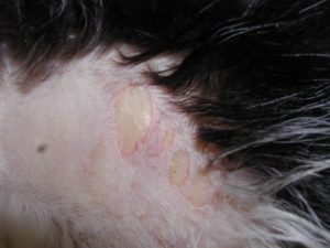
- Fragile skin – This sign occurs in about 15% to 50% of the cases. The skin shows extreme fragility and tears very easily. This clinical sign is unique to cats with HAC but skin fragility also occurs in cats treated with glucocorticoids and with other systemic diseases.
-
-
-
- Skin bruising: Occurs in about 12% to 25% of the cases.
- Many cases show clinical signs about 10 months before the diagnosis.
- A clinical sign that appears to be unique to iatrogenic HAC is the medial curling of the ear tips.
-
Important Facts
- Polyuria, polydipsia, and polyphagia are common signs and they may or may not be associated with diabetes mellitus.
- Alopecia occurs in 34% to 83% of the cases.
- Fragile skin develops in 15% to 50% of cats with HAC.
- Most cases show clinical signs about 10 months before the diagnosis.
-
Diagnosis
- Diagnosis should be based on the following:
- A characteristic history.
- Characteristic clinical signs.
- Supportive clinicopathological abnormalities.
- Diagnostic endocrine test results.
- Clinicopathological abnormalities – They can be supportive but are not specific.
- Hyperglycemia and glucosuria are the most common laboratory abnormalities and reported to occur in about 80% to 90% of the cases. However, a report of six cats with HAC describes diabetes mellitus in only three of the cats.
- Hypercholesterolemia occurs in about 30% of the cases.
- Because cats lack the steroid-induced isoenzyme, elevation in alkaline phosphatase (ALP) is present in only about 7% of cats compared to nearly 90% of dogs with HAC. Alanine aminotransferase (ALT) may also be elevated due to hepatopathy related to diabetes mellitus or HAC.
- ALP and ALT often normalize with adequate regulation of diabetes.
- In contrast to dogs, cats only rarely show low urine specific gravity (i.e. <1.020).
- Lymphopenia, eosinopenia and neutrophilia (i.e. stress leukogram) occur inconsistently in cats with HAC.
- Diagnosis should be based on the following:
Important Facts
- Hyperglycemia and glucosuria are the most common laboratory abnormalities associated with feline HAC.
- Elevation in ALP is present in only about 7% of cats compared with nearly 90% of dogs with HAC.
- In contrast to dogs, cats with HAC rarely show low urine specific gravity (i.e. <1.020).
-
- Endocrine evaluation:
- Screening tests:
- ACTH stimulation test:
- It has a diagnostic accuracy of only about 30-35%. The ACTH stimulation test is clearly less sensitive as a diagnostic test than the dexamethasone suppression test. Most clinicians do not use this test for the diagnosis of feline HAC because of its high frequency of false negative results.
- A post-ACTH cortisol concentration of ≥ 19.0 µg/dl (190 ng/ml) has been considered consistent with HAC.
- Laboratories are encouraged to establish their own reference values to avoid using canine or incorrect reference values.
- Stressed cats and cats with non-adrenal diseases may have an exaggerated response to ACTH.
- The ACTH stimulation test is the only test that can differentiate spontaneous from iatrogenic HAC and is the test used to monitor response to medical therapy.
- Protocol:
- Collect a pre sample and post samples at 1 and 2 hours after the intramuscular administration of 2.0 U/kg of ACTH gel.
- Alternatively, plasma is obtained before and 30 to 60 minutes after 0.125 mg (or 125 µg) of synthetic ACTH is administered intramuscularly per cat.
- ACTH stimulation test:
- Screening tests:
- Endocrine evaluation:
Important Facts
- The ACTH stimulation test has an accuracy of only about 30-35%.
- Some authors recommend not using this test for the diagnosis of feline HAC.
- Stressed cats and cats with nonadrenal diseases may also have an exaggerated response to ACTH.
- The ACTH stimulation test is considered less sensitive as a diagnostic test than the dexamethasone suppression test.
-
-
-
- Low-dose-dexamethasone-suppression test:
- It is considered the most reliable diagnostic test for feline HAC.
- Protocol:
- The test is performed as in dogs but a higher dose of dexamethasone is used.
- Intravenous dexamethasone at the dose of 0.1 mg/kg is more reliable to diagnose feline HAC than the dog’s dose of 0.01 mg/kg; therefore, use this higher dose in cats.
- Determine plasma cortisol levels before and 4 and 8 hours after dexamethasone administration.
- Test interpretation:
- Post-dexamethasone plasma cortisol levels of 1.0 µg/dl (10 ng/ml) or less at 4 and 8 hours are considered normal.
- Concentrations of 0.9 µg/dl to 1.3 µg/dl (9 to 13 ng/ml) at 4 and 8 hours are considered borderline.
- Concentrations of 1.4 µg/dl (14 ng/ml) or higher at both 4 and 8 hours are consistent with a diagnosis of HAC.
- Urine cortisol:creatinine ratio:
- As for the dog, this test has a good negative predictive value. In other words, if it is normal it is unlikely that the patient has HAC; however, if it is elevated it will require further testing as the ratio will also be elevated in nonadrenal disorders.
- It is recommended to collect at least two morning urine samples at home by the owner to avoid in-hospital spurious elevation of cortisol due to stress.
- Low-dose-dexamethasone-suppression test:
-
-
Important Facts
- The low-dose-dexamethasone-suppression test is more accurate than the ACTH stimulation test to diagnose feline HAC.
- Intravenous dexamethasone at the dose of 0.1 mg/kg is more reliable to diagnose feline HAC than the dog’s dose of 0.01 mg/kg, which is 10 times lower.
- Post-dexamethasone cortisol values equal or higher than 1.4 µg/dl (14 ng/ml) at both 4 and 8 hours are consistent with a diagnosis of HAC.
- The cat should be kept quiet and not disturbed during the 8-hour testing period.
- Urine cortisol:creatinine ratio can be used as a screening test. If it is normal, it is unlikely that the cat has HAC. If is abnormal (i.e. elevated), further testing needs to be done.
-
-
- Localizing tests:
- High-dose-dexamethasone-suppression test:
- The protocol is the same used in the low-dose-dexamethasone-suppression test; however, the dose is 1.0 mg/kg given intravenously and only a 4-hour sample is needed. Studies have shown that the results of 4 and 8 hours are similar.
- As with the low-dose test, the cat should be kept quiet and not disturbed during the 4-hour testing period.
- Arbitrarily, suppression of post-dexamethasone plasma cortisol level is defined as a value less than 50% of baseline. Suppression can also be defined as plasma cortisol less than 1.4 µg/dl (14 ng/ml) at 4 hours.
- Most cats with HAC fail to suppress plasma cortisol level below 1.4 µg/dl (14 ng/ml) at 4 or 8 hours after 1.0 mg/kg of dexamethasone.
- Lack of suppression, either on a percentage basis or using an absolute value is consistent with a diagnosis of HAC but should not be thought to confirm a diagnosis of an adrenal tumor because most cats with pituitary-dependent disease do not suppress after a high dose of dexamethasone.
- Diagnosis of adrenal tumor is usually confirmed using abdominal ultrasonography.
- Suppression of serum cortisol level using both percentage decrease and an absolute decrease after intravenous administration of dexamethasone at the dose of 1.0 mg/kg, is documented in a few cats with pituitary-dependent HAC but not in those with adrenal-dependent disease.
- High-dose-dexamethasone-suppression test:
- Localizing tests:
-
Important Facts
- The high dose protocol is the same used in the low-dose-dexamethasone test; however, the dose is 1.0 mg/kg (i.e. 10 times higher).
- Compared to dogs, most cats with HAC do not suppress the post-dexamethasone cortisol levels at 8 hours.
- These cases confirm a diagnosis of HAC but should not be considered as adrenal-dependent HAC.
- Few cats will suppress using percentage (>50% of baseline) and an absolute cortisol decrease (bellow 1.4 µg/dl (14 ng/ml) cortisol value at 8 hour). These cases have PDC.
- Diagnosis of adrenal-dependent HAC is usually suspected or confirmed using abdominal ultrasonography.
-
-
-
- Plasma endogenous ACTH:
- As in dogs, this test can be interpreted reliably only after the diagnosis has been confirmed with acceptable screening test results.
- Endogenous ACTH concentration in healthy cats ranges from 0 to 110 pg/ml.
- Three cats reported in the literature with adrenal-dependent HAC had undetectable to low plasma endogenous ACTH concentrations, and 13 cats diagnosed with pituitary-dependent HAC had ACTH concentrations that ranged from 9 to greater than 1000 pg/ml (mean 281 pg/ml).
- Plasma endogenous ACTH:
-
-
Important Facts
- As in dogs, the plasma endogenous ACTH concentration can only be interpreted reliably after the HAC diagnosis has been confirmed with acceptable screening test results.
- Undetectable to very low plasma ACTH concentration is suggestive of adrenal-dependent HAC and high concentration is indicative of pituitary-dependent HAC.
-
-
- Abdominal ultrasonography:
- Ultrasonography can be extremely reliable in differentiating adrenal-dependent HAC (i.e. unilateral adrenomegaly) from pituitary-dependent HAC (i.e. bilaterally normal-sized to bilaterally enlarged adrenal glands).
- Abdominal ultrasonography:
-
Important Facts
- Pituitary-dependent function tests need to be interpreted in conjunction with historical, clinical and clinicopathologic findings before any conclusions can be drawn.
- No single test is infallible.
-
Treatment
- HAC is very debilitating in cats and the prognosis is guarded because most cats will have concurrent diabetes mellitus, which is typically a challenge to control.
- Although therapy is difficult and the prognosis is guarded, an attempt is usually made to control the disease because of the deteriorating clinical condition of affected cats.
- Adrenalectomy, unilateral in cats with adrenal-dependent HAC or bilateral in cats with pituitary-dependent HAC, has provided the best results in managing cats with HAC.
- The surgical protocol and medical management of cats during and after the procedure are similar to those used in dogs.
- Post-surgical complications contributing to death or euthanasia include sepsis, pancreatitis, thromboembolic phenomena, wound dehiscence, and adrenal insufficiency.
- The longest surviving cats are those that have had an adrenocortical adenoma or carcinoma removed surgically.
- The most important determinant of long-term prognosis in cats undergoing adrenalectomy for pituitary-dependent HAC is the ability of the owner and clinician to successfully manage the iatrogenic adrenal insufficiency.
- Medical therapy has had limited success in the management of feline HAC. However, it should be considered as a pre-surgical preparation option because most cats with HAC are at high risk for surgery because of their diabetes mellitus and fragile skin.
- The non-surgical treatment modalities that could be used are include (i) the adrenolytic drug o,p’-DDD or mitotane (Lysodren®), (ii) drugs that block the cortisol synthesis such as, trilostane, ketoconazole and metyrapone and, (iii) destruction of the pituitary source of ACTH via radiation therapy.
- Lysodren® has been reported to control the disease well in one cat (see reference for additional information– Schwedes CS, 1997). The induction dose was 37.5 mg/kg/day and the maintenance dose was 50 mg/kg per week.
- Trilostane (Vetoryl®) is an inhibitor of 3-ß hydroxysteroid dehydrogenase, an enzyme that participates in the synthesis of cortisol and aldosterone. Its effect is reversible. Very few cats with HAC have been treated with trilostane and the results are variable. However, trilostane has been currently considered the first treatment option for cats with HAC but more experience is needed before any conclusion can be made. Reported protocols include (i) 10-30 mg/cat, orally, every 12-24 hours and, (ii) 1-2 mg/kg every 8-12 hours. Give trilostane with food to improve absorption. The licensed product may be reformulated if doses significantly lower than 5 mg per dose are required. The treatment goal is to improve the clinical signs and the cat’s quality of life. Adverse effects of trilostane include anorexia, lethargy, weight loss, pancreatitis and hypoadrenocorticism. Treatment response should be based primarily on clinical signs but the ACTH stimulation test or urine cortisol:creatinine ratio should be performed to monitor for hypoadrenocorticism. The suggested timing for the ACTH stimulation test is 2-4 hours post trilostane administration. Follow the dog protocol for frequency of ACTH testing. Dose adjustments should be based primarily on the patient’s clinical signs.
Important Facts
- Adrenalectomy, unilateral in cats with adrenal-dependent HAC or bilateral in cats with pituitary-dependent HAC, has provided the best results in managing cats with HAC. However, trilostane, a reversible inhibitor of cortisol (primarily) and aldosterone synthesis, has been currently considered as the first treatment option for feline HAC,
- The longest-surviving cats have been those that have had an adrenocortical adenoma or carcinoma removed surgically.
- The most important determinant of long-term prognosis in cats undergoing adrenalectomy is the ability of the owner and clinician to successfully manage the iatrogenic adrenal insufficiency.
- Medical therapy has had limited success in the management of feline HAC. However, it should be considered as a pre-surgical preparation option. Moreover, when more experience with trilostane is gained, it may show to be the best treatment option for feline HAC.
References
Boland LA, Barrs VR. Peculiarities of feline hyperadrenocorticism: update on diagnosis and treatment. J Feline Med Surg 2017; 19:933-947.
Bugbee A, Renee Rucinsky R, Cazabon S et al. 2023 AAHA selected endocrinopathies of dogs and cats guidelines. J Am Anim Hosp Assoc 2023; 59: 113–135. DOI 10.5326/JAAHA-MS-7368
Feldman EC. Hyperadrenocorticism in cats. In: Canine and Feline Endocrinology. 4th ed. St. Louis: Elsevier; 2015; 452-484.
Hoenig M. Feline hyperadrenocorticism-where are we now? J Feline Med Surg 2002; 4:171-174.
Schwedes CS. Mitotane (o,p’DDD) treatment in a cat with hyperadrenocorticism. J Small Anim Pract 1997; 38:520.
Watson PJ and Herrtage ME. Hyperadrenocorticism in six cats. J Small Anim Pract 1998; 39: 175-184.
