3.5 Schnauzer Comedo Syndrome
-
General Considerations
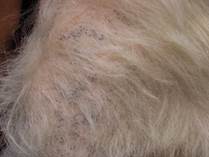
- As you probably know, comedones constitute hair follicles filled with disorganized keratin. These follicles fail to produce hair. Clinically, comedones appear as small black lesions associated with the opening of hair follicles. They are widely known as blackheads.
-
- Schnauzer comedo syndrome is suspected to be a hereditary follicular keratinization defect of miniature schnauzers characterized by multiple comedones on the dorsal midline.
- It is suggested to occur due to a developmental dysplasia of hair follicles.
- Lesions are usually first noticed during grooming in the young adult as small comedones on the dorsum.
- Lesions are asymptomatic unless a secondary infection is present or the affected hair follicles rupture. Then pruritus and/or discomfort may be observed.
Important Facts
- Schnauzer comedo syndrome is suspected to be a hereditary follicular keratinization defect of miniature schnauzers characterized by multiple comedones along the dorsal midline.
- Lesions are asymptomatic unless a secondary infection is present or affected hair follicles rupture (i.e. furunculosis). In this case, pruritus and/or discomfort may be observed.
-
Clinical Signs
- This is a disease of miniature schnauzers.
- Clinical signs may be subtle initially and may not be noticed until worsening.
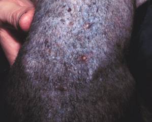
- Papular comedones associated with the openings of hair follicles are present along the dorsal midline and can extend from the neck to the tail. These lesions can become crusty if follicles rupture and/or secondary bacterial infection develops.
- Secondary superficial bacterial folliculitis is common in chronic cases.
-
- Occasionally, the comedonic and inflamed hair follicles will rupture (i.e. furunculosis), which will result in erythematous papules and nodules that will eventually evolve to form draining tracts. Deep pyoderma may occur if the ruptured follicles become infected.
- The condition can be controlled but not cured.
Important Facts
- Papular comedones (blackheads) are present along the dorsal midline and can extend from the neck to the tail.
- Secondary superficial or deep bacterial folliculitis are common, especially in chronic cases.
- When hair follicles eventually rupture (i.e. furunculosis), large papules and nodules will form initially and evolve to draining tracts exuding a sanguineous-purulent exudate. The exudate will dry and form dark crusts.
- The secondary infection leads to hypotrichosis and alopecia with a moth-eaten appearance of the hair coat.
- The condition can be controlled but not cured.
-
Diagnosis
- The presence of comedones along the dorsum of a miniature schnauzer is strongly suggestive of this condition.
- The diagnosis is often based on the characteristic clinical signs and the patient’s signalment. However, in the presence of unusual cases, skin biopsy is recommended to confirm a presumptive diagnosis and rule out diseases in the list of differentials diagnoses.
- Differential diagnoses include demodicosis, hypothyroidism, hyperadrenocorticism, and superficial bacterial folliculitis. Ideally, all suspected cases should be scraped to rule out demodicosis, which is a curable disease. In addition, cytology of crusty and/or draining lesions should be performed to determine if a secondary bacterial infection is present. Tests to diagnose hyperadrenocorticism and hypothyroidism may be indicated if systemic and other cutaneous signs correlating with these diseases are present.
Important Facts
- The presence of comedones in a miniature schnauzer is strongly suggestive of this condition.
- Skin biopsy is recommended in the face of an unusual case.
- Skin scrapings and cytology should be performed in all cases to rule out demodicosis and secondary bacterial folliculitis.
- Diagnostic tests for hyperadrenocorticism and hypothyroidism are indicated if systemic and other cutaneous signs correlating with these diseases are present.
-
Treatment
- This condition is usually controlled; however, it cannot be cured.
- There might be variability of response among different patients. Mild cases may not require therapy.
- Topical therapy involves the periodic use of anti-seborrheic shampoos (e.g. benzoyl peroxide, salicylic acid, sulfur, and tar) once to twice weekly. Frequency may be reduced for maintenance use according to the clinical response.
- Benzoyl peroxide gels are helpful to remove tightly adherent comedones. However, they may be irritating to the skin with frequent use.
- If deep pyoderma is present, systemic antibiotic based on culture and susceptibility may be recommended until complete resolution of the secondary infection. Topical antimicrobials should be considered prior to systemic antibiotic for cases of superficial pyoderma and mild cases of deep pyoderma. If systemic antibiotic is required, combine it with topical therapy.
- Cases refractory to the mentioned topical therapy may benefit from topical and/or systemic retinoids.
Important Facts
- Topical therapy involves the periodic but long-term use of anti-seborrheic shampoos (e.g. benzoyl peroxide, salicylic acid, sulfur, and tar).
- Systemic antibiotic may be indicated for cases complicated by deep pyoderma. If the secondary infection is superficial, consider treating with topical antimicrobials before using systemic antibiotic.
- Topical antimicrobials should be considered for localized/mild cases of deep pyoderma.
- If a decision to use systemic antibiotic was made, make sure to add topical antimicrobials to the treatment regimen and choose the antibiotic based on culture and susceptibility.
References
Kwochka KW: Primary Keratinization Disorders of Dogs. In: Griffin CE, Kwochka KW, MacDonald JM (eds). Current Veterinary Dermatology. St Louis, Mosby Year Book, 1993; p 176-190.
Kwochka KW: Overview of normal keratinization and cutaneous scaling disorders of dogs. In: Griffin CE, Kwochka KW, MacDonald JM (eds). Current Veterinary Dermatology. St Louis, Mosby Year Book, 1993; p 167-175.
Miller, Griffin and Campbell. Chapter 14. Keratinization defects. In: Muller & Kirk’s Small Animal Dermatology. 7th ed., W.B. Saunders, Missouri, 2013; p 630-646.
Power HT, Ihrke PJ. Synthetic retinoids in veterinary dermatology. Vet Clin North Am: Small Anim Pract, Philadelphia, WB Saunders, 1990; p 1525.
