3.3 Ichthyosis – Dogs
-
General Considerations
- Ichthyosis is a rare hereditary keratinization disorder of dogs characterized by mild to severe scaling of the skin and rarely thickening (i.e. hyperkeratosis) of footpads.
- Based on electron microscopy (EM), ichthyosis is subdivided into epidermolytic and nonepidermolytic forms.
- Ichthyosis has been reported in the Norfolk terrier, Jack Russel terrier, golden retriever, Labrador retriever, American bully, American bulldog, Great Dane, German shepherd, Cavalier King Charles spaniel, and the Chihuahua dog. These breeds have been shown to have different forms of ichthyosis with different genetic mutations affecting lipids and structural proteins but to have similar clinical phenotypes characterized by excessive scaling.
- In the Norfolk terrier, epidermolytic ichthyosis is autosomal recessive and caused by a mutation in the gene that encodes for the epidermal keratin 10 (KRT10). It is the only epidermolytic ichthyosis reported in the dog to this date.
- In the Jack Russell terrier, nonepidermolytic ichthyosis is caused by a loss of function mutation in transglutaminase 1 (TGM1). This is an enzyme involved in the formation of the cornified cell envelop, which is an important component of the epidermal barrier.
- In the golden retriever, nonepidermolytic ichthyosis is autosomal recessive and associated in most reported cases with a mutation in PNPLA1. This gene is believed to be involved in the organization and metabolism of lipids within the outer epidermis. A genetic test is currently available and is useful to assess for a carrier state in breeding dogs. Another genetically distinct form of ichthiosis has been reported in golden retrievers. The ABHD5 gene was shown to be mutated in all 14 affected dogs. This gene encodes an acyltransferase enzyme required for the metabolism of lipids. The authors of the paper describing this new genetic mutation in golden retriever with ichthyosis propose to designate this disease as golden retriever ichthiosis type 2.
- In the Labrador retriever, nonepidermolytic ichthyosis is autosomal recessive and associated with an intragenic genomic duplication of the PNPLA1 gene, which is also involved in the golden retriever ichthyosis.
- In the American bulldog and American bully (crossing between the American Staffordshire and the American pit bull terrier), nonepidermolytic ichthyosis is autosomal recessive and caused by a mutation in NIPAL4. This gene encodes for the protein ICHTHYN, which is likely related to lipid metabolism in the epidermis.
- In the Great Dane, nonepidermolytic ichthyosis is autosomal recessive and associated with a mutation in SLC27A4. The SLC27A4 protein has been shown to be indirectly involved in the uptake of fatty acids at the plasma membrane supporting its role in the formation of the skin barrier.
- In the German shepherd, nonepidermolytic ichthyosis is associated with a de novo heterozygous mutation of ASPRV1, which encodes an enzyme known as “skin aspartic protease” (SAS-Pase). This enzyme is involved in profilaggrin-to-filaggrin processing, thus it plays an important role in skin barrier formation. It is important to mention that this mutation was reported in only one German shepherd dog at this time. The mode of transmission was reported to be autosomal dominant.
- In the Chihuahua, nonepidermolytic ichthyosis is associated with a mutation in SDR9C7, which encodes an enzyme involved in the formation of a crucial component of the corneocyte lipid envelope, which is part of the epidermal barrier.
- In Cavalier King Charles spaniel dogs, a congenital and familial form of itchthyosiform dermatosis and keratoconjunctivitis sicca has been documented. A single base-pair deletion in the FAM83H gene was reported in this condition.
- Due to the inheriting nature of canine ichthyosis, breeding of affected dogs is not recommended.
Important Facts
- Ichthyosis is a rare hereditary keratinization defect of dogs characterized by mild to severe scaling of the skin and rarely hyperkeratosis of footpads.
- Ichthyosis has been reported in the Norfolk terrier dog, Jack Russel terrier, golden retriever, Labrador retriever, American bulldog, American bully, Great Dane, German shepherd, Chihuahua, and the Cavalier King Charles dog.
- Genetic mutations have been identified in various breeds and genetic tests are available for some of the affected breeds.
- The only epidermolytic form of ichthyosis is the one reported in the Norfolk terrier dog.
- The mode of transmission in all reported cases, with the exception of the ASPRV1 mutation in the German shepherd dog, is autosomal recessive.
- Breeding of affected dogs is not recommended.
-
Clinical Signs
- The onset of clinical signs is usually before 1 year of age; however, adult-onset cases have been reported.
- Norfolk terriers usually present multifocal areas of pigmented scales with alopecia and roughening of the skin.
-
- Jack Russell terrier ichthyosis is usually severe presenting with large, thick, adherent parchment paper-like scales.
-
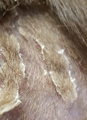
- The disease is generally considered phenotypically mild in both genetic types of the golden retriever ichthyosis. Affected dogs develop large, white to greyish, adherent to loose scales. Scales are present in many parts of the body but they are often prominent along the trunk. Scales tend to be more adhered on ventrum where they are often associated with hyperpigmentation. The disease may have periodic episodes of exacerbation and remission. The clinical signs of the Labrador retriever ichthyosis are similar to the signs of the disease in golden retrievers.
-
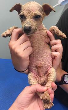
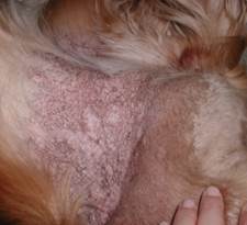
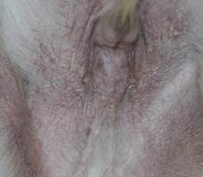
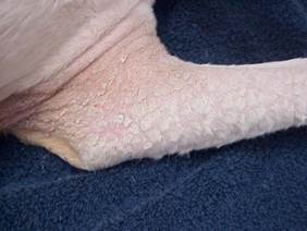
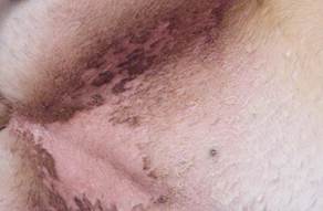
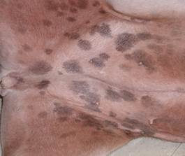
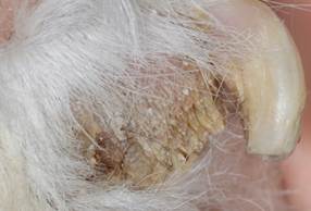
- In the American bulldog and American bully dogs, ichthyosis is a more severe disease than that of golden retrievers. The affected dogs typically develop signs before weaning and present with a scruffy/disheveled hair coat. The non-haired skin is erythematous and associated with adherent brownish scales, giving the affected skin a wrinkled appearance. In the adult dog, the entire abdomen, axilla and inguinal regions have a reddish-brown discoloration. The scales in these areas also acquire a reddish-brown discoloration. Large white to light tan adhered to loose scales are distributed throughout many parts of the body. Footpad hyperkeratosis may be a feature of some adult dogs. Unlike golden retrievers, the skin lesions do not wax or wane.
-
- In more severe forms, particularly in the Jack Russel terrier, American bulldog, and American bully, Malassezia overgrowth and bacterial folliculitis may be present and may be associated with pruritus that can be misinterpreted as an allergic skin disease. The Malassezia overgrowth in these dogs contributes to the brown-reddish discoloration of their skin.
-
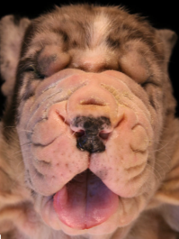
- In Great Danes, the clinical signs are unique and characterized by prominent and thicken skin wrinkles associated with white to yellow scales. Clinical signs in 15 reported cases developed a few days to 5 weeks after birth. Lesions were localized to various parts of the body but were more pronounced around the eyes and muzzle. Non-haired skin sites, especially axillae and legs, appeared dry and had a leathery texture. All affected dogs were reported to be of the black/harlequin color type. The poor prognosis led to all 15 dogs being euthanized.
A. Hoffman, J. Metzger, A. Wohlke, M. Peters, J. Junginger, R. Mischke, O. Distl and M. Hewicker-Trautwein. Congenital ichthyosis in 14 Great Dane puppies with a new presentation. Vet Pathol 2015; DOI: 10.1177/0300985815595516
-
- The only reported case in the German shepherd dog was characterized by generalized and severe grayish and loose to adhered scaling. The affected areas were also mildly erythematous and associated with hypotrichosis to alopecia. Comedones were present on ventral abdomen and inguinal area. The dog was mildly pruritic. Apparently, the disease was present since birth.
Bauer A et al. PLoS Genet 2017; DOI:10.1371/journal.pgen.1006651.
Bauer A et al. PLoS Genet 2017; DOI:10.1371/journal.pgen.1006651.
-
- In the only reported Chihuahua case, clinical signs were characterized by generalized, typically large and thick scales that adhered to the skin surface and hair shafts. The dog had mild to moderate pruritus that resolved with treatment for a secondary Malassezia overgrowth. Lesions were present at the time of adoption, when the dog was 3 months old.
Kiener S et al. Anim Genet 2023; 54:562-565.
-
- In Cavalier King Charles spaniel dogs, the clinical signs include keratoconjuntivitis sicca, a hair coat that is curly and coarsened, scaling associated with hyperpigmentation of the abdomen, hyperkeratosis of footpads, and nail dystrophy. The disease is often known as “dry eye curly coat syndrome” or “congenital keratoconjunctivitis sicca and ichthyosiform dermatosis”. The disease manifests at birth or few days thereafter as small body size and inability to open the eyes. Further clinical signs will develop later.
Important Facts
- Onset of clinical signs typically occurs before 1 year of age.
- The clinical presentation and disease severity will vary according to the genetic mutation and affected breed but, in all forms, the disease hallmark is excessive scaling.
- In some forms, Malassezia overgrowth and bacterial folliculitis can be present and may be associated with pruritus that can be misinterpreted as allergic skin disease. However, keep in mind that some dogs will have an associated allergic condition.
-
Diagnosis
- The clinical diagnosis of ichthyosis is based on the characteristic signs developing at a young age in the breeds reported to be affected. The hallmark sign is generalized excessive scaling and/or footpad hyperkeratosis.
- Differential diagnoses include other keratinization or seborrheic disorders, sebaceous adenitis, allergic dermatitis, parasitic disorders (e.g. cheyletiellosis), hypothyroidism, cutaneous T-cell lymphoma, and zinc-responsive dermatosis.
- Histopathological changes support the clinical diagnosis and help rule out other diseases in the differential diagnosis list. However, abnormal findings are generally non-specific and characterized by moderate to marked basket weave to lamellar, orthokeratotic hyperkeratosis, which often extends to the follicular infundibulum. Dermal inflammation when present is minimal if secondary infection/overgrowth is not present. Some forms of ichthyosis will have unique histopathological changes. For example, in Grate Danes, keratinocytes of the stratum spinosum have vacuoles containing a lightly eosinophilic amorphous material. Amorphous eosinophilic material was also present in the sebaceous glands and the dilated deep portion of hair follicles. This amorphous material stained with Alcian blue and Sudan red III stains in the reported cases indicating an alcianophilic and lipid-rich material.
- Genetic tests to identify the mutated genes are currently available for the golden retriever type 1 and type 2 ichthyosis (UC Davis Veterinary Genetics Laboratory and PennGen, University of Pennsylvania School of Veterinary Medicine and others), and for the American bulldog/American bully (PennGen, University of Pennsylvania School of Veterinary Medicine).
Important Facts
- The clinical diagnosis of ichthyosis is based on the presence of excessive generalized scaling with or without footpad hyperkeratosis, particularly at a young age in the breeds reported to be affected.
- Histopathological changes support the clinical diagnosis and help rule out other diseases in the differential diagnoses list. However, the changes are non-specific.
-
Treatment
- The goal of therapy is to control scaling and improve the stratum corneum barrier function to decrease the adaptive responses (i.e. hyperplasia, hyperkeratosis, inflammation) and to reduce the frequency of infections.
- The treatment regimen must be tailored to each patient and severity of clinical signs and requires good owner compliance to be effective.
- Topical therapy is the mainstay treatment for canine ichthyosis and includes (i) keratolytic agents to help reduce the excessive scaling, and, (ii) moisturizers and emollients to improve the altered function of the skin barrier (i.e. reduce transepidermal water loss). Examples include warm water soaks to help remove scales, anti-seborrheic/keratolytic shampoos (e.g. sulfur, salicylic acid), anti-seborrheic gels for locally severe lesions (e.g. tretinoin, Retin-A®, and urea and salicylic acid containing products), and humectants. Ceramides and essential fatty acids-enriched products can also be beneficial. A study showed that topical treatment of Jack Russell terries ichthyosis with omega-O-acylceramide resulted in decreased transepidermal water loss and normalization of the skin pH of affected dogs.
- Topical antimicrobials (e.g. chlorhexidine and miconazole) can be used for prevention and treatment of secondary bacterial and/or yeast overgrowth/infections.
- The frequency of the topical therapy should be tailored to the severity of clinical signs and then tapered based on the clinical response.
- Care should be exercised to avoid further damage to the skin barrier with harsh chemicals.
- High doses of oral omega-3 and omega-6 fatty acids may also be beneficial.
-
Prognosis
- Ichthyosis long-term prognosis is considered guarded to poor not because scale formation cannot be controlled but because continual therapy will be needed for the entire life of the patient because there is no cure for this disease. In additional, the quality of life of severe cases, such as the Great Dane disorder, is poor.
Important Facts
- Treatment will be needed for the entire life of the patient and it is directed at controlling the scaling.
- Topical therapy includes anti-seborrheic shampoos and gels to help remove the scales, and moisturizers or humectants to hydrate the skin.
References
Bauer A, Waluk DP, Galichet A et al. A de novo variant in the ASPRV1 gene in a dog with ichthyosis. PLoS Genet 2017; DOI:10.1371/journal.pgen.1006651.
Briand A, Cochet-Faivre N, Reyes-Gomez E et al. NIPAL4 deletion identified in an American bully with autosomal recessive congenital ichthyosis and response to topical therapy. Vet Med Sci 2019; DOI: 10.1002/vms3.149.
Credille KM, Barnhart KF, Minor JS et al. Mild recessive epidermolytic hyperkeratosis associated with a novel keratin 10 donor splice-site mutation in a family of Norfolk terrier dogs. Br J Dermatol 2005; 153:51-58.
Forman OP, Penderis J, Hartley C et al. Parallel mapping and simultaneous sequencing reveals deletions in BCAN and FAM83H associated with discrete inherited disorders in a domestic dog breed. PLoS Genet 2012; 8(1):doi:10.1371/journal.pgen.1002462.
Hoffmann A, Metzger J, Wihlke A et al. Congenital ichthyosis in 14 Great Dane puppies with a new presentation. Vet Pathol 2015; DOI: 10:1177/0300985815595516.
Kiener S, Wiener DJ, Hopke K, et al. ABHD5 frameshift deletion in golden retrievers with ichthyosis. G3 (Bethesda) 2022; 12(2), jkab397.
Kiener S, Castilla E, Jagannathan V et al. SDR9C7 missense variant in a Chihuahua with non-epidermolytic ichthyosis. Anim Genet 2023; 54:562-565.
Mauldin EA. Canine ichthyosis and related disorders of cornification. Vet Clin North Am: Small Anim Pract 2013; 43(1): 89-97.
Mauldin EA et al. Autosomal recessive congenital ichthyosis in American Bulldogs is associated with NIPAL4 (ICHTHYIN) deficiency. Vet Pathol 2015; 52(4):654-662.
Mauldin E, Bradley C, Casal M et al. Skin barrier, phenotypic and genotypic characterisation of autosomal recessive ichthyosis in TGM1-deficient Jack Russell Terriers and response to topical ceramide. Vet Dermatol 2023; DOI: 10.1111/vde.13285.
Metzger J, Wohlke A, Mischke R et al. A novel SLC27A4 splice acceptor site mutation in Great Danes with ichthyosis. PLoS One 2015, DOI:10.1371/journal.pone.0141514.
Miller, Griffin and Campbell. Chapter 12. Congenital and hereditary defects. In: Muller & Kirk’s Small Animal Dermatology. 7th ed., W.B. Saunders, Missouri, 2013; p 573-617.
Nett CS et al. Epidermal dysplasia and Malassezia infection in two West Highland WhiteTerrier siblings: an inherited skin disorder or reaction to severe Malassezia infection? Vet Dermatol 2001; 12(5): 285-290.
Power HT, Ihrke PJ. Synthetic retinoids in veterinary dermatology. Vet Clin North Am: Small Anim Pract, Philadelphia, WB Saunders, 1990; p 1525.
Rietmann SJ, Clegg JL, Jagannathan V et al. Intragenic PNPLA1 duplication in Labrador retrievers with
nonepidermolytic ichthyosis. Vet Dermatol 2025; DOI: 10.1111/vde.13341.
