3.4 Familial Nasal Parakeratosis and Footpad Hyperkeratosis – Dogs
-
General Considerations
- Hereditary nasal parakeratosis and footpad hyperkeratosis are hereditary keratinization disorders characterized by an accumulation of tightly adhered keratin on the nasal planum or footpads.
- Hereditary nasal parakeratosis without footpad involvement:
- An autosomal recessive nasal parakeratosis caused by a mutation in the SUV39H2 gene has been reported in Labrador retrievers and their crosses and in Greyhound dogs.
- Hereditary footpad hyperkeratosis without nasal planum involvement:
- A monogenic autosomal recessive footpad hyperkeratosis has been reported in Irish terrier and Kromfohrländer dogs. A missense mutation in the FAM83G gene has been identified in these breeds. The protein encoded by this gene has been reported to be involved in BMP and WNT signaling pathways, which are important in pre and postnatal hair follicle morphogenesis and differentiation. Irregular hair morphology was associated with the syndromic phenotype of one of the Kromfohrländer dogs in a report.
- Hereditary footpad hyperkeratosis reported in the Dogue de Bordeaux is associated with a KRT16 frameshift variant with focal nonepidermolytic footpad hyperkeratosis.
- A DSG1 frameshift variant was reported in a Rottweiler dog with footpad hyperkeratosis.
Important Facts
- Familial nasal parakeratosis and footpad hyperkeratosis are hereditary keratinization disorders characterized by an accumulation of tightly adhered keratin on the nasal planum or footpads.
- A hereditary nasal parakeratosis, without footpad involvement, has been reported in Labrador retrievers and their crosses and in Greyhound dogs.
- Hereditary footpad hyperkeratosis, without nasal involvement, has been reported in several dog breeds including the Irish terrier, Kromfohrländer, Dogue de Bordeaux and Rottweiler.
-
Clinical Signs
- Hereditary nasal or footpad hyperkeratosis usually occurs in young dogs by 12 months of age.
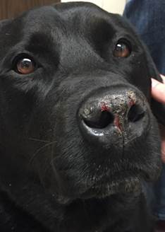
- Hereditary nasal parakeratosis is characterized by the presence of adhered crusts, scales, and fissures at the nasal planum. Lesions typically develop at a young age.
- This inherited condition has only been reported in the Labrador retriever and Greyhound dog.
- Planum nasal lesions may bleed if traumatized.
-
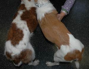
- Hereditary footpad hyperkeratosis has been reported in the Irish terrier, Kromfohrländer, Dogue de Bordeaux, and Rottweiler breeds.
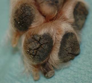
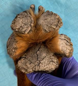
- Lesions typically develop before or at one year of age and are characterized by various degrees of thickening and hyperkeratosis of the footpads.
- Lesions can be focal or diffuse. In severe cases, prominent keratinous proliferations and fissures will develop.
- All footpads are usually involved and the metacarpal/metatarsal pads can also be affected (reported in the Rottweiler dog).
- Pain when walking is often associated with this disease.
- In addition to the footpad lesions, the nail plates are very hard and appear to grow faster in the Irish terrier and Kromfohrländer dogs with the FAM83G mutation. Moreover, one of the 13 reported Kromfohrländer cases also had an abnormal hair coat.
- Hereditary footpad hyperkeratosis has been reported in the Irish terrier, Kromfohrländer, Dogue de Bordeaux, and Rottweiler breeds.
Drogemuller M. et al. PLoS Genet; doi.org/10.1371/journal.pgen.1004370.
Backel KA et al. Genes 2020; 11: 469.
Important Facts
- Hereditary nasal parakeratosis or footpad hyperkeratosis usually occurs in young dogs by 12 months of age.
- Lesions may be focal or diffuse and characterized by tightly adherent thick accumulations of keratin on the nasal planum or footpads, according to the breed predisposition.
- The affected areas may be accompanied by fissures, erosions, and ulcers.
- Nasal lesions may bleed if traumatized.
- Irregular hair coat appearance was reported in one of the 13 Kromfohrlände dogs diagnosed with hereditary footpad hyperkeratosis.
- Pain when walking is usually associated with hereditary footpad hyperkeratosis.
-
Diagnosis
- Compatible signalment, history, and clinical signs are important components of the diagnostic equation.
- If other clinical signs, either systemic or dermatologic are present, a thorough workup to identify an underlying etiology is necessary.
- All diseases that cause lesions on the nasal planum or footpads should be considered as differential diagnoses and include canine distemper, pemphigus foliaceus, pemphigus erythematosus, facial discoid lupus erythematosus, zinc responsive dermatosis, and superficial necrolytic dermatitis. Keep in mind that most of these diseases develop lesions in other parts of the body.
- Skin biopsy of the affected area(s) for histopathology is an important diagnostic test, to confirm the presumptive clinical diagnosis or rule out diseases in the list of differential diagnoses.
Important Facts
- If nasal planum lesions start by 12 months of age in Labrador retrievers and their crosses or Greyhounds, hereditary nasal parakeratosis should be suspected.
- If footpad lesions start by 12 months of age in Irish terriers, Kromfohrländers, Dogue de Bordeaux, and Rottweilers, hereditary footpad hyperkeratosis should be suspected.
- Keep in mind that Irish terriers and Kromfohrländers may also have an abnormal hair coat.
- Histopathology of affected areas can confirm the clinical diagnosis or can rule out other diseases affecting the nasal planum or footpads.
-
Treatment
- Nasal and digital hyperkeratotic lesions are non-curable and treatment is long-term and symptomatic.
- In mild cases, it is mostly a cosmetic disease and may not require therapy.
- Treatment may be required in severe cases and when dogs show discomfort.
- The excess keratin needs to be softened and removed by trimming.
- Initially, hydrating the lesions with water soaks or wet dressings may be sufficient to allow the physical removal of excessive keratin.
- In many cases, a topical keratolytic agent in a gel formulation is applied every 12 hours initially and then as needed to keep the condition under control. Examples: KeraSolv Gel® (salicylic acid, lactic acid, urea), topical 0.01% or 0.025% tretinoin gel (Retin-A®).
- If fissures are present, topical corticosteroid and topical antibiotic may be needed to control secondary inflammation and infection.
- Continued application of hydrating agents, such as propylene glycol, petroleum jelly, and salicylic acid, or products containing essential oils, are usually required at varying frequencies.
Important Facts
- Nasal parakeratosis and digital hyperkeratotic lesions are non-curable diseases and treatment is long-term and symptomatic.
- Initially, hydrating the lesions with water soaks or wet dressings may be sufficient to allow the physical removal of excessive keratin.
- A topical keratolytic agent should be initially applied every 12 hours and then as needed to keep the condition under control.
- Areas of fissures and erosions/ulcerations are often complicated by secondary bacterial infection and should be topically treated with corticosteroid and antibiotic ointments or creams. Make sure to perform cytology of affected areas to determine if infection is present.
- Continued application of hydrating agents, such as propylene glycol, petroleum jelly, and salicylic acid, or products containing essential oils, are usually required at varying frequencies.
References
Kwochka KW: Primary Keratinization Disorders of Dogs. In: Griffin CE, Kwochka KW, MacDonald JM (eds). Current Veterinary Dermatology. St Louis, Mosby Year Book, 1993; p 176-190.
Kwochka KW: Overview of normal keratinization and cutaneous scaling disorders of dogs. In: Griffin CE, Kwochka KW, MacDonald JM (eds). Current Veterinary Dermatology. St Louis, Mosby Year Book, 1993; p 167-175.
Miller, Griffin and Campbell. Chapter 14. Keratinization defects. In: Muller & Kirk’s Small Animal Dermatology. 7th ed., W.B. Saunders, Missouri, 2013; p 630-646.
Power HT, Ihrke PJ. Synthetic retinoids in veterinary dermatology. Vet Clin North Am: Small Anim Pract, Philadelphia, WB Saunders, 1990; p 1525.
