2.14 Epidermolysis Bulhosa Acquisita – Dogs
-
General Considerations
- Epidermolysis bulhosa acquisita (EBA) is a rare autoimmune subepidermal blistering disease reported in dogs but not cats.
- It is the second most common autoimmune subepidermal blistering diseases of dogs, following mucous membrane pemphigoid.
- It is a severe disease that affects haired skin, mucosae, and mucocutaneous junctions.
- Autoantibodies (mostly IgG) are generally directed against collagen VII, an adhesion molecule and main component of anchoring fibrils.
Important Facts
- Epidermolysis bullosa acquisita, although rare, is the second most common subepidermal blistering autoimmune disease reported in dogs.
- The disease is severe and affects haired skin, mucosae, and mucocutaneous junctions.
- Lesions are deep because autoantigens target collagen VII, which is a protein component of the basement membrane zone located deeply in the dermal-epidermal junction.
-
Clinical Signs
- Signalment:
- Epidermolysis bullosa acquisita generally affects young dogs, median age 1.2 years (range: 4 months to 8 years). Almost half of the dogs develop lesions before 1 year of age.
- Male dogs are 2.3 times more likely to develop the disease than female dogs.
- The Great Dane appears to be predisposed, representing 58% of the reported cases. German shorthair pointer is the second most commonly affected breed, accounting for 13% of the cases.
- A large breed representation and young age at disease onset, strongly suggest a genetic predisposition for disease development.
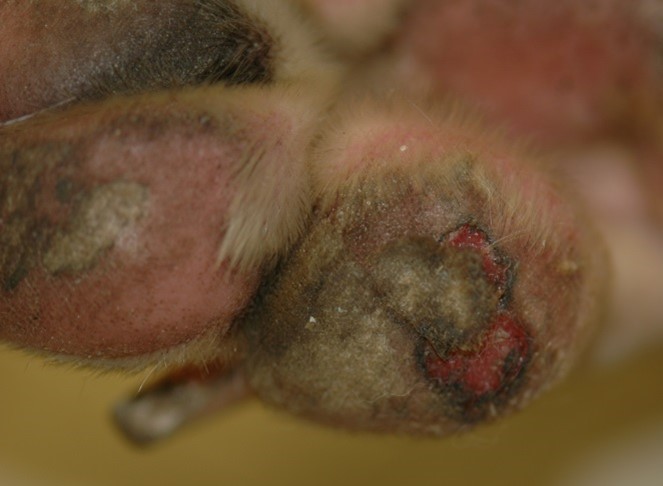
- Lesions:
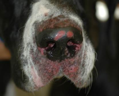
- Lesions typically start as erythematous macules/patches, erythematous papules, and urticarial plaques that evolve to form the characteristic tense vesicles and bullae, which can be hemorrhagic. These lesions are transient and rarely seen.
- Signalment:

-
-
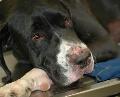
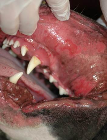
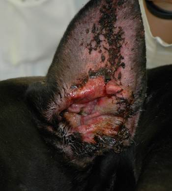
- Once vesicles and/or bullae rupture, they are replaced with deep erosions, ulcers, crusts, alopecia, and scarring if the disease becomes chronic.
-
-
-
- Systemic signs of pyrexia, depression, lethargy, anorexia, and lymphadenopathy are common. Anemia and thrombocytopenia may be seen. Pruritus has been reported in 38% of the cases and pain in 86%.
-
-
- Lesion distribution:
- Most commonly affected areas include the oral cavity (92%), lips (79%), concave pinnae (79%), and the haired skin such as muzzle, axillae, and inguinal areas. In contrast to other autoimmune subepidermal blistering diseases the footpads (sloughing) are often affected (71%). Epidermolysis bullosa acquista can be generalized.
- Lesion distribution:
Important Facts
- Great Dane dogs and German shorthair dogs are overrepresented.
- More than half of the dogs develop the disease before 1 year of age.
- The male to female ratio is 2.3.
- Primary lesions (i.e. erythematous macules, erythematoys plaques, tense vesicles, and bullae) and/or secondary lesions (i.e. deep erosions, ulcers, crusts, alopecia, and scarring) are present at the mucosae and/or mucocutaneous junctions and/or haired skin (i.e. concave pinnae, muzzle, axilla, inguinal area, and footpads).
- Oral cavity and footpads are typically severely affected.
- Systemic signs are frequently present.
-
Diagnosis
- Differential diagnoses:
- All diseases that cause predominantly vesicles and ulcers and that oral mucosae and footpads may be involved including pemphigus vulgaris, bullous pemphigoid, erythema multiforme, toxic epidermal necrolysis, drug reaction, and cutaneous T-cell lymphoma.
- Cytology:
- Direct smears prepared of the exudate collected from intact vesicles or from the exudate obtained from an ulcer, deep erosion, or underneath crusts. These samples will not reveal acantholytic keratinocytes.
- Cytology is not diagnostic but is important to investigate the presence of secondary infections.
- Histopathology:
- Subepidermal cleft and acellular vesicle formation (may contain fibrin, erythrocytes, variable numbers of neutrophils +/- eosinophils). Subepidermal vacuolation may be present just below the basement membrane zone. Superficial dermis may have neutrophilic infiltrate and microabscesses, +/- eosinophils.
- Biopsy an intact vesicle/bulla if possible; otherwise, sample the active border of an erosion or ulceration.
- Aim to submit the biopsy samples to a pathologist with interest in dermatohistopatholgy. Make sure to include a detailed history and clinical signs description. If possible, also submit good quality photos.
- Ask the pathologist to perform periodic-acid Schiff (PAS) staining of the sample or immunohistochemistry for collagen IV. The PAS stain will color the lamina densa of the basement membrane zone magenta. In the case of epidermolysis bullosa acquisita, the roof of the vesicle/bulla should stain. This test will help differentiate epidermolysis bullosa acquisita cases from other autoimmune subepidermal blistering diseases, which stain the floor of the cleft/vesicle. Keep in mind that the sensitivity of this test is low.
- Subepidermal cleft and acellular vesicle formation (may contain fibrin, erythrocytes, variable numbers of neutrophils +/- eosinophils). Subepidermal vacuolation may be present just below the basement membrane zone. Superficial dermis may have neutrophilic infiltrate and microabscesses, +/- eosinophils.
- Direct immunofluorescence or immunohistochemistry:
- Presence of linear deposition of immunoglobulins along the basement membrane zone is diagnostic.
- Remember! Skin biopsy is submitted for these tests and samples should be fixed in Michel’s fixative for immunofluorescence and formalin for immunohistochemistry.
- Direct immunofluorescence or immunohistochemistry is positive in about 80% of the cases.
- Indirect immunofluorescence:
- Best results with salt-split canine gingiva as substrate. It is positive in 92% of the cases tested.
- Most commonly, the fluorescence is present on the dermal side of the split. However, in rare cases the fluorescence is present on both sides of the split.
- Serum sample is used for this test.
- CBC, chemistry profile, urinalysis:
- Used as baseline for monitoring potential side effects associated with therapy.
- Differential diagnoses:
Important Facts
- The patient’s history and clinical signs are helpful diagnostic information.
- Oral cavity and footpads are typically severely affected.
- Histopathology is the most important diagnostic test and reveals subepidermal cleft and acellular vesicle formation. However, the vesicle may contain erythrocytes, variable numbers of neutrophils, +/- eosinophils.
- Indirect immunofluorescence using salt-split canine gingiva is positive in 92% of the tested cases; however, it is not widely available limiting its use in clinical practice.
-
Treatment
- A complete disease remission occurred within 203 months in all dogs treated with one or more of the following treatment options: (See “Therapy for Autoimmune Diseases” for dose and specifics on treatment regimens).
- Glucocorticoids.
- Colchicine.
- Azathioprine.
- Mycophenolate mofetil.
- Chlorambucil.
- Human intravenous immunoglobulin (IVIG) might be a good option for refractory cases.
- A complete disease remission occurred within 203 months in all dogs treated with one or more of the following treatment options: (See “Therapy for Autoimmune Diseases” for dose and specifics on treatment regimens).
-
Prognosis
- The prognosis is good to guarded.
- In almost half of the reported dogs, immunosuppressive drugs could be discontinued without recurrence of clinical signs.
References
Bizikova P, Linder KE, Anderson JG. Erosive and ulcerative stomatitis in dogs and cats: which immune-mediated diseases to consider? J Am Vet Med Assoc 2023; doi.org/10.2460/javma.22.12.0573.
Bizikova P, Olivry T, Linder K et al. Spontaneous autoimmune subepidermal blistering diseases in animals: a comprehensive review. BMC Vet Res 2023; https://doi.org/10.1186/s12917-023-03597-1.
Medleau L, Hnilica KA. Chapter 8. Autoimmune and immune-mediated skin disorders. In: Small Animal Dermatology: A color Atlas and Therapeutic Guide 2006. 2nd ed. W.B. Saunders, Missouri, 189-227.
Miller, Griffin and Campbell. Chapter 9. Autoimmune and immune-mediated dermatoses. In: Muller & Kirk’s Small Animal Dermatology 2013. 7th ed., W.B. Saunders, Missouri; 432-462.
Olivry T. Auto-immune skin disease in animals: time to reclassify and review after 40 years. BMC Vet Res 2018; https://doi.org/10.1186/s12917-018-1477-1.
Olivry T, Chan LS. Autoimmune blistering dermatoses in domestic animals. Clin Dermatol 2001; 19(6):750-760.
Olivry T., Jackson HA. Diagnosing new autoimmune blistering skin diseases of dogs and cats. Clin Tech Small Anim Pract 2001; 16: 225-229.
