3.1 Disorders of Keratinization – Learning Objectives, General Consideration and Classification
Learning Objectives
- Remember! Keratinization disorders is a term used to describe clinical signs associated not only with abnormalities in the process of formation of the epidermis (epidermogenesis) and, particularly stratum corneum (keratinization), but also with abnormalities in apocrine and sebaceous gland functions, intercellular lipid formation, cell cohesion, and cell desquamation.
- Know! There are relatively few primary feline keratinization disorders. Only primary seborrhea and feline acne are reported in cats.
- Keep in mind! Scaling is a clinical sign always present.
- Know! Secondary Malassezia and bacterial infections are frequently observed in many keratinization disorders due to alterations in the surface microenvironment and alterations in the stratum corneum, which are important components of the skin barrier.
- Keep in mind! The diagnostic challenge when managing a scaling disorder is to determine if the clinical signs are secondary to an underlying dermatosis or are the result of a primary keratinization defect. Most cases that you will be dealing with in practice will be secondary to an underlying skin disorder.
- Know! Keratinization disorders can be classified based on two factors: clinical signs and etiology. Based on clinical signs, the keratinization disorders can be classified in seborrhea sicca, seborrhea oleosa, seborrheic dermatitis, and seborrheic otitis externa. Based on etiology the disorder of keratinization can be classified in primary and secondary.
- Know how to differentiate clinically each type of seborrhea.
- Remember! Secondary Malassezia and bacterial infections can occur with seborrhea oleosa or sicca but are most common with seborrhea oleosa.
- Know! Secondary keratinization disorders typically occur with most types of skin diseases; therefore, is seen much more often than primary keratinization disorders. Scaling with or without excessive sebum secretion is observed in areas affected by the underlying dermatosis.
- Know! Primary keratinization disorders can be hereditary, idiopathic, or nutritional.
- Know! Hereditary keratinization disorders include follicular dysplasia/dystrophy, epidermal dysplasia, ichthyosis, lichenoid-psoriasiform dermatosis, primary idiopathic seborrhea, Schnauzer comedo syndrome, and nasal and footpad hyperkeratosis.
- Know! Idiopathic keratinization disorders include acne, ear margin dermatosis, and nasodigital hyperkeratosis.
- Know well the clinical presentation, diagnosis, and treatment of the following: primary idiopathic seborrhea, Schnauzer comedo syndrome, acne, ear margin dermatosis, and zinc responsive dermatosis. You will be seeing these conditions!
-
General Considerations
- Keratinization disorder is a term used to describe the clinical signs associated with abnormalities in the process of epidermopoiesis, keratinization, apocrine or sebaceous gland function, intercellular lipid formation, cell cohesion, and cell desquamation.
- Keratinization disorders are classified into primary and secondary causes. In primary causes, the excessive scaling is due to a direct defect in one or more steps in the formation of the stratum corneum. Known defects include mutations in genes which encode the structural proteins that form the corneocyte (e.g. transglutaminases), or enzymes involved in lipid formation or lipid transport. Secondary disorders are those where excessive scaling develops due to an underlying condition (e.g. allergic skin diseases, sarcoptic mange, hypothyroidism, epitheliotropic lymphoma, etc.). Most keratinization disorders arise from secondary causes.
- Excessive scaling is the characteristic clinical sign of keratinization disorders.
- Alterations in glandular secretions will result in either excessive sebum production (seborrhea oleosa) or deficient sebum secretion (seborrhea sicca).
- Alterations in hair follicle function will result in follicular casts (excessive keratin produced in the follicle canal adheres to and surrounds the hair shafts) and comedones (black heads – the upper portion of the hair follicle (infundibulum) is dilated and filled with keratin).
- Malassezia and/or bacterial overgrowth and bacterial infection are frequently observed in many keratinization disorders due to alterations in the surface microenvironment, the stratum corneum, and other components of the skin barrier.
- The diagnostic challenge when managing a scaling disorder is to determine if clinical signs are secondary to an underlying dermatosis or are the result of a primary keratinization defect.
- Therapeutic and prognostic factors for keratinization disorders are variable.
- In general, if the keratinization disorder is secondary to an underlying disease and the underlying dermatosis is identified and treated successfully, the keratinization disorder has an excellent prognosis for complete cure and/or control.
- If there is a primary keratinization defect, life-long therapy is usually required.
Important Facts
- Keratinization disorders are classified into primary and secondary causes.
- Most keratinization disorders arise from secondary causes.
- Excessive scaling is the hallmark sign of any disorder of keratinization.
- Alterations in glandular function can also occur resulting in seborrhea oleosa, if excessive sebum secretion accompanies the scaling or seborrhea sicca if decreased sebum secretion is associated with scaling.
- Comedones (i.e. black heads) and follicular casts (i.e. keratin surrounding groups of hair shafts) are also signs of disorders of keratinization.
- Secondary pyoderma and Malassezia and/or bacterial overgrowth are common complications of keratinization disorders!
- In general, if the keratinization disorder is secondary to an underlying disease and the underlying dermatosis is identified and well controlled, the keratinization disorder has an excellent prognosis for complete cure and/or control.
- If there is a primary keratinization defect, life-long therapy is usually required.
-
Classification
- Keratinization disorders can be classified based on:
- Clinical signs.
- Etiology: primary and secondary causes.
- Classification based on clinical signs:
- Seborrhea sicca:
- It is characterized by white and loose scales. These signs are typical of the “dry” form. The hair coat is typically dull and is not oily.
- Odor is usually not present.
- Seborrhea oleosa:
- It is characterized by greasy yellow/brown scales. This color results from sebum accumulation on the scale surface. The hair coat is oily. Scales adhere to hairs.
- “Rancid fat” odor may be present.
- Secondary Malassezia and bacterial overgrowth or infection are common.
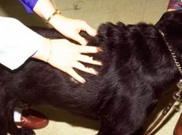
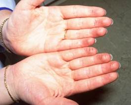
- In some cases, you need to touch the animal to differentiate a seborrhea oleosa from a seborrhea sicca. If the seborrhea is oleosa, your fingers become coated with a thin layer of oil.
- Seborrhea sicca:
- Keratinization disorders can be classified based on:
-
-
- Seborrheic dermatitis:
- It is the inflammatory form.
- Scaling (with or without greasiness) and clinical evidence of inflammation characterize this form.
- Multifocal hyperkeratotic plaques may be present.
- Investigate the presence of Malassezia overgrowth and bacterial overgrowth/infection because they are common problems in greasy skin.
- Ceruminous otitis externa:
- Excessive waxy debris in the ear canal.
- Odor and erythema are typically present.
- Secondary Malassezia and bacterial overgrowth in the external ear canals causing inflammation are common.
- The external ear canal is a modified extension of the skin; therefore, seborrheic otitis externa is common in seborrheic dermatitis especially the cases associated with atopic dermatitis.
- REMEMBER! Secondary Malassezia and bacterial overgrowth and bacterial infection are common.
- Seborrheic dermatitis:
-
Important Facts
- Based on clinical signs, keratinization disorders can be classified in seborrhea sicca, seborrhea oleosa, seborrheic dermatitis, and seborrheic otitis externa.
- Seborrhea sicca is characterized by white and loose scales. The coat is dry and dull but not oily.
- Seborrhea oleosa is characterized by greasy yellow-brown scales which can adhere to the hair, by oily coat, and “rancid fat” odor.
- Seborrheic dermatitis is characterized by scaling and inflammation.
- Secondary Malassezia overgrowth and bacterial infection or overgrowth are commonly present.
-
- Classification based on etiology:
- Secondary keratinization disorders:
- External and internal causes with effect on keratinization and/or sebum production will lead to the keratinization disorder.
- Inflammation associated with the primary disease and/or self-trauma result in the keratinization disorder. Therefore, scaling with or without excessive sebum secretion is observed in areas affected by the underlying disease.
- Secondary keratinization disorders may occur with most types of skin diseases including:
- Parasitic: cheyletiellosis, demodicosis, scabies.
- Allergic: canine atopic dermatitis, feline atopic skin syndrome, food allergy, fleabite allergy, contact allergy.
- Endocrine: hypothyroidism, hyperadrenocorticism, sex hormone dermatoses.
- Pyoderma: epidermal collarettes associated with superficial pyodermas.
- Dermatophytosis
- Neoplastic: epitheliotropic T-cell lymphoma
- Secondary keratinization disorders:
- Classification based on etiology:
Important Facts
- Secondary keratinization disorders may occur with most types of skin diseases and, as a result, they are seen much more often than primary keratinization disorders.
- Scaling, with or without excessive sebum secretion, is observed in areas affected by the underlying dermatosis.
-
-
- Primary keratinization disorders:
- The diseases classified as primary keratinization defects are either hereditary, idiopathic, or nutritional in origin.
- Hereditary (confirmed or presumed) keratinization disorders:
- Ichthyosis.
- Familial nasal and footpad hyperkeratosis.
- Schnauzer comedo syndrome.
- Idiopathic keratinization disorders:
- Primary.
- Acne.
- Ear margin seborrhea.
- Nasodigital hyperkeratosis.
- Nutritional keratinization disorders:
- Zinc responsive dermatosis
- Fatty acid deficiency.
- Primary keratinization disorders:
-
Important Facts
- Primary keratinization disorders can be hereditary, idiopathic or nutritional.
- We will limit our discussion to primary keratinization disorders.
