1.11 Canine Follicular Dysplasias
-
General Considerations
- In general, follicular dysplasia is a breed specific condition associated with alopecia and/or various coat changes such as dry, frizzy and poor hair coats, changes in coat color, loss of primary hairs, and curly or twisted hair shafts.
- Clinical signs may vary to some extent according to the breed.
- It is possible that we are dealing with different diseases and the grouping under “follicular dysplasias” based on histopathological findings may not be ideal.
- The diseases we will discuss under the category of follicular dysplasias include color dilution alopecia, black hair follicular dysplasia, and non-color-linked follicular dysplasias.
-
Color Dilution Alopecia
- Pathogenesis and clinical signs:
- Color dilution alopecia will develop in some dogs with blue (dilution of black) or fawn (dilution of brown) color.
- The pathogenesis is currently unknown. The presence of large melanin clumps in basal keratinocytes, hair matrix cells, hair shafts, hair follicle canal, and dermis surrounding the follicles suggests an alteration in melanin transfer in the affected breeds. Why not all dogs with coat color dilution develop alopecia is currently unknown and indicates a complex pathomechanism. Only allele d is known in the dog’s D locus. If this gene was solely responsible for the disease development, all dogs with diluted coat color would develop alopecia.
- The disease is inherited as an autosomal recessive trait.
- The incidence of color dilution alopecia varies according to the breed. It occurs in about 93% and 73% of blue and fawn Doberman pinchers, respectively, while most Weimaraners will not develop the disease.
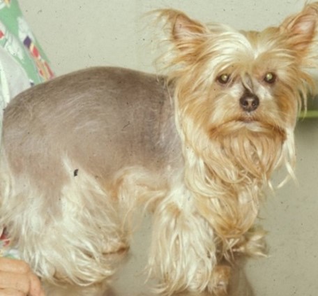
- Breeds that have been reported with color dilution alopecia include Doberman, dachshund, Great Dane, whippet, Italian greyhound, chow chow, Yorkshire terrier, miniature Doberman pincher, standard poodle, silky terrier, Chihuahua, Boston terrier, saluki, Newfoundland, German shepherd dog, schnauzer, Shetland sheepdog, schipperke, Bernese mountain dog, Labrador retriever, and mongrels with diluted coat colors.
- The reported age range at start of clinical signs is 3 months to 3 years and dogs with more diluted coat colors (e.g. gray) will develop alopecia earlier than dogs with less diluted coat colors (e.g. steel blue). Most light-colored dogs are completely alopecic by 2 to 3 years of age.
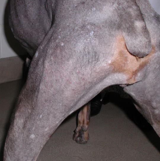
- The disease may start with recurrent bacterial folliculitis along the dorsum or hypotrichosis. Alopecia eventually and progressively develops and only affects the areas of dilute coat color. In some cases, the alopecia will precede the bacterial folliculitis.
- Pathogenesis and clinical signs:
-
-
- The extensive melanin clumping in the hair shaft and associated distortion of the cuticular-cortical structure of the hair are thought to be the initial cause of alopecia because it leads to fragility and breakage of hair shafts..
-
-
-
- Comedones (i.e. black head) and scales may be present at the alopecic sites.
-
Important Facts
- Color dilution alopecia is an inherited disease that affects some dogs with blue or fawn coat color.
- The fact that not all dogs with dilute coat color develop alopecia suggests a complex pathomechanism. The fact that not only the d gene is involved support this hypothesis.
- Dogs with lighter coat color (e.g. gray) develop alopecia earlier than dogs with darker coat color (e.g. steel blue).
- Various breeds can be affected but Doberman pinchers appear to be over-represented with about 93% and 73% of blue and fawn Dobermans, respectively, developing the disease between 3 months and 2-3 years of age.
-
- Diagnosis:
- Differential diagnosis includes follicular demodicosis, bacterial folliculitis, dermatophytosis, idiopathic seborrhea, and hypothyroidism in the older dog.
- Development of alopecia between 3 months to 3 years of age that affects exclusively the diluted coat suggests color dilution alopecia.
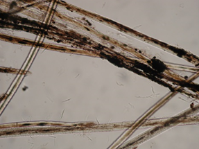
- Trichoscopy findings are characteristic and include large clumps of melanin (macromelanosomes) within the hair shaft cortex and medulla, which many result in distortion of the hair shaft. Breakage of the hair cortex or cuticle absence may also be seen. Microscopic examination of the hair shaft of diluted coats from non-affected dogs also show melanin clumping but they are smaller and do not alter the hair shaft structure.
- Histopathologic changes include the presence of large melanin clumps in basal keratinocytes of the epidermis, follicular wall, hair matrix cells, hair shafts, hair follicle canal, and dermis surrounding the follicles. With time, the follicles become dilated and cystic.
- Diagnosis:
Important Facts
- The development of alopecia limited to areas of dilute coat color between 3 months to 3 years of age highly suggests color dilution alopecia.
- Microscopic examination of hair shafts and histopathology will show large clumps of melanin that will distort their normal structures.
-
- Treatment:
- Treatment is palliative and should focus on treating and preventing secondary skin infections and avoiding sun exposure of the alopecic areas.
- Moisturizing shampoos and essential fatty acids may help reduce the excessive scaling that often accompanies this disease.
- Make sure to tell pet owners that the hair will not regrow in areas of diluted coat color.
- Treatment:
-
Black Hair Follicular Dysplasia
- Pathogenesis and clinical signs:
- Similar to color dilution alopecia, this condition is associated with abnormal melanization of back hairs of bicolor or tricolor coats.
- The abnormality is partially related to one or more mutations within or near the MLPH gene but, other changes are required for the alopecia to develop. The mode of transmission is believed to be autosomal recessive.
- Black hair follicular dysplasia has been recognized in mongrels and in various breeds, including border collie, bearded collie, basset hound, Papillion, saluki, beagle, Jack (Parson) Russell terrier, American cocker spaniel, schipperke, Cavalier King Charles spaniel, dachshund, Gordon setter, New Zealand huntaway dog, pointer, and large Munsterlander.
- The initial clinical sign occurs as early as 4 weeks of age and is characterized by loss of luster of black hairs. This hair coat change gradually evolves to complete to almost complete alopecia by 6 to 9 months of age.
- Pathogenesis and clinical signs:
-
- Diagnosis:
- The presence of alopecia limited to areas of black hair coat in a young dog is highly suggestive of this condition.
- In general, clumps of melanin cannot be seen when hair shafts are examined under the microscope because of the black pigmentation of the hair cortex. However, multiple bumps can be noted in some cases and they likely correspond to the large clumps of melanin (macromelanosomes).
- The histopathological findings are similar to the ones present in color dilution alopecia and characterized by clumps of melanin in the basal cell of the epidermis and follicular walls, in addition to the hair matrix cells. Melanophages are noted around the hair follicle bulb. Overall, the pigmentary abnormality is less pronounced compared to color dilution alopecia.
- Treatment:
- There is no treatment for black hair follicular dysplasia. Pet owners should be advised to not breed the dog as genetic plays a role in disease development.
- Diagnosis:
Important Facts
- Black hair follicular dysplasia is associated with hair loss limited to the black coat of bicolor or tricolor dogs.
- It starts at about 1 month of age and it has been reported in various breeds.
- The diagnosis is based on the characteristic history, typical clinical signs, and findings of trichoscopy and histopathology.
- The melanin clumps present in the skin and hairs are not as pronounced as they are in color dilution alopecia.
-
Non-color-linked Follicular Dysplasias
- Pathogenesis and clinical signs:
- The pathogenesis is unknown but, the high incidence of this condition in certain breeds suggests that genetics play a role. In the Irish water spaniel, a dominant mode of inheritance is suggested. Mutations in the R-spondin 2 gene are likely the cause of the hair loss in Portuguese water dogs.
- Some colleagues include post-clipping alopecia and flank alopecia under the sub-category of non-color-linked follicular dysplasia. However, these two disorders are discussed somewhere else under “miscellaneous alopecias”.
- The breeds reported to develop non-color-linked follicular dysplasias include Siberian huskies, Alaskan malamutes, Doberman pinchers, miniature pinchers, Manchester terriers, Irish water spaniels, Portuguese water dogs, curly-coated retrievers, Weimaraners, and anecdotally other breeds.
- Clinical signs and age of onset vary among breeds:
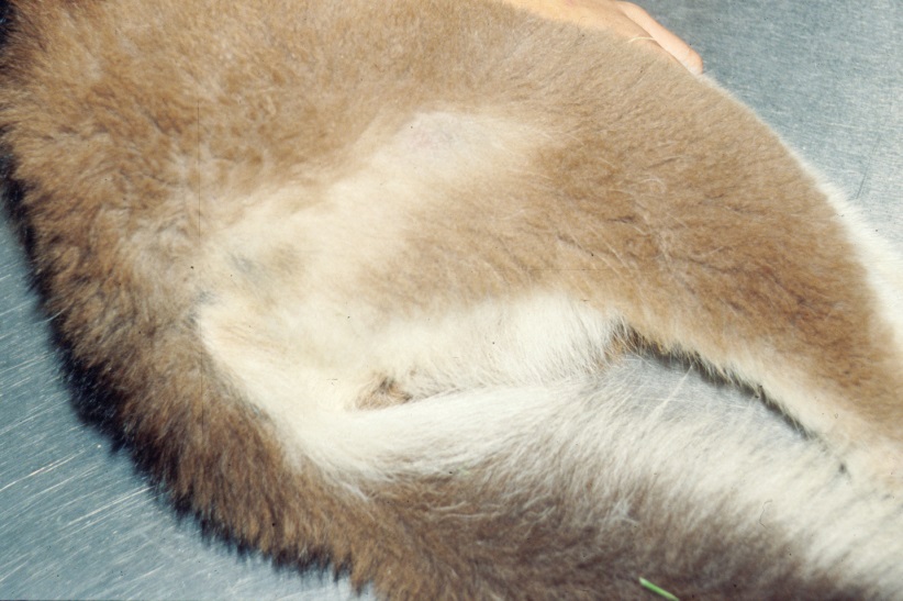
- Siberian huskies – Guard hairs along the trunk are progressively lost between 3 to 4 months of age and the coat acquires a reddish tinge. The hair coat changes may not start until 3 to 4 years of age in malamutes.
- Pathogenesis and clinical signs:
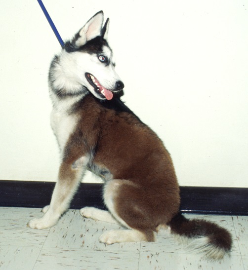
Courtesy: Rosario Cerundolo
-
-
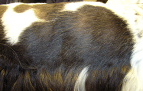
- Doberman pinchers, miniature pinchers, and Manchester terriers – Dogs start to loose hair in the flanks and the alopecia progresses slowly to involve the caudal aspect of the dorsum. The hair loss typically starts between 1 and 4 years of age.
- Irish water spaniels, Portuguese water dogs, and curly-coated retrievers – At 2 to 4 years of age the alopecia first starts on the caudal dorsum and progresses slowly to involve the tail and trunk. Hair regrowth may occur temporarily but eventually the hair falls off again and the alopecia becomes permanent.
-
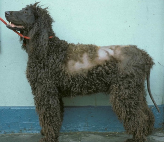
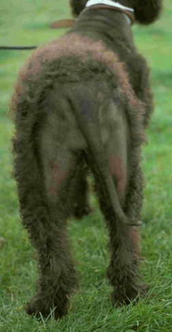
dysplasia. The non-inflammatory alopecia affecting the dorsal and lateral trunk, tail and caudal thighs. Courtesy: Rosario Cerundolo.
-
-
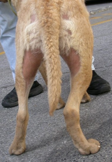
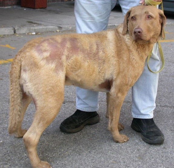
- Chesapeake Bay retriever – In a case series of 10 dogs, the reported age of onset was 5 months and 2 years. The alopecia typically involves the lateral, dorsal and ventral aspects of the trunk, axillae and caudal thighs. The hair loss does not cycle.
-
-
-
- Weimaraners – Diffuse truncal alopecia starting between 1 and 3 years of age was reported in five Weimaraners. The histopathological findings were similar to color dilution alopecia but less pronounced.
- Diagnosis:
- Considering the relatively tardive age of onset, the main differential diagnoses are sex hormone imbalance and hypothyroidism.
- The presence of non-inflammatory alopecia and/or coat quality alteration in predisposed breeds is suggestive of this condition if endocrine alopecia has been ruled out.
- Histopathological findings may vary to some extent among the breeds. The findings include infundibular hyperkeratosis, telogenization of hair follicles, various degrees of melanin clumping (typically less severe than color dilution alopecia), and variable degrees of dysplastic hair follicles. Some breeds may have specific abnormalities such as the vacuolization of the hair follicle matrix in the Portuguese water dog.
- Treatment: There is no specific treatment for follicular dysplasias. Owners have to be made aware of the genetic influence in the development of this condition and advised to not breed their affected pets
- The following non-specific measures should be instituted as indicated: (i) prevent sun exposure, (ii) treat appropriately secondary skin infections, and (iii) apply skin moisturizers to minimize excessive scaling as needed.
-
Important Facts
- The pathogenesis is unknown but genetics likely play a role.
- The breeds reported to develop non-color-linked follicular dysplasias include Siberian huskies, Alaskan malamutes, Doberman pinchers, miniature pinchers, Manchester terriers, Irish water spaniels, Portuguese water dogs, curly-coated retrievers, Weimaraners, and anecdotally other breeds.
- The age of onset varies according to the breed but most cases develop alopecia between 1 and 4 years of age.
- Clinical signs vary among the breeds.
- Histopathology shows follicular dysplasia with melanin clumping but it is less prominent than in color dilution alopecia.
References
Bond R, Varjonen K, Hendricks A, et al. Clinical and pathological features of hair coat abnormalities in curly coated retrievers from UK and Sweden. J Small Anim Pract 2016; 57: 659–667.
Brunner MAT, Rüfenacht S, Bauer A, et al. Bald thigh syndrome in sighthounds—revisiting the cause of a well-known disease. PLoS One 2019;14:e0212645.
Cerundolo R, Lloyd DH, McNeil PE, et al. An analysis of factors underlying hypotrichosis and alopecia in Irish Water Spaniels in the United Kingdom. Vet Dermatol 2000; 11: 107–122
Cerundolo R, Mauldin EA, Goldschmidt MH et l. Adult-onset hair loss in Chesapeake Bay retrievers: a clinical and histopathological study. Vet Dermatol 2005; 16: 39-46.
DeClementi C, Bailey K.L., Goldstein SC et al. Suspected toxicosis after topical administration of minoxidil in 2 cats. J Vet Emerg and Crit Care 2004; 14(4):287-292.
LaFort-Dassot C, Beco L, Carlotti NE. Follicular dysplasia in five Weimaraners. Vet Derm 2002; 13:253-260.
Miller WH, Griffin GE, Campbell KL. Muller & Kirk’s Small Animal Dermatology. 7th ed. St. Louis: Elsevier Inc., 2013; 537-540.
Miller WH, Griffin GE, Campbell KL. Muller & Kirk’s Small Animal Dermatology. 7th ed. St. Louis: Elsevier Inc., 2013; 554-572.
Miller WH, Griffin GE, Campbell KL. Muller & Kirk’s Small Animal Dermatology. 7th ed. St. Louis: Elsevier Inc., 2013; 593-599.
Miller WH Jr, Scott DW. Follicular dysplasia of the Portuguese water dog. Vet Derm 1995; 6:67-74.
Miller MA, Dunstan RW. Seasonal flank alopecia in boxers and Airedale terriers: 24 cases (1885-1992). J Am Vet Med Assoc 1993; 203:1567-1572.
Muntener T, Schuepbach-Regula G, Frank L, et al. Canine noninflammatory alopecia: a comprehensive evaluation of common and distinguishing histological characteristics. Vet Dermatol 2012; 23: 206-e44. DOI: 10.1111/j.1365-3164.2012.01049.x
Parker HG, Chase K, Cadieu E, et al. An insertion in the RSPO2 gene correlates with improper coat in the Portuguese water dog. J Hered 2010; 10:612-617.Parker WM and Scott DW. Growth hormone responsive alopecia in the mature dog: A discussion of 13 cases. J Am Anim Hosp Assoc 1980; 16(6): 824-828.
Welle MM. Canine noninflammatory alopecia: An approach to its classification and a diagnostic aid. Vet Pathol 2023; 60: 748-769.
Welle M, Philippe U, Rüfenacht S, et al. MLPH genotype—melanin phenotype correlation in dilute dogs. J Hered 2009;100: S75–S79.
