2.1 Autoimmune Skin Diseases – Dogs and Cats – Learning Objectives and Summary
Learning Objectives
- Remember! Pemphigus foliaceus and facial lupus erythematous are the most common autoimmune skin disease of dogs and cats.
- Know that autoimmunity is a specific humoral or cell-mediated immune response against the body’s own tissue.
- Autoimmunity occurs through the exposure of previously hidden antigens with a change in T cell activity, antigen mimicry, and genetic influences.
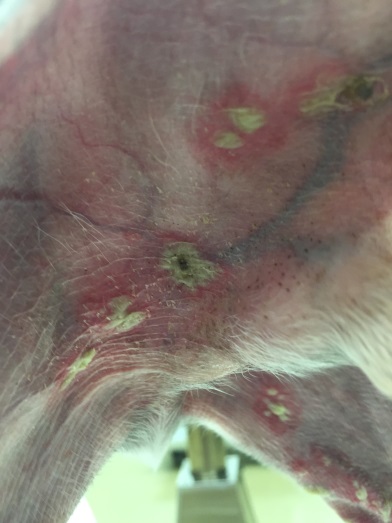
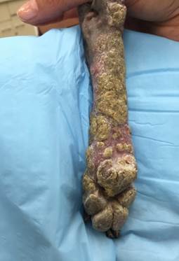
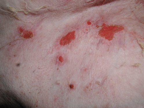
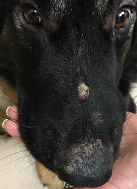
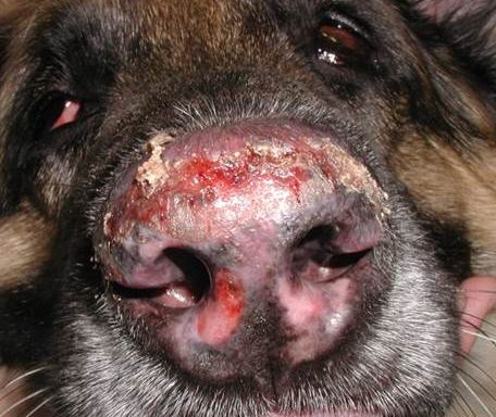
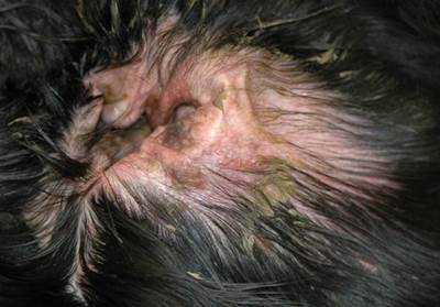
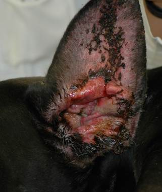
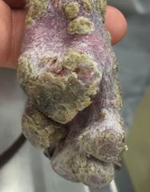
- Know that crusts, erosions, ulcers, pustules, vesicles, bullae, and/or papules are lesions often present in autoimmune skin disorders. Also, know that the face, mainly the nasal planum and dorsal muzzle, are often affected in pemphigus foliaceus/erythematosus and facial discoid lupus erythematosus. Footpads are commonly affected in pemphigus foliaceus and epidermolysis bullosa acquisita. Pinnae are affected in pemphigus foliaceus/erythematosus and epidermolysis bullosa acquisita. Mucocutaneous junctions (e.g. oral, ocular, and genital) are often affected in pemphigus vulgaris, epidermolysis bullosa acquisita, mucous membrane pemphigoid, mucocutaneous lupus erythematosus, and bullous pemphigoid. Ventral abdomen and inguinal areas are commonly affected in vesicular cutaneous lupus erythematosus.
- Know that history and clinical signs (type and distribution of lesions) are very helpful to obtain an accurate diagnosis. Histopathology is essential and it is diagnostic most of the time if the samples are collected properly. Cytological exam of direct smears from intact pustules, vesicles, bullae, or from recent erosions or crusted lesions can be diagnostic of pemphigus if the samples demonstrate nondegenerate neutrophils, +/- eosinophils and numerous acantholytic cells. Direct immunofluorescence and immunohistochemistry can be helpful if histopathology is not diagnostic. However, keep in mind that these tests are not routinely available and their sensitivity is generally low (i.e. many false negative results can occur). False positive results often occur when nasal planum and footpads are sampled, thus, avoid these sites if pursing these diagnostic tests. CBC, chemistry profiles, and urinalyses are important to be performed to check for systemic involvement (i.e. SLE), to monitor for potential side effects associated with immunosuppressive therapies and to have baseline data before starting systemic therapy with immunosuppressants.
- ANA is highly sensitive (positive in 90% of SLE cases) but has low specificity (many false positive results). Moreover, the sensitivity of this test is low for cases of cutaneous lupus erythematosus.
- Review the important details involved on taking biopsy samples for histopathology, immunofluorescence, and immunohistochemistry.
- Remember! Ideally, you should submit your samples to a dermatohistopathologist or someone highly interested in skin. Be sure to provide a good history, detailed description of clinical signs, body sites sampled, good quality photos, and diagnostic test results if available.
-
General Considerations
- Pemphigus foliaceus and facial discoid lupus erythematosus are the most common autoimmune skin diseases of dogs and cats.
- Mucous membrane pemphigoid, although rare, is the most common autoimmune sub-epidermal blistering skin disease in dogs.
- Autoimmune skin diseases usually occur in middle-aged to older dogs and cats, except for epidermolysis bullosa acquisita and exfoliative cutaneous lupus erythematosus, which generally affect young dogs.
- Autoimmunity is a condition characterized by a specific humoral or cell-mediated immune response against the body’s own tissue.
- How does the loss of tolerance to self-antigens occur (autoimmunity)?
- Exposure of previously hidden antigens.
- Change in T cell activity:
- Loss of suppression.
- Activation of auto-reactive response.
- Antigen mimicry – triggers immune response:
- Infectious agents – viral, bacterial, fungal.
- Environmental agents – insect vectors.
- Genetic influences:
- Familial links.
- MHC antigen associations.
Important Facts
- Pemphigus foliaceus and facial discoid lupus erythematous are the most common autoimmune diseases of dogs and cats.
- Mucous membrane pemphigoid is the most common autoimmune subepidermal blistering disease in dogs.
- Autoimmunity is a condition characterized by a specific humoral or cell-mediated immune response against the body’s own tissue.
-
Clinical Signs
- Autoimmune skin diseases can show great variation in cutaneous signs. However, certain lesion patterns are consistently observed:
- Papules, pustules, vesicles, and bullae.
- Autoimmune skin diseases can show great variation in cutaneous signs. However, certain lesion patterns are consistently observed:
-
-
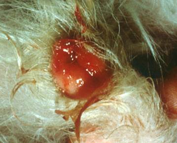
- Crusts, erosions, and ulcers.
-
-
-
- Face:
- Nasal planum and dorsal muzzle (e.g. pemphigus foliaceus and pemphigus erythematosus [dorsal muzzle], facial discoid lupus erythematosus [nasal planum]).
- Face:
-
-
-
-
- Pinnae (pemphigus foliaceus, pemphigus erythematosus, and epidermolysis bullosa acquisita).
-
-
-
-
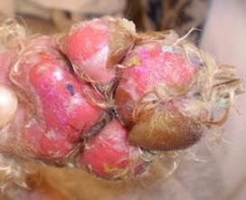
- Footpads are typically affected in pemphigus foliaceus and epidermolysis bullosa acquisita.
-
-
- Mucous membranes – oral, ocular, and/or genital mucosae are typically affected in pemphigus vulgaris, mucocutaneous lupus erythematosus, bullous pemphigoid, mucous membrane pemphigoid, and epidermolysis bullosa acquisita.

-
-
- Ventral abdomen and inguinal areas are typically affected in vesicular cutaneous lupus erythematosus.
-

-
-
- Other patterns:
- Total body.
- Atypical distribution patterns.
- Other patterns:
-
Important Facts
- Crusts, erosions, ulcers, papules, pustules, vesicles, and bullae are lesions often present in autoimmune skin diseases.
- The dorsal muzzle and nasal planum are frequently affected in pemphigus foliaceus and pemphigus erythematosus (dorsal muzzle) and facial discoid lupus erythematosus (nasal planum).
- Footpads are often affected in pemphigus foliaceus and may also be affected in bullous pemphigoid and epidermolysis bullosa acquisita.
- Mucocutaneous junctions (e.g. oral, ocular and genital) are typically affected in pemphigus vulgaris, bullous pemphigoid, mucous membrane pemphigoid and epidermolysis bullosa acquisita.
- Ventral abdomen and inguinal areas are often affected in vesicular cutaneous lupus erythematosus.
- Total body can be affected in some cases.
-
Diagnosis
- History and clinical signs (type of lesions and distribution) are important to obtaining an accurate diagnosis.
- Histopathology is helpful and can be diagnostic in many cases.
- Cytological exam of direct smears from intact pustules, vesicles, or from recent erosions and crusted lesions can be a helpful diagnostic test for the pemphigus complex. In the case of recent crusted lesions, remove the crust gently and sample the fresh exudate covering the surface underneath the crust or covering the underneath lesion.
- The presence of nondegenerate neutrophils, occasionally eosinophils, and acantholytic keratinocytes are strongly suggestive of pemphigus.
- Few acantholytic keratinocytes may be seen in an occasional high-power microscopic field in suppurative conditions such as pyodermas. However, when acantholytic keratinocytes are present in clusters or large numbers in several microscopic fields you should be suspicious of pemphigus. Be aware that dermatophyte infections caused by Trichophyton spp. can clinically and histologicaly resemble pemphigus foliaceus.
- Immunofluorescence or immunoperoxidase can be used to further classify the disease if histopathology is not definitive. However, the sensitivity and specificity of these tests are not good and they are generally not available in commercial laboratories.
- Direct immunofluorescence: the patient’s affected skin is tested for the presence of autoantibodies. Biopsies taken from the nasal planum of dogs and cats and footpads of dogs will often result in false positive reactions (immunoglobulins are often present in normal tissue at these sites). Therefore, results should be interpreted with caution if these areas are sampled.
- Indirect immunofluorescence: the patient’s serum is tested for autoantibodies.
- Complete blood cell count, serum chemistry profile, and urinalysis:
- Systemic lupus erythematosus (SLE): Multiple organ systems affected. Below are some examples.
- Renal disease (glomerular nephropathy).
- Blood cell abnormalities (hemolytic anemia, thrombocytopenia).
- Bone marrow abnormalities.
- Joint inflammation.
- Hepatitis, pleuritis.
- These tests are essential to determine overall health.
- They help identify problems associated with immunosuppressive therapies.
- Example: liver disease that could develop during treatment.
- These tests will serve as baseline values for monitoring side effects associated with treatment.
- Systemic lupus erythematosus (SLE): Multiple organ systems affected. Below are some examples.
- Anti-Nuclear antibody assay (ANA):
- The sensitivity of ANA is low in cutaneous forms of lupus erythematosus such as facial discoid lupus erythematosus, generalized discoid lupus erythematosus, vesicular cutaneous lupus erythematosus, mucocutaneous lupus erythematosus, and exfoliative cutaneous lupus erythematosus.
- It is usually positive in SLE and in some cases of pemphigus erythematosus.
- About 10% of SLE cases have negative ANA results.
- Low specificity (i.e. many false positive results).
Important Facts
- The patient’s history and clinical signs (type and distribution of lesions) are important diagnostic elements.
- Histopathology is essential and is diagnostic most of the time.
- Cytological exam of direct smears from intact pustules, vesicles, bullae, or from recent erosions or crusted lesions can be a supportive diagnostic test for pemphigus if it demonstrates nondegenerate neutrophils, +/- eosinophils, and numerous acantholytic keratinocytes. Remember, however, that acantholytic keratinocytes can also be seen in some cases of dermatophytosis and severe pyoderma.
- Direct immunofluorescence and immunoperoxidase can be helpful if histopathology was not diagnostic. However, many false negative results can occur. Moreover, false positive results often occur when nasal planum and footpads are sampled.
- CBC, chemistry profile, and urinalysis are important to check for systemic involvement (SLE) and to monitor for potential side effects associated with immunosuppressive therapies.
- ANA sensitivity for the various forms of cutaneous lupus erythematosus is low.
- ANA is highly sensitive if the dog has SLE (positive in 90% of cases) but has low specificity (many false positive results).
-
Biopsy Technique
- Histopathology:
- Biopsy primary lesions such as intact pustules, vesicles, and/or bullae when possible.
- If these lesions are not present, biopsy an early papule, the inflamed border of a fresh erosion/ulceration, and/or crusted lesions (biopsy through the crust and make sure to include the crust as part of your sample).
- The so called “blister watch” is not always possible. It entails examining the patient every 2 to 3 hours for the development of primary lesions (i.e. pustules, vesicles, and/or bullae) and biopsying the lesion immediately.
- Obtain several samples (three to five). It is always helpful to biopsy different stages of disease development (i.e. different lesions such as papules, pustules, vesicles, crusts, erosions, etc.).
- Do not perform surgical preparation or scrub the affected skin before sampling because this will destroy or damage the lesions.
- Do not pinch, squeeze, or grab the skin sample with forceps because this will damage the tissue and affect the histopathologic diagnostic features. You can use your fingers or a 22-25 gauge needle to grab the sample instead of using forceps.
- There is no need to include non-affected skin as part of the sample.
- Remove the excess blood and quickly place the samples in 10% formalin solution.
- If possible, use a dermatohistopathologist or a pathologist experienced experienced in analyzing skin diseases. Make sure to provide the pathologist with the following:
- Detailed patient’s history.
- Detailed description of clinical signs.
- Body sites sampled.
- Good quality photos of the patient’s lesions.
- Diagnostic test results if available.
- Note – Before sampling, contact the laboratory to determine the maximal number of samples that can be collected for the same price (e.g. our lab charges the same for five samples)
- Histopathology results:
- Read the histopathology description carefully and contact the pathologist if you have any questions.
- Avoid treating without a diagnosis because the animal will be treated for life with medications that potentially can cause severe side effects.
- Histopathology:
Important Facts
- When possible, biopsy primary lesions such as intact pustules, vesicles (blisters), and/or bullae.
- If these lesions are not present, biopsy an early papule, recent erosion/ulceration, and/or crusted lesion (make sure to biopsy through the crust and include it as part of the sample).
- Obtain several biopsy samples (three to five) at different stages of disease development, whenever possible. However, check with the lab the maximal number of samples that can be collected for the same price.
- Do not perform surgical preparation or scrub the affected skin because this could destroy or damage diagnostic lesions.
- Do not pinch, squeeze, or grab the skin sample with forceps because this will damage the tissue and affect the histopathologic diagnostic features.
- Use a dermatohistopathologist or a pathologist highly interested in skin, if possible.
- Make sure to provide the pathologist with a thorough history and detailed clinical signs, good quality photos of the lesions, diagnostic test results if available, and body sites sampled.
- Avoid treating without a diagnosis because the animal will be treated for life with medications that potentially can cause severe side effects.
-
- Immunofluorescence and immunohistochemistry:
- Biopsy intact vesicles (blisters) and pustules and the inflamed border of eroded and ulcerated lesions. Do not sample normal skin.
- No surgical preparation of the skin is allowed! You can damage the skin or destroy diagnostic lesions if you scrub the skin before sampling.
- Avoid sampling old and chronic lesions.
- Media:
- Immunofluorescence:
- Use fresh Michel’s fixative – Mail the sample at ambient temperature.
- Immunohistochemistry:
- Formalin fixed tissue with no other special preparations.
- Immunofluorescence:
- Immunofluorescence and immunohistochemistry:
Important Facts
- For immunofluorescence and immunohistochemistry, biopsy intact vesicles and pustules and the inflamed border of lesions. Do not sample normal skin.
- Do not surgically prepare the skin.
- Avoid sampling old and chronic lesions.
- For immunofluorescence, place your sample in Michel’s fixative and mail them at room temperature.
- For immunohistochemistry, place the samples in 10% formalin.
-
- Failure to confirm the diagnosis on histopathology, immunofluorescence, and immunohistochemistry can be due to one or more of the following:
- Absence of recent lesions.
- Improper selection and handling of biopsies.
- Loss of antigenicity of immunoglobulin deposits.
- Prior glucocorticoid therapy.
- Failure to confirm the diagnosis on histopathology, immunofluorescence, and immunohistochemistry can be due to one or more of the following:
Important Facts
- Common reasons for false negative results on histopathology and/or immunofluorescence or immunohistochemistry include absence of recent lesions; improper selection and handling of biopsies; loss of antigenicity of immunoglobulin deposits, and prior steroid therapy.
References
Bizikova P., Linder EL, and Anderson JG. Erosive and ulcerative stomatitis in dogs and cats: which immune-mediated diseases to consider? J Am Vet Med Assoc 2023; 261: S48-S57.
Bizikova P, Olicry T, Linder K et al. Spontaneous autoimmune subepidermal blistering diseases in animals: a comprehensive review. BMC Vet Res 2023; 19:55.
Medleau L, Hnilica KA. Chapter 8. Autoimmune and Immune-mediated Skin Disorders. In: Small Animal Dermatology: A color Atlas and Therapeutic Guide. 2nd ed. W.B. Saunders, Missouri, 2006; p. 189-227.
Miller, Griffin and Campbell. Chapter 9. Autoimmune and Immune-mediated Dermatoses.. In: Muller & Kirk’s Small Animal Dermatology. 7th ed., W.B. Saunders, Missouri, 2013; p 432-440.
Olivry T, Linder KE and Banovic F. Cutaneous lupus erythematosus in dogs: a comprehensive review. BMC Vet Res 2018; 14:132.
Olivry T. A review of autoimmune skin diseases of domestic animals I – superficial pemphigus. Vet Dermatol 2006; 17: 291-305.
Tham HL, Olivry T, Linder KE et al. Mucous membrane pemphigoid in dogs: a retrospective study of 16 new cases. Vet Dermatol 2016; 27: 376-e94.
Tham HL, Linder KE and Olivry T. Deep pemphigus (pemphigus vulgaris, pemphigus vegetans and paraneoplastic pemphigus) in dogs, cats and horses: a comprehensive review. BMC Vet Res 2020; 16: 457.
